The Determination of Enzyme Specific Activity
Summary
TLDRThis video script details an experiment on determining enzyme specific activity, focusing on glutathione S-transferase (GST). It covers the preparation of cytosolic fractions from mice, measuring GST activity using the CDNB method, creating a BSA standard curve with the Biuret method, and calculating protein content and enzyme specific activity. The script also discusses the importance of GST in detoxification and its relevance in cancer treatment, where inhibiting GST can enhance the effectiveness of chemotherapy.
Takeaways
- 🔬 The experiment aims to determine the specific activity of glutathione S-transferase (GST) enzyme and protein concentration using the Bradford method.
- 🧬 The activity of GST enzyme in protein mixtures is dependent on the amount of protein used, necessitating the knowledge of protein content in the mixture.
- 📚 Monomer and polymer concepts are introduced, with monomers being the building blocks like amino acids, and polymers being long chains like proteins.
- 🌟 Amino acids, the monomers of proteins, are differentiated by their R groups and can polymerize to form linear chains, such as peptides.
- 🔍 Three types of protein quantitative analysis are discussed: volumetric, gasometric, and spectrophotometry, with the experiment using visible light spectrophotometry.
- 📈 The Bradford method is chosen for protein quantification, which involves a color reaction with Coomassie Brilliant Blue G-250 dye, measured at 550 nm.
- 🛠️ The preparation of cytosolic fraction containing GST from mice involves several steps of centrifugation to isolate the enzyme.
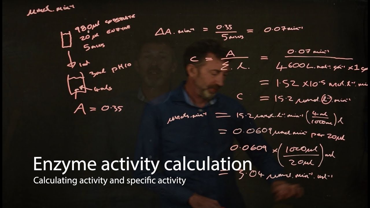
- 🧪 The enzyme activity of GST is determined using the CDNB method, which measures the formation of a conjugate product at 340 nm over time.
- 📊 A standard curve using bovine serum albumin (BSA) is created to measure the concentration of the GST enzyme in the sample.
- ⚖️ The specific activity of the enzyme is calculated by dividing the enzyme units by the protein concentration, indicating the enzyme's purity and efficiency.
- 💊 The importance of measuring enzyme activities is highlighted, particularly in pharmaceutical fields, where GST enzymes play a role in detoxification and cancer treatment.
Q & A
What is the main objective of the experiment described in the script?
-The main objective of the experiment is to determine the specific activity of the GST enzyme, measure the protein concentration using the Biuret method, and understand the fundamentals of enzyme activity in relation to protein content.
What is the difference between a monomer and a polymer as described in the script?
-A monomer is a single small molecule that can be joined with other identical molecules to form a polymer. In the context of proteins, amino acids are the monomers, and proteins are the polymers formed by the linkage of these amino acids.
What are the elements that typically make up amino acids, the monomers of proteins?
-Amino acids, which are the monomers of proteins, typically contain elements such as hydrogen (H), nitrogen (N), oxygen (O), and carbon (C).
What are the different types of protein quantitative analysis mentioned in the script?
-The script mentions three types of protein quantitative analysis: volumetric analysis, gasometric analysis, and spectrophotometry, with the latter including UV and visible light spectrophotometry.
Why is it necessary to measure the protein content in a sample before determining enzyme activity?
-Measuring the protein content is necessary because the activity of enzymes like GST in a protein mixture depends on the amount of protein present. This information is crucial for accurately determining the specific activity of the enzyme.
What is the Biuret method used for in the experiment?
-The Biuret method is used in the experiment to determine the protein concentration in the cytosolic fraction containing the GST enzyme by measuring the color complex formed between the protein and the Biuret reagent at a wavelength of 550 nanometers.
What are the advantages and disadvantages of using the Biuret method for protein quantification?
-The advantages of the Biuret method include its simplicity and speed, and the fact that the protein concentration measured is comparable to the polypeptide bond. Disadvantages include lower sensitivity and sometimes unstable color development.
What is the significance of measuring enzyme activities and protein concentrations in pharmaceutical fields?
-Measuring enzyme activities and protein concentrations is significant in pharmaceutical fields because it helps in understanding the detoxification processes, such as the role of GST enzymes in the excretion of xenobiotics and chemotherapeutic agents, which can be crucial in the treatment of diseases like cancer.
What is the purpose of creating a standard curve using the BSA method in the experiment?
-The purpose of creating a standard curve using the BSA (Bovine Serum Albumin) method is to measure the concentration of the GST enzyme in the cytosolic fraction by comparing the absorbance values obtained from the sample to the known concentrations of BSA.
How is the specific activity of an enzyme calculated in the experiment?
-The specific activity of an enzyme is calculated by dividing the total enzyme units, which is the amount of enzyme that catalyzes one micromole of substrate reaction per minute, by the protein concentration in milligrams per milliliter.
Outlines
🔬 Introduction to Enzyme Specific Activity Experiment
The video introduces an experiment aimed at determining the specific activity of the glutathione S-transferase (GST) enzyme. It explains the importance of knowing the protein content in a sample to accurately measure enzyme activity. The video uses monomers and polymers as an analogy to explain proteins and their building blocks, amino acids. It further discusses the quantification of proteins through various methods including volumetric, gasometric, and spectrophotometry, with a focus on the visible light spectrophotometry using the Biuret method. The Biuret reaction, which measures the protein content by the color complex formed with copper ions, is highlighted for its simplicity and speed, despite being less sensitive.
🧪 Understanding GST Enzyme and Its Role in Detoxification
This section delves into the role of the GST enzyme in phase II detoxification reactions, which are crucial for the excretion of xenobiotics from the body. It explains the involvement of three different enzymes in this process, with a focus on GST. The video describes the types of substrates that GST can act upon and the different mechanisms of GST conjugation. It also discusses the various types of GST enzymes and their substrates, including CDNB, which is used in the experiment. The preparation steps for the cytosolic fraction containing GST from mice are outlined, including the use of male rats, fasting, sacrifice, and centrifugation techniques to isolate the enzyme.
📊 Measuring Enzyme Activity and Protein Content
The video script outlines the steps for measuring the GST enzyme activity using the CDNB method and preparing a standard curve using the Biuret method. It details the procedure for preparing different fractions and the importance of using distilled water as a blank. The process of mixing reagents, measuring absorbance at specific time intervals, and calculating the enzyme activity is explained. The script also covers the creation of a BSA standard curve for determining protein concentration and the steps for measuring the protein content in the cytosolic fraction containing GST.
📈 Creating a Standard Curve and Calculating Enzyme Specific Activity
This part of the script focuses on the creation of a standard curve using the Biuret method to measure the concentration of the GST enzyme. It describes the preparation of a series of BSA concentrations and the procedure for reading absorbance to plot the standard curve. The video explains how to use the standard curve to determine the protein concentration in the sample. It also discusses the calculation of enzyme specific activity, emphasizing its importance in assessing enzyme purity and understanding the efficiency of the GST enzyme in detoxification processes.
💊 Significance of GST Enzyme in Pharmaceutical Research
The final paragraph highlights the significance of the GST enzyme in pharmaceutical research, particularly in the context of cancer treatment. It discusses how GST enzymes play a role in the excretion of chemotherapeutic agents and how inhibiting these enzymes can enhance the effectiveness of cancer treatments. The video concludes by emphasizing the importance of understanding and measuring enzyme activities, especially in relation to detoxification and the development of antitumor agents.
Mindmap
Keywords
💡Enzyme Specific Activity
💡Protein Concentration
💡Monomer
💡Polymer
💡Amino Acid
💡Peptide Bond
💡Spectrophotometry
💡Glutathione S-Transferase (GST)
💡1-Chloro-2,4-dinitrobenzene (CDNB)
💡Bovine Serum Albumin (BSA)
💡Xenobiotics
Highlights
Experiment aims to determine the specific activity of Glutathione S-Transferase (GST) enzyme and protein concentration using the Bradford method.
GST enzyme activity in protein mixtures depends on the amount of protein used, necessitating protein content measurement.
Proteins are built from amino acids, which are the monomers, and understanding their structure is crucial for the experiment.
Different methods for protein quantitative analysis are discussed, including volumetric, gasometric, and spectrophotometry.
The Bradford method, a type of spectrophotometry, is chosen for its simplicity and speed in measuring protein concentration.
The Bio-Rad protein assay is highlighted for its advantages in measuring protein-peptide chain complexes.
GST enzyme plays a critical role in phase 2 detoxification processes in the body.
The experiment involves five steps: preparation of cytosolic fraction, enzyme activity determination, standard curve preparation, protein content determination, and specific activity calculation.
Cytosolic fraction containing GST is prepared from mice liver using a series of centrifugation steps.
GST enzyme activity is measured using the CDNB method, which involves a reaction with GSH and monitoring absorbance.
A BSA standard curve is essential for determining the concentration of the GST enzyme in the sample.
The Bradford method is used to measure the protein concentration in the cytosolic fraction containing GST.
Calculating enzyme specific activity is crucial for understanding the enzyme's purity and efficiency.
The significance of measuring enzyme activities in pharmaceutical fields, especially in cancer treatment, is discussed.
GST enzyme's role in detoxifying chemotherapeutic agents and its potential as a target for cancer treatment is highlighted.
Transcripts
hello and welcome to the second
experiment called the determination of
enzymes specific activity
the objectives of these experiments are
determine the gst enzyme activity and
determine the protein concentration by
the right method and the last one is
determine determine the specific
activity of dsd enzyme
the fundamental of these experiments are
the activity of the gst enzyme in the
protein mixtures
depends on the amounts of protein used
in the reaction therefore it is
necessary to know that the protein
content in the edit folio
so as you can see here on the picture
there is a monomer and polymer here
so the monomer is a small molecule let's
say this monomer contains a two white
circle and
one black circle
while the polymer is
is a long chain molecule made up of
repeated patterns of monomers for
example here there are four
uh different uh four repeated patterns
of monomer containing white white and a
black circle
so a monomer is a single molecule that
can be joined together with other same
molecules to form a polymer
the building blocks of the proteins are
amino acid which contain elements such
as
hnoc
and more
they are the monomers of the proteins
when hundreds or thousands of amino
acids join together they create the
proteins which are then used for many
tasks in organisms such as doing work in
cells help with that with dna
replication and so on
so we can
say that monomer is an amino acid itself
and the polymer is a protein
so amino acid there are 20 kinds of
amino acid that differentiate by the r
chain here
there is an amine chain and the carboxyl
chain here and the r can be anything so
it can be a non-polar it can be
uncharted polar or a chart polar
so amino acid itself can polymerize to
form a linear chains for example like d
peptide here at the bottom one
so these two
amino acids are
joined together with the peptide bond
here
so it can be said that this is a d
peptide
so moving on so uh
how to quantify the protein so and
protein quantitative analysis there are
three types of it so there is a
volumetric and the gastometry and sp
spectrum for dometry and the volumetric
analysis containing there is a dou haul
which is uh the determination of the
total end and then formal titration
there so they are using the
uh alkali metric titration and a
gasoline while the gasometric analysis
they used uh converting the an
amine group to an n gas
while the spectrophotometry here so
there are two types of spectrophotometry
the uv and the visible light the uv is
used used for
uh quantified proteins with aromatic
groups such as phenylalanine thyrosine
and riptophan while the visible light
such as your buret falling and lorry
in
the
way
so in our
in our experiment here we are going to
use the visible light spectrophotometry
we are using the bio-red uh
analysis so the bare analysis is measure
[Music]
the protein complex between the peptide
chains uh that
making a complex with the
buret
ion such as
co2 plus here
and their protein complex uh between the
protein and by red reaction here co2
plus will be performed in a blue purple
color which is can be measured under the
wavelength 550 nanometer
so the biored reaction gives a
plus reaction to a compound containing
ch2 and h2 or chn hn h2 rcs and h2
the advantages of using the biored
method is that the concentration of a
protein is a comparable to the
polypeptide bone
and then the method is uh
simple and fast all the disadvantages of
the bird method here is they are less
sensitive
and then the color is sometimes an
unstable yeah
so why this method is less sensitive to
others because
many a sample that containing like this
uh
here chains ch2 and several this for
this kind of chain so they can be
measured by this method
so
it can be protein it can be others uh
sample so that's why it's less sensitive
so moving on why
a big questions
why we need to measure the protein
concentration and why we need to measure
the enzyme activities is that really
important
so for our experiment we are going to
[Music]
measure the gst enzyme so as you can see
here in the picture the phase two enzyme
reaction
there uh involves three different
enzymes such as
glucoronaceal transferase or ugt
in glucoronidation
while the second one is
enzyme sulfur transferases or st
involving in sulfation and the last one
is enzyme glutathione as transversal
involved in glutathione conjugation
so we're going to
explain about the
dst here or glutathione s transparency
this phase 2 enzyme reaction are
involved
in the excretion of some xenobiotics in
our body
so including the gst one
so as we can see here in the picture the
glutathione and enzyme biotic
helped by the enzyme of tst will
transform or make a glutathione s
conjugate to be ex
to be excluded
from our body
for the glutathione conjugation as i
already explained to you that there are
three enzymes there so the glutathione
conjugation
uh include and
several uh
xenobiotics such as electrophilics
antibiotics or the synovitics that
already uh transform bio biologically to
electrophilic trophies
while the substrate for the gst itself
share three common features
such as the
the substrate must be hydrophobic and
the electrophilic and react to
non-enzymatically with potassium
at a measurable rate
and so moving on to the next slide there
are
three types of uh
gst
conjugation or
there is three different processes
for making the godathion s conjugate or
conjugation the first one is a direct
conjugation by displacement of an
electron uh withdrawal
uh withdrawing group so
sorry it's covered by this screen here
so for example there is a four nitro
quinoline one oxide here on the left
so this
nitro oxide
group
will be
displaced by the gs minus one so that
this position will be displaced by the
gs while the direct conjugation by
adding of glutathione here means that
for example the
j ethyl malaya here
the the h plus ion or the double bond
here in the c
will be adding uh or will be
broken down uh
by the gs so the c uh double bond will
be
replaced by the c with the gs ion here
while the conjugation of a strain ring
system uh format metabolically for
example this is a chlorobenzene or
p450 will be
a
form
or uh
will be transformed into
a 3 4 oxide which is a structurally
this is
more
uh
unstable
uh by p 450 so that the gs minus or
glutathione minus here can be
displaced or can be
replace
the h
ion here by the
gs ion
as you can see here there are several
types of gst
detoxification or types there is a alpha
and fee in mu and tether and fee and
zeta and so on
and the substrate and the gst enzyme
firstly published in 1974
including the cdnb that you are going to
use in your experiment
so uh moving on uh the preparation upon
determination of enzyme specific
activity measurement so in this
experiment you are going to do uh five
different things or five uh continue
i mean related uh experiment the first
one is prepare the cyto cytosolic
fraction containing the gst
enzyme from mice this is already
prepared by
the
uh by our staff and the second one is
the determination of enzyme activity or
gst in a red liver by the cdnb method
and the third one is a preparation of
provine serum albumin standard curve
using the buyer method and the fourth
one is determination of the protein
content of cytosolic fraction containing
gst by the bird method and the last one
you're going to determine the enzyme
specific activity calculation so overall
there are five steps to
finally can
measure the specific activity of the dst
enzyme
so for the first preparation cytosolic
fraction of the gst from mice
if we can
read here
we have
we we need to fit the male rats which is
the strain sprocket only one with the
weight 200 and up to 250 with the pellet
and water until it full and let the red
fast for
24 hours prior to the next step then
after that you need to sacrifice the red
with the neck and leave with the neck
dislocation
and for the cytosolic fraction
preparation containing dst by a
centrifuge method so after you
did the dislocation so
the liver will be uh
chop up and then will be uh
will be uh separate between the enzyme
and the the other fraction by using the
centrifuge centrifugation method
and then after that the red liver take
the red lever and immediately
uh put and phosphate
buffer uh in the cold one and add ph
seven then measure the weight and cross
the red liver in a phosphate buffer
using a coat blender
for five minutes
and after that centrifuge the refined
homogeneity at 10 000 g for 30 minutes
at 44 degrees
and take an end transfer the supernatant
to a new tube and discharge the
precipitated layer
and centrifuge the supernatant at
105
g 1000g for 90 minutes at 4 degree
it's like a
several step of centrifugation to take
the tst enzyme
and the supernatant layer is cytosolic
fraction containing the gst enzyme as i
already explained before
after the centrifugation there's
supposed to be two layers there so here
we need to take and out the supernatant
and store the cytosolic fraction at
minus 80 until for the use for the cdnb
conjugation with gsh
and the first step that already prepared
by our laboratories so the second step
is we're going to determine the enzyme
activity or gst right liver by the cdnb
method cdnp is stand for one chloro 2.4
d nitrobenzene
so here as we can see on the table there
are
two different fraction or two different
type one is a
dst
cytosol fraction one is a an arc or it's
a distilled water
so
why we need to use the
distilled water because it's going to be
used as a blank here
so please prepare two types of of lust
there
[Music]
as you can see on the explanation of the
experiment
you are going to prepare two flags one
is cytosol fraction
and then the other one is distilled
water and put 920 for split buffer and
then
put 20 microliter of gsh and 20
microliter of cdnb
and then the last one is add the
cytocytosol fraction for example and
distill water for blank the gsdnb
conjugate when
when after you mix it up all of this
reagent you you will have a gsd and b
conjugate product form and then measure
at 340 nanometer from minute 0 to minute
3 using a spectrum photometric as a
simple kinetic program so it means after
you
mix all the reagent there so you need to
uh measure
the zero minute and
minute three so you have two different
uh
measurement
results for each sample so you will you
will have
four different measurement there
uh the one two for example and two for
plain
so please make a note that the
measurement time which is considered as
the 0 minute the same as the other
treatment it means that if you are mixed
all the reaction
so the zero minute it means the the last
time when you edit the cdnb for the
sample also similar for the blank one
so starting time is calculated yeah
after you add
at the city and b so the city and b1
should be added
the last after you put all the reaction
there
such as first cytosol fraction phosphate
buffer gsh and the last one you need to
put the cdnb and
make it as zero minute
so the end of product will be rate or
observation absorbent absorption per
minute so
after that the gst enzyme activity is
then measured in micro mole product per
minute so the observation you need to
measure there is reaction speed so it
means the absorbance the delta
absorbance so
before uh so as you can see here there
is a two uh measurement
the zero minute and the minute three so
the delta minute it means that the
minute three uh minus minute zero so you
can got the delta absorbance there per
minute
and then
the other calculation here is you need
to
calculate the gst activity and speed
reaction in b here divided by eight
point five hundred uh
[Music]
micro mole percent per micro mole
centimeter so you can got the micro mole
gstnb per minute is
stated as c
the result of this calculation is needed
for
further calculation the activity of the
enzyme so you need to
measure all of this step
and the third
one after you
measure the
speed of
speed of reaction so the third one is
making a standard curve why you need to
make a standard curve because you need
to
measure the concentration of the gst
enzyme so
here is how to measure
how to make the bsa standard curve or a
bsa stand for the bovine serum albumin
using the biored method
so the first materials is biored reagent
and the standard solution of bsa and the
phosphate buffer you can read by
yourself and then the procedure is very
simple so wait
the albumin and put it in a
volumetric flask and add little quartz
and make an albumin standard curve with
the series concentration ranging from
100 to 700 microgram per milliliter so
the procedure in c here is very
important because you need to make a
range of concentration that you're going
to use to make a standard curve
so here it is the standard curve table
here
so there is one up to six and the other
one as
a sample so it's going to be a seven
different uh flask or seven different
solution you need to prepare
the first one is a albumin solution
which is contained the bsa one microgram
per milliliter and the second plus you
is the vsa concentration of 1.5 and the
other one is 2 and 3 and 4 and for the
last one
there is no bsa there because it's going
to be your blank
and then the last one is your sample
of gst sample that you are going to
figure out how many uh how many
concentration is there
so
the standard curve
will be
making this uh
six
uh
six absorbance uh reading
and you need after you read the
absorbance there so you need to make a
linear graph equation
uh like this is the concentration this
is the absorbance there so you you're
going to find out
the the concentration of the sample you
are added
uh you are going to measure so for
example
uh
or after you mix all of the reaction to
make the standard curve and then you got
the equation like for example
y
uh
y equal to b x plus a
and for example if
the
absorbance is y so if the absorbance is
0.234
then the
0.234 equals bx plus a
and the x1 stands for your concentration
your sample concentration so it means
that
0.234 which is y minus a divided by b
so the four
step is the determination of protein
concentration of cytosol fraction
containing the gst
so here
also uh all you need to do is just mix
mix all of the reagent here cytosol
fraction phosphate buffer and
bioreaction
and incubate those solution in the room
temperature for 10 minutes and measure
the absorbance absorption of the mixed
solution at 550 nanometer
against a blank
which is containing no cytosol fraction
and then
after you read all of the this
absorbance here this this this one this
absorbance here
you're going to
measure the cytosol protein content
which is the total uh
the total volume per cytosol fraction
milligram per microliter
and this one is you can get this one by
uh measuring this one
first
and within within the form microliter
there were going to be
some milligram protein so it means after
you
you
measure the eggs
uh protein concentration which is shown
here you can put the concentration as a
cytosol protein contained by dividing
the
ten thousand uh the thousand
one
times the total volume uh divided
cytosol fraction volume
and then within the four microliter
there they are going to be
d the cytoso protein contain within a
four microliter they're going to be a
milligram protein
so please make a note the one unit of
enzyme is the total of enzyme that
catalyze one microliter of substrate
reaction per minute and an enzyme
specific activity is the total of enzyme
units per milli divided by the protein
concentration in milligram per milli or
the total enzyme unit over milligram
protein therefore the unit for specific
enzyme activity is unit per milligram
and then the purity of enzyme is
proportional to the value of its
specific activity meaning that the
greater value of the specific activity
the higher the purity of the enzyme
so the next question is why we need to
measure the enzyme activities regarding
to the pharmaceutical fields why
um ensuring the gst enzyme is so
important
for example here
there is a
one
scientific report
here
with the title advance in antitumor
agents start getting glutathione as
transversal
so the dst is as already explained to
you that yes it's a family office 2
detoxification enzyme
so
[Music]
the this enzyme
catalyzed the conjugation of glutathione
including the chemotherapetic agents
so
the
tst is
an enzyme that will be excluded
or catalyze
or will be ex will be will will help the
excretion of some semibiotics
so
the gst in a
[Music]
cancer patient that as you can as you
can as you know that the cancer is
such a cell which is uh
have a super
growth that they can uh divide the cell
can divide into a multiple or trill
multiple
uh produce multiple cell so
you can imagine that the tst enzyme is
often produced there so it means
the excretion of some xenobiotics will
be totally also
uh
multiple times ex
doing excretion
so it means that
whenever the cancer patients
they receive some chemotherapy
so it means
the cell will be
expressed or will be exclude
the chemotherapetics drugs also in huge
amounts
so the gst enzyme
can be
can be targeted as an
uh
xenobiotic target
so
whenever the gst is inhibited so it
means that the chemotheraphatic
drug can be used or can be treated the
cancer patient successfully because the
gst is inhibited and the gst cannot
express or exclude
any
chemotherapeutic drugs outside our body
so it means that the chemotheraphatic
drugs
can
can give any benefit a maximum benefit
to our body
so thank you for listening
浏览更多相关视频
Praktikum Biokimia Farmasi : Penentuan Aktivitas Spesifik Enzim
Session 5 - 08 What is a BAS
Liver and Catalase makeup lab video
54th GST Council Meeting highlights: What's getting cheaper and what’s getting pricier
Session 2 - 05 ABA files + Other Bank and Economic terms
ADVANCE TALLY & G S T | IMPORTANT QUESTION 💯 | 2023 BCOM SEM 2
5.0 / 5 (0 votes)