Type IV Hypersensitivity |T- Cell mediated Hypersensitivity |Mechanism | Examples
Summary
TLDRThis tutorial delves into type four hypersensitivity reactions, focusing on cell-mediated immunity. It explains how CD4+ T cells produce cytokines leading to chronic inflammation. The video outlines the pathogenesis of delayed-type hypersensitivity, highlighting T helper 1 and T helper 17 responses. It also covers clinical examples like tuberculin reactions and contact dermatitis, and discusses T cell-mediated cytotoxicity in diseases like type 1 diabetes and rheumatoid arthritis. The tutorial encourages active learning through practice tests on Visia, an engaging platform for medical knowledge.
Takeaways
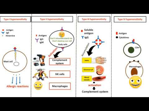
- 🧬 Type Four Hypersensitivity, also known as cell-mediated hypersensitivity, is primarily caused by inflammation resulting from cytotoxic T cells.
- 🔬 CD4 positive T cells produce cytokines that lead to chronic inflammation, often in response to environmental or self-antigens.
- 🌟 Delayed Type Hypersensitivity (DTH) is a prototype of T-cell mediated inflammation, detectable within 24 to 48 hours post-antigen exposure.
- 🔍 T helper 1 (Th1) and T helper 17 (Th17) cells are key contributors to DTH, with Th1 promoting macrophage activation and Th17 recruiting neutrophils.
- 🛡 The pathogenesis of DTH involves the activation of CD4 positive T cells, differentiation into effector T cells, and resulting inflammation and tissue injury.
- 🔄 Upon re-exposure to an antigen, memory T cells, which are long-lived, respond quickly to prevent further inflammation.
- 💉 Clinical examples of CD4 positive T cell-mediated inflammation include tuberculin reactions and contact dermatitis.
- 🛑 In persistent antigen scenarios, such as with Mycobacterium tuberculosis, T helper one cells can lead to granuloma formation, a hallmark of granulomatous inflammation.
- 🚨 CD8 positive cytotoxic T lymphocytes kill antigen-expressing target cells through apoptosis, a key mechanism in cell-mediated immunity.
- 🏥 Diseases mediated by T cell cytotoxicity include type 1 diabetes, graft rejection, and responses against viruses and tumor cells.
- 📚 The script concludes with a summary of type four hypersensitivity, its mechanisms, and examples, encouraging active learning through practice tests and feedback on the Visia platform.
Q & A
What is type four hypersensitivity?
-Type four hypersensitivity, also known as cell-mediated hypersensitivity, is primarily caused by inflammation resulting from cytotoxic T cells, which are produced by CD4 positive T cells.
What are the main cells involved in type four hypersensitivity reactions?
-The main cells involved in type four hypersensitivity reactions are CD4 positive T cells, which produce cytokines leading to inflammation.
What type of inflammation is typically associated with type four hypersensitivity?
-Type four hypersensitivity is most often associated with chronic inflammation.
Which T helper cells contribute to delayed type hypersensitivity?
-T helper 1 (Th1) and T helper 17 (Th17) cells contribute to delayed type hypersensitivity.
What is the role of interferon gamma in T helper 1 cells?
-Interferon gamma, produced by T helper 1 cells, promotes further helper cell development and activates other immune cells, such as macrophages.
How do T helper 17 cells respond to extracellular pathogens?
-T helper 17 cells produce interleukin-17 and other cytokines and chemokines, which recruit more neutrophils and monocytes to promote inflammation against extracellular pathogens.
What happens to effector T cells after the inflammation is cleared?
-After the inflammation is cleared, effector T cells transform into memory T cells, which are long-lived and respond quickly to the same antigen if encountered again.
What are the clinical examples of CD4 positive T cell-mediated inflammatory reactions?
-Clinical examples of CD4 positive T cell-mediated inflammatory reactions include tuberculin reaction and contact dermatitis.
What is the difference between T helper 1 and T helper 17 in terms of the pathogens they target?
-T helper 1 cells target intracellular pathogens, such as some bacteria and viruses, while T helper 17 cells handle extracellular bacteria and fungi.
How do activated CD8 positive cytotoxic T lymphocytes kill target cells?
-Activated CD8 positive cytotoxic T lymphocytes kill target cells by inducing apoptosis, either through the Fas ligand and Fas receptor interaction or by forming a perforin and granzyme complex.
What are some diseases associated with T cell-mediated cytotoxicity?
-Diseases associated with T cell-mediated cytotoxicity include type 1 diabetes, graft rejection, reactions against various viruses, and destruction of some tumor cells.
Outlines

This section is available to paid users only. Please upgrade to access this part.
Upgrade NowMindmap

This section is available to paid users only. Please upgrade to access this part.
Upgrade NowKeywords

This section is available to paid users only. Please upgrade to access this part.
Upgrade NowHighlights

This section is available to paid users only. Please upgrade to access this part.
Upgrade NowTranscripts

This section is available to paid users only. Please upgrade to access this part.
Upgrade NowBrowse More Related Video

Hipersensitivitas Imun: Respon Imun Tubuh Yang Berlebihan | Kata Dokter
Immunology of Hypersensitivity
Hypersensitivity types in 4 minutes

Hipersensibilidades (Parte II - Hipersensibilidades dos tipos II, III e IV)

Hypersensitivity, Overview of the 4 Types, Animation.

Hipersensitivitas Tipe 3 (Immune complex-mediated Hypersensitivity), Immunology
5.0 / 5 (0 votes)