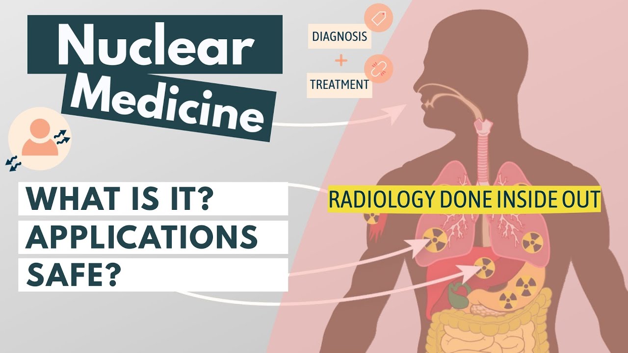
Bone Scan , A Technical Perspective #bnms #bonescan #nuclearmedicine #patientcare
Summary
TLDRThis beginner's guide to bone scans explains the most common nuclear medicine procedure, used to detect bone abnormalities such as fractures, infections, arthritis, and metastases. The video details the role of the radioactive tracer Tc-99m HDP, how it binds to bone during remodeling, and the steps of both standard and three-phase bone scans. Viewers learn about patient preparation, injection, waiting periods, imaging techniques including whole-body and SPECT/CT scans, and the importance of clinical history. Practical tips for positioning, image acquisition, and safety measures are highlighted, providing a clear and engaging overview for aspiring nuclear medicine professionals.
Takeaways
- 😀 The most common procedure in nuclear medicine is a bone scan, used to detect conditions or pathologies in bones.
- 😀 A radioactive tracer, typically Tc-99m HDP (technetium 99m dihydroxyphosphate), is injected to highlight bone activity during remodeling or repair.
- 😀 The tracer attaches to hydroxyapatite crystals in bones and is absorbed during bone remodeling, which allows detection of abnormalities.
- 😀 Bone scans can show increased or decreased tracer uptake, which may indicate fractures, arthritis, infections, or bone metastasis.
- 😀 Bone scans are classified into two types: standard bone scans and three-phase bone scans, each with specific protocols.
- 😀 A standard bone scan involves injecting the tracer and performing a whole-body scan after a few hours, with optional static or SPECT images.
- 😀 A three-phase bone scan is used for conditions like arthritis or infections, with flow, blood pool, and delayed phases to track tracer distribution over time.
- 😀 Patient identity verification, clinical history review, and explanation of the procedure are critical steps before scanning.
- 😀 During scanning, patient positioning, removal of metal objects, and body contouring on the gamma camera are important for accurate imaging.
- 😀 SPECT or SPECT/CT can provide detailed 3D images for specific areas, enhancing diagnostic accuracy for localized conditions.
- 😀 Drinking water and frequent urination between injection and scan improves image quality by reducing background tracer in the bladder.
- 😀 The final images are processed and reviewed by consultants to help with diagnosis, prognosis, and clinical decision-making.
Q & A
What is the most common procedure performed in nuclear medicine?
-The most common procedure performed in nuclear medicine is a bone scan.
What is the primary purpose of a bone scan?
-A bone scan detects conditions or pathologies in the bones, such as fractures, arthritis, bone infections, or metastases from cancer.
Which radiopharmaceutical is most commonly used for a bone scan, and what does it do?
-The most commonly used radiopharmaceutical is Tc-99m HDP (Technetium-99m Dihydroxyphosphate). It attaches to hydroxyapatite crystals in the bones and highlights areas undergoing repair or remodeling, allowing abnormal uptake to be detected with a gamma camera.
What are the two main classifications of bone scans?
-Bone scans can be classified as standard bone scans and three-phase bone scans.
What differentiates a three-phase bone scan from a standard bone scan?
-A three-phase bone scan includes the flow phase (dynamic imaging during injection), blood pool phase (static imaging after 10 minutes), and delayed phase (whole-body imaging after a few hours). Standard bone scans only involve injection and delayed imaging.
Why is patient history important before performing a bone scan?
-Patient history helps match clinical details to scan findings, ensuring accurate interpretation. It is especially important because bone scans are sensitive but not highly specific.
How is the radiopharmaceutical administered, and what is the typical dose?
-The radiopharmaceutical is administered intravenously through a cannula or butterfly needle. The typical dose for Tc-99m HDP is 600 megabecquerels (MBq).
What instructions are given to patients after the tracer injection before scanning?
-Patients are advised to drink plenty of water and empty their bladder frequently to improve image quality. They typically wait about 3 hours before returning for the delayed scan.
What is the purpose of additional static images or SPECT/CT during a bone scan?
-Static images focus on specific body parts for better detail. SPECT/CT combines 3D imaging with CT for precise localization and enhanced visualization of abnormalities.
For what clinical situations is a three-phase bone scan typically used?
-Three-phase bone scans are used for conditions like arthritis, bone infections, unexplained joint pain, or suspected infected prostheses, where detailed blood flow and tissue activity information is needed.
How are patients positioned during a bone scan to ensure accurate imaging?
-Patients lie on the scanning couch, with the head and feet properly aligned, often in a pigeon-toe position with straps for stability. Metal objects like belts, watches, and keys are removed to prevent interference.
Why is it necessary to perform identity checks and clinical justification before a bone scan?
-Identity checks prevent scanning the wrong patient, and clinical justification ensures the scan is medically appropriate, authorized, and matches the patient’s condition for accurate interpretation.
Outlines

This section is available to paid users only. Please upgrade to access this part.
Upgrade NowMindmap

This section is available to paid users only. Please upgrade to access this part.
Upgrade NowKeywords

This section is available to paid users only. Please upgrade to access this part.
Upgrade NowHighlights

This section is available to paid users only. Please upgrade to access this part.
Upgrade NowTranscripts

This section is available to paid users only. Please upgrade to access this part.
Upgrade NowBrowse More Related Video
Nuclear medicine explained in 2 minutes
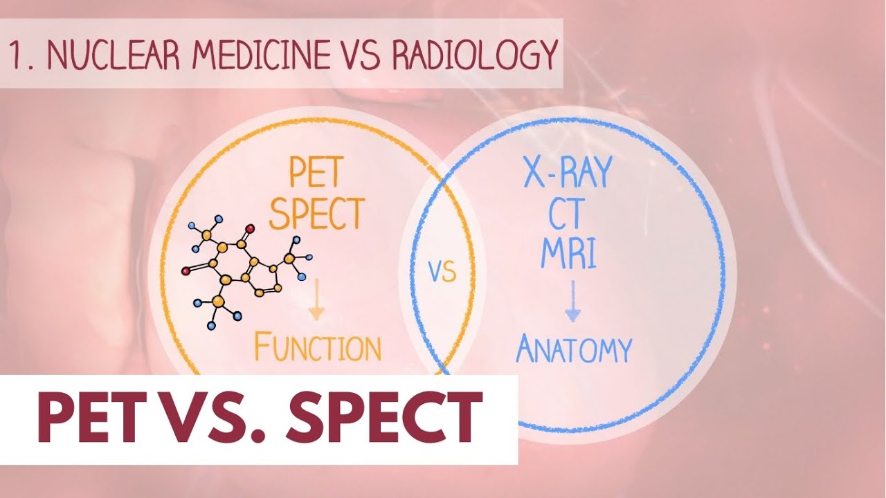
PET vs. SPECT scan | Dr. Paulien Moyaert

PET vs SPECT | The basics (Updated video)

Gangguan dan Kelainan Sistem Gerak Manusia || Upaya Mencegah Serta Mengatasi gangguan sistem gerak

Nuclear Chemistry Medical Applications

Disorders and Disease of the Skeletal System
5.0 / 5 (0 votes)