Muscles of Mastication - Anatomy Tutorial
Summary
TLDRThis tutorial explains the four muscles of mastication—temporalis, masseter, medial pterygoid, and lateral pterygoid—and clarifies they are innervated by the mandibular branch (V3) of the trigeminal nerve (CN V), unlike facial expression muscles supplied by CN VII. It describes each muscle’s location, origin and insertion (e.g., temporalis on the coronoid process; masseter on the lateral ramus; pterygoids on the pterygoid plates and condyle), their actions—elevation, depression, protrusion, retraction and lateral movements—and the lateral pterygoid’s two heads. The video also demonstrates clinical testing by palpating muscles during jaw clenching.
Takeaways
- 😀 The muscles of mastication are innervated by the mandibular branch (V3) of the trigeminal nerve.
- 😀 The trigeminal nerve (cranial nerve V) controls both the muscles of mastication and facial sensation.
- 😀 The muscles of facial expression are controlled by the facial nerve (cranial nerve VII).
- 😀 There are four main muscles involved in mastication: temporalis, masseter, medial pterygoid, and lateral pterygoid.
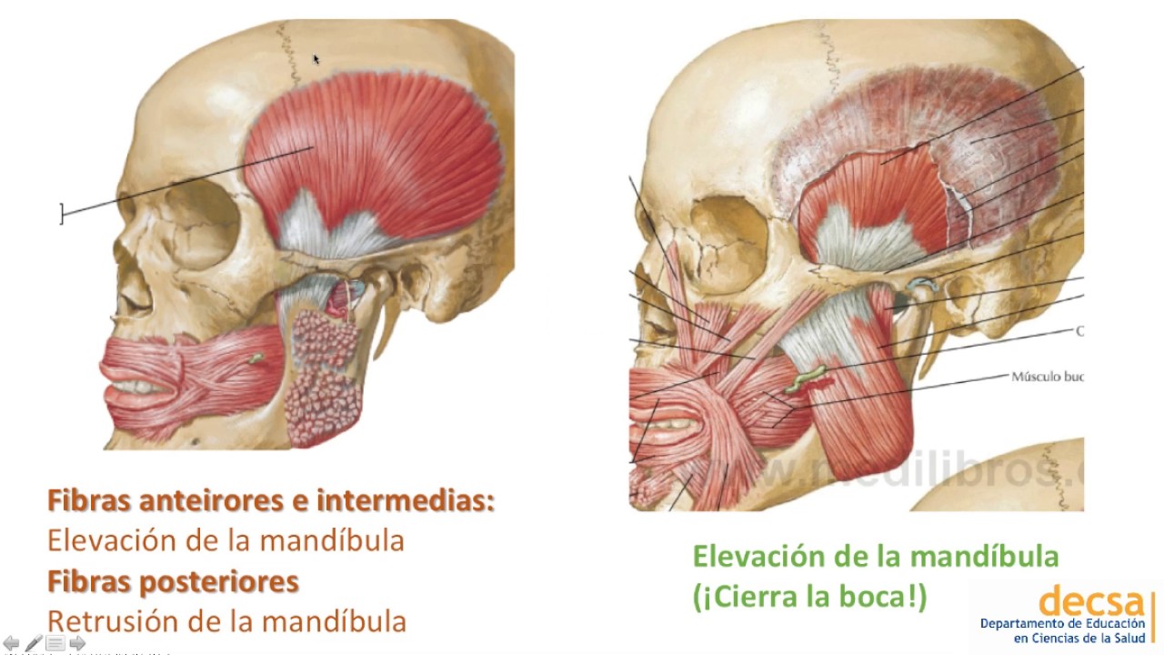
- 😀 The temporalis muscle originates in the temporal fossa of the skull and inserts onto the coronoid process of the mandible.
- 😀 The temporalis muscle helps elevate and retract the mandible, and its fibers can also cause retraction.
- 😀 The masseter muscle has both a superficial and deep part, originating from the zygomatic arch and inserting onto the lateral surface of the ramus of the mandible.
- 😀 The masseter muscle is primarily responsible for elevating the mandible but does not contribute to retraction.
- 😀 The pterygoid muscles (medial and lateral) originate from the pterygoid process of the sphenoid bone.
- 😀 The lateral pterygoid muscle is involved in protrusion and side-to-side movements of the mandible, while the medial pterygoid muscle contributes to elevation and side-to-side movements.
- 😀 The lateral pterygoid muscle has two parts: superior and inferior, with the superior part inserting into the condylar process and the inferior part into the neck of the mandible.
Q & A
What nerve innervates the muscles of mastication?
-The muscles of mastication are innervated by the mandibular branch of the trigeminal nerve, also known as V3.
What are the main movements involved in mastication?
-The four main movements of the mandible during mastication are: retraction (movement posteriorly), protrusion (movement anteriorly), elevation (moving upwards), and depression (moving downwards).
Which cranial nerve is responsible for facial expression muscles?
-The facial nerve, also known as cranial nerve VII, is responsible for the innervation of the muscles of facial expression.
Where does the temporalis muscle insert, and what is its function?
-The temporalis muscle inserts on the coronoid process of the mandible. It functions to elevate and retract the mandible.
How can you palpate the temporalis muscle?
-To palpate the temporalis muscle, place your fingers over the temporal region of the face and clench your teeth. You will feel the muscle contracting.
What is the origin and insertion of the masseter muscle?
-The masseter muscle originates from the zygomatic arch and inserts on the lateral surface of the ramus of the mandible.
What is the role of the masseter muscle in mastication?
-The masseter muscle's primary role in mastication is to elevate the mandible, facilitating the chewing motion.
Where do the lateral and medial pterygoid muscles insert?
-The lateral pterygoid muscle inserts onto the condylar process of the mandible, while the medial pterygoid muscle inserts on the medial surface of the mandible's angle.
What is the distinguishing feature of the lateral pterygoid muscle?
-The lateral pterygoid muscle is distinguished by having two parts: the superior lateral pterygoid and the inferior lateral pterygoid. These parts have different points of origin and insertion on the mandible.
How do the lateral pterygoid and medial pterygoid muscles differ in their actions?
-The lateral pterygoid muscle is involved in protrusion and side-to-side movements of the mandible, while the medial pterygoid muscle is involved in elevation and side-to-side movements.
Outlines

This section is available to paid users only. Please upgrade to access this part.
Upgrade NowMindmap

This section is available to paid users only. Please upgrade to access this part.
Upgrade NowKeywords

This section is available to paid users only. Please upgrade to access this part.
Upgrade NowHighlights

This section is available to paid users only. Please upgrade to access this part.
Upgrade NowTranscripts

This section is available to paid users only. Please upgrade to access this part.
Upgrade NowBrowse More Related Video

The Mandible: Anatomy and Muscles (3D Animation)

Muscles of Mastication (Origin, Insertion, Function) | Anatomy

MÚSCULOS DA ATM #medicina #anatomia #anatomy #medico #fisioterapia #anato #odontologia #odonto
Intra-Oral NeuroMuscular Therapy for TMJ demonstrated by Stew Wild

Muscles of Facial Expression
ATM y Músculos de la Masticación - Anatomía
5.0 / 5 (0 votes)