MÚSCULOS DA ATM #medicina #anatomia #anatomy #medico #fisioterapia #anato #odontologia #odonto
Summary
TLDRThis video offers an in-depth exploration of the muscles involved in the temporomandibular joint (TMJ). It covers the TMJ's unique characteristics, including its structure, the two distinct joints, and the functional role of the articular disc. The four primary muscles—temporalis, masseter, medial pterygoid, and lateral pterygoid—are examined in detail, with a focus on their origin, insertion, and functions like occlusion, retraction, protrusion, and lateral movement. The video emphasizes the coordinated movement of the TMJ and the neural innervation that governs these processes, offering valuable insights for understanding this critical joint's anatomy and functionality.
Takeaways
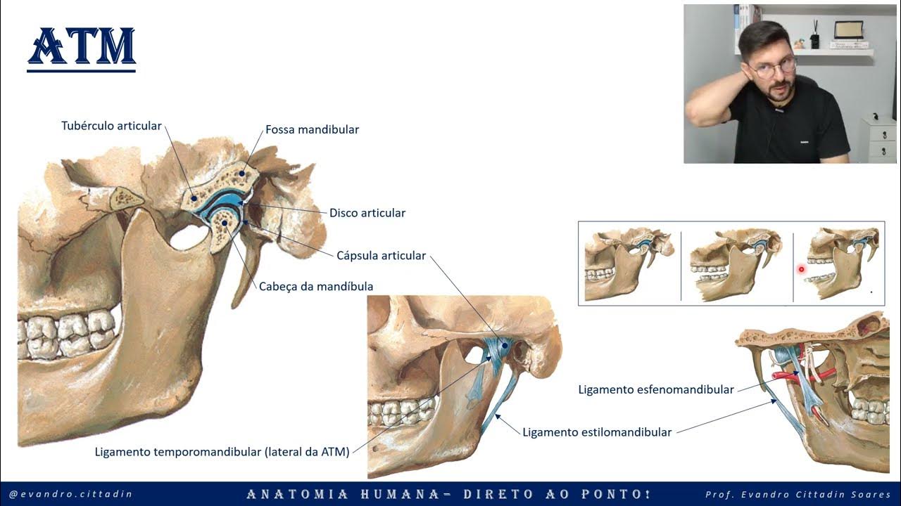
- 😀 The temporomandibular joint (TMJ) is a unique synovial joint consisting of the condyle of the mandible, the articular tubercle of the temporal bone, and the mandibular fossa.
- 😀 The TMJ operates as two distinct joints (right and left), each separated by a disc, creating an upper and lower joint for movement.
- 😀 The movements of the TMJ, including protrusion, retraction, elevation, and depression, are controlled by the muscles of mastication.
- 😀 The TMJ is unique because both joints work simultaneously, meaning any movement in one joint happens in the other as well.
- 😀 The TMJ plays a crucial role in dental health, as it is subject to similar pathologies as other synovial joints in the body.
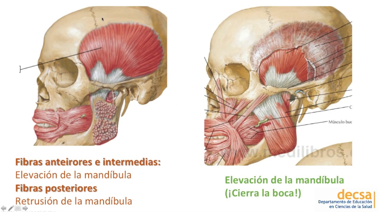
- 😀 The four main muscles involved in TMJ movement are the temporalis, masseter, medial pterygoid, and lateral pterygoid.
- 😀 The temporalis muscle originates from the temporal bone and inserts into the coronoid process of the mandible, responsible for occlusion, retraction, and lateralization of the mandible.
- 😀 The masseter muscle, originating from the zygomatic arch, is one of the strongest muscles in the body, responsible for mandibular occlusion and protrusion.
- 😀 The medial pterygoid muscle, originating from the sphenoid bone, assists with mandibular occlusion and protrusion, while also playing a key role in stabilizing the mandible.
- 😀 The lateral pterygoid muscle, consisting of two heads, plays a key role in opening the jaw (depression) and moving the condyle of the mandible anteriorly during TMJ opening.
- 😀 The TMJ muscles are innervated by branches of the trigeminal nerve (cranial nerve V), including the temporal, masseteric, medial pterygoid, and lateral pterygoid nerves.
- 😀 A mnemonic, such as associating the medial pterygoid with 'M' and the lateral pterygoid with 'L', can help students differentiate between these two muscles.
Q & A
What are the three basic structures that make up the TMJ?
-The three basic structures of the TMJ are the condyle of the mandible, the articular tubercle of the temporal bone, and the mandibular fossa.
What role does the disc play in the TMJ?
-The disc in the TMJ separates the mandibular fossa from the jaw structure, creating two joints: an upper joint and a lower joint, which allows the TMJ to perform movements like protrusion, retraction, elevation, and depression.
How are the two TMJ joints on the left and right sides coordinated?
-The movements of the TMJ on both sides are coordinated, meaning when one joint moves, the other must also move simultaneously, reflecting the unique functionality of the TMJ.
What are the four main muscles involved in TMJ movement?
-The four main muscles involved in TMJ movement are the temporalis, masseter, medial pterygoid, and lateral pterygoid muscles.
What are the primary functions of the temporalis muscle?
-The temporalis muscle primarily performs occlusion (closing the jaw), retraction (pulling the mandible backward), and lateralization (moving the mandible side to side).
Where does the masseter muscle originate and insert, and what are its key functions?
-The masseter muscle originates from the zygomatic arch and inserts into the angle and ramus of the mandible. Its key functions include mandibular occlusion (closing the jaw) and protrusion (moving the jaw forward).
How can one differentiate between the medial and lateral pterygoid muscles?
-The medial pterygoid makes one of the legs of the letter 'M' and inserts on the internal aspect of the mandible, while the lateral pterygoid forms the letter 'L' and inserts on the condyle of the mandible and the articular disc.
What are the primary functions of the medial pterygoid muscle?
-The medial pterygoid muscle is responsible for occlusion (closing the jaw) and protrusion (moving the jaw forward).
What is the key function of the lateral pterygoid muscle?
-The lateral pterygoid muscle plays a crucial role in opening the jaw by depressing and anteriorly moving the mandible. It also helps with protrusion of the mandible.
Which nerve innervates the muscles involved in the TMJ, and what is its origin?
-The muscles involved in the TMJ are innervated by the mandibular nerve, which is a branch of the trigeminal nerve (cranial nerve V).
Outlines

This section is available to paid users only. Please upgrade to access this part.
Upgrade NowMindmap

This section is available to paid users only. Please upgrade to access this part.
Upgrade NowKeywords

This section is available to paid users only. Please upgrade to access this part.
Upgrade NowHighlights

This section is available to paid users only. Please upgrade to access this part.
Upgrade NowTranscripts

This section is available to paid users only. Please upgrade to access this part.
Upgrade NowBrowse More Related Video

Dentacademy - Anatomi Umum - Sistem Mastikasi - Part 1
ATM y Músculos de la Masticación - Anatomía

Temporomandibular Joint 😲 | Biomechanics Part 2/2
ATM - Articulação temporomandibular - Sistema articular - Anatomia direto ao ponto!

Anatomia da Articulação Temporomandibular (ATM) PARTE 1
Анатомия височно-нижнечелюстного сустава - meduniver.com
5.0 / 5 (0 votes)