Respiratory System of the Human Body - How the Lungs Work! (Animation)
Summary
TLDRThis animation takes viewers on a detailed journey through the human respiratory system, showcasing how oxygen and other gases are inhaled while protective mechanisms in the upper airways filter out particles and insects. Highlighting the role of the diaphragm and other respiratory muscles in lung inflation and deflation, it explains the gas exchange process in alveoli where oxygen is absorbed into the bloodstream and carbon dioxide is released. The script also describes the pleural cavity's function in facilitating lung movement.
Takeaways
- 🌬️ Oxygen and other gases, as well as water and small particles, are part of the air we breathe, with the respiratory system playing a crucial role in filtering and utilizing these elements.
- 👃 The upper respiratory system, including the nasal cavity, provides protective mechanisms to filter out large dirt particles and insects, using nasal hair and cilia to trap small particles.
- 🕊️ Nucociliary clearance is the process by which cilia move mucus, trapping particles, towards the pharynx for swallowing, thus preventing unwanted particles from entering the lower respiratory tract.
- 🔥 The upper airways warm and moisten the air we breathe, which is essential for the proper functioning of the respiratory system.
- 🗣️ The epiglottis plays a vital role during swallowing, closing the larynx to prevent food or liquid from entering the lower respiratory tract.
- 🌿 The trachea and bronchi form the main branches of the bronchial tree, leading to the lungs and facilitating the transport of air.
- 💓 The heart pumps deoxygenated blood to the lungs via the pulmonary arteries, and oxygenated blood returns to the heart through the pulmonary veins to be distributed to the body's organs.
- 🌿 Each lung is divided into lobes and segments, with the right lung having three lobes and the left lung having two, each with its own air and blood supply.
- 💨 Alveoli, tiny air sacs at the ends of the bronchial tree, are the primary sites for gas exchange, with an estimated 300 to 400 million present in humans.
- 🔄 Type 1 pneumocytes, which make up the walls of the alveoli, are extremely thin, allowing for efficient gas exchange between oxygen and carbon dioxide.
- 🩸 Erythrocytes, or red blood cells, are responsible for binding and transporting oxygen in the bloodstream, with iron ions in hemoglobin binding to oxygen molecules.
- 💪 Breathing involves various muscles, including the diaphragm, which is the primary muscle for respiration, and chest muscles that assist in inflating and deflating the lungs.
Q & A
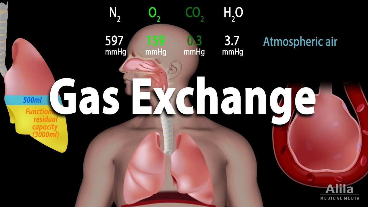
What are the main components of the air we breathe that are important for the human respiratory system?
-The main components of the air we breathe that are important for the human respiratory system include oxygen, which is vital for us, and water vapor. Additionally, other gases like krypton and even radioactive radon are present, though not essential for our respiratory function.
How does the upper respiratory system protect against unwanted particles entering the lower respiratory tract?
-The upper respiratory system provides protective mechanisms such as nasal hair, which stops large dirt particles or insects, and the nasal mucosa, which traps smaller particles. Ciliated cells on the nasal mucosa move in a wave-like manner to push mucus towards the pharynx, a process known as mucociliary clearance.
What is the role of the epiglottis during swallowing, and how does it interact with the respiratory system?
-The epiglottis, connected to the tongue, plays a crucial role during swallowing by closing the larynx. This ensures that no food or liquid can enter the lower respiratory tract, allowing the bolus to slide into the esophagus instead.
How does the structure of the trachea support its function in the respiratory system?
-The trachea is reinforced by incomplete rings of hyaline cartilage, which provides structural support and helps maintain an open passage for air to flow into the lungs.
What is the difference in the number of lobes between the left and right lungs, and what is the significance of this difference?
-The left lung has two lobes, an upper and a lower lobe, while the right lung has three lobes. This difference accommodates the space occupied by the heart and allows for efficient gas exchange in both lungs.
How does the pulmonary circulation differ from the systemic circulation in terms of the blood it carries?
-The pulmonary circulation carries deoxygenated blood from the heart to the lungs via the pulmonary arteries, where it picks up oxygen and becomes oxygenated. Oxygenated blood then returns to the heart through the pulmonary veins, which is different from the systemic circulation that carries oxygenated blood to the body and returns deoxygenated blood to the heart.
What is the bronchial tree, and how does it facilitate the respiratory process?
-The bronchial tree is a tubular system of the lungs that starts from the main bronchi and branches into finer and finer tubes. It facilitates the respiratory process by distributing air into progressively smaller airways until it reaches the alveoli, where gas exchange occurs.
How many alveoli are estimated to be in the human lungs, and what is their role?
-It is estimated that humans have 300 to 400 million alveoli. These tiny air sacs are the primary sites for gas exchange, allowing oxygen to diffuse into the bloodstream and carbon dioxide to be released from the blood into the alveoli.
What are type 1 pneumocytes, and how do they contribute to the gas exchange process in the alveoli?
-Type 1 pneumocytes are extremely thin cells that line the walls of the alveoli. Their thinness allows for efficient diffusion of oxygen and carbon dioxide, facilitating the gas exchange process.
How do erythrocytes, or red blood cells, participate in the gas exchange within the alveoli?
-Erythrocytes move through the blood vessels surrounding the alveoli, where they pick up oxygen that binds to the iron in hemoglobin. At the same time, they release carbon dioxide from the bloodstream into the alveoli, facilitated by the process of diffusion.
What is the role of the diaphragm in the breathing process, and how does it contribute to inhalation and exhalation?
-The diaphragm is a muscle located near the abdomen and is responsible for most of the work of breathing. During diaphragmatic breathing, when the diaphragm contracts and lowers, it allows the lungs to expand, facilitating inhalation. When the diaphragm relaxes and rises, the lungs deflate, leading to exhalation.
What is the pleura, and how does it assist in the movement of the lungs?
-The pleura is a double-layered serous membrane that covers the lungs (inner pleura) and lines the rib cage and diaphragm (outer pleura). It reduces friction between the lungs and the chest wall, allowing for smooth expansion and contraction of the lungs during breathing.
Outlines
🌪️ Upper Respiratory System Protection and Gas Exchange
This paragraph delves into the intricacies of the human respiratory system, emphasizing the role of the upper airways in filtering and conditioning the air we breathe. It explains how oxygen, water, and other gases, along with small and large particles, are part of the air we inhale. Protective mechanisms in the upper respiratory tract, such as nasal hair and ciliated cells, prevent unwanted particles from reaching the lower respiratory system. The process of nucociliary clearance is highlighted, where mucus traps particles and moves them towards the pharynx for swallowing. The paragraph also covers the anatomy of the trachea, bronchi, and lungs, including the distinction between the left and right lungs and the role of the heart in pumping blood to and from the lungs. The bronchial tree and its branches are described, leading to the alveoli, which are the sites of gas exchange. The alveoli are composed of thin type 1 pneumocytes that facilitate the diffusion of oxygen and carbon dioxide.
🛡️ Gas Exchange and Respiratory Muscles in Action
The second paragraph focuses on the process of gas exchange within the alveoli and the role of erythrocytes in transporting oxygen. It describes how deoxygenated blood is enriched with oxygen from the alveoli and releases carbon dioxide through the process of diffusion. The importance of iron in erythrocytes for oxygen binding is also mentioned. The paragraph then shifts to explain the mechanics of breathing, highlighting the various muscles involved in inflating and deflating the lungs. It details the movements of the chest muscles, including the neck and rib muscles, and the diaphragm's crucial role in respiration. The diaphragm's action in creating negative pressure for inhalation and positive pressure for exhalation is described, along with the concepts of chest breathing and diaphragmatic breathing. The paragraph concludes with a visual representation of the inhalation and exhalation process.
💧 The Pleural System: Lubrication for Lung Movement
The final paragraph introduces the pleural system, which serves as a lubricant to facilitate the lungs' movement against the chest wall. It explains the dual-layer structure of the pleura, with the inner pleura covering the lungs and the outer pleura lining the rib cage and diaphragm. This arrangement allows for the smooth shifting of the inner pleura against the outer pleura during breathing. The pleural cavity, a potential space between the two pleural layers, is also mentioned, emphasizing its role in reducing friction and allowing for efficient lung expansion and contraction during the respiratory cycle.
Mindmap
Keywords
💡Respiratory System
💡Nasal Cavity
💡Cilia
💡Epiglottis
💡Trachea
💡Bronchi
💡Alveoli
💡Pneumocytes
💡Erythrocytes
💡Diaphragm
💡Pleura
Highlights
The animation provides an unprecedented illustration of the human respiratory system.
Oxygen, water, and other gases like krypton and radioactive radon are part of the air we breathe.
Protective mechanisms in the upper airways prevent large dirt particles and insects from entering the lower respiratory tract.
Small particles are trapped by the nasal mucosa and ciliated cells move in a wave-like manner to push mucus towards the pharynx.
Nasal mucus captures small particles that are then swallowed, a process known as nucociliary clearance.
Upper airways warm and moisten inspired air, crucial for respiratory health.
The epiglottis prevents food or liquid from entering the lower respiratory tract during swallowing.
The trachea is reinforced by incomplete rings of hyaline cartilage and branches into two main bronchi.
The left lung has two lobes, while the right lung has three lobes.
The heart pumps deoxygenated blood to the lungs through the pulmonary arteries.
Oxygenated blood returns to the heart through the pulmonary veins and is distributed to all organs.
The bronchial tree starts with the main bronchi and branches into finer and finer tubes.
Humans have an estimated 300 to 400 million tiny air sacs called alveoli.
Alveoli are largely composed of thin type 1 pneumocytes that facilitate gas exchange.
Gas exchange occurs in the alveoli, where oxygen diffuses into the blood and carbon dioxide is released.
Erythrocytes (red blood cells) bind oxygen and transport it throughout the body.
The diaphragm is the most important muscle for respiration, inflating and deflating the lungs.
Chest breathing involves the movement of the ribs and sternum, increasing lung volume.
Diaphragmatic breathing is the natural way of breathing at rest, with the diaphragm rising and lowering.
The pleura serves as a lubricant, allowing the lungs to move against the chest wall during breathing.
Transcripts
this animation illustrates the human
respiratory system in an unprecedented
way
let's do this fantastic journey together
only oxygen which is important for us is
part of the air we breathe water also
enters our respiratory system as well as
other gases like Krypton
or even radioactive radon
[Music]
in addition small and large particles
are part of the air which shouldn't
enter our lower respiratory tract
for this reason the upper Airways
provide certain protective mechanisms
large dirt particles or insects are
stopped in the nasal cavity by our nasal
hair
small particles are trapped by the nasal
mucosa
ciliated cells are located on the nasal
mucosa these cilia move in a wave-like
manner
this allows the Cilia to push the nasal
mucus produced by the goblet cells
towards the pharynx
[Music]
small particles that have been caught by
the nasal mucus are swallowed this
process is called nucociliary clearance
furthermore the upper Airways such as
the nasal and oral cavities warm and
moisten inspired air
during the act of swallowing the
epiglottis which is connected to the
tongue ensures that no food or liquid
can enter the lower respiratory tract
to do this the epiglottis closes the
larynx during swallowing so that the
bolus slides into the esophagus
[Music]
the trachea right below the larynx is
surrounded by incomplete rings of
hyaline cartilage for reinforcement
near the heart the trachea branches into
two main bronchi
the main bronchi lead to the left and
right lungs the left lung has only two
lobes
upper and lower
whereas the right lung has three lobes
the centrally located heart pumps
deoxygenated blood to the lungs through
the pulmonary arteries
oxygenated blood returns to the heart
through the pulmonary veins and
eventually to all organs
the main bronchi form the trunks for the
bronchial tree like the branches of a
tree the tubular system of the lungs
starts here into finer and finer
branches the right lung has three lobes
with ten lung segments each lung segment
receive its own air and blood supply
[Music]
at the ends of the bronchial tree are
tiny air sacs called alveoli it is
estimated that humans have 300 to 400
million alveoli
[Music]
large branches with cartilage are called
bronchi thin branches at the end of a
bronchial tree are called bronchioles
instead of cartilage bronchioles are
surrounded by elastic fibers and smooth
muscle
the exchange of gases occurs with the
help of the alveoli individual alveoli
are found on the walls of the bronchioli
respiratory
several alveoli are grouped together at
the end of a bronchioli respiratory to
form an alveolar Sac which is supplied
by one alveolar duct
alveoli are approximately 50 micrometers
in size
they enlarge considerably during
inhalation
alveoli are largely composed of thin
cells called type 1 pneumocytes these
cells are extremely thin allowing oxygen
and carbon dioxide to diffuse
air with the required oxygen enters the
alveoli via the alveolar duct this is
where gas exchange begins
all alveoli are covered with a network
of thin blood vessels
deoxygenated blood can thus be enriched
with oxygen and at the same time release
unneeded carbon dioxide into the alveoli
this is done with the help of red blood
cells the erythrocytes
[Music]
erythrocytes move through the blood
vessels and take up the oxygen shown
here in blue
iron ethy in these blood cells is
responsible for binding the oxygen
an iron ion can bind to one oxygen
molecule so that the oxygen can be
transported
thank you
at the same time the carbon dioxide in
the bloodstream and emitted by red blood
cells diffuses into the alveoli
this is made possible by diffusion
[Music]
in the alveoli we see a lot of oxygen
whereas in the blood vessels there is a
lot of carbon dioxide
this difference in concentration is
eventually balanced by diffusion
this is how the differences in
concentration are equalized
[Applause]
[Music]
let's now look at how air and therefore
oxygen is transported into our lungs
our body has various muscles that
inflate and deflate the lungs the
muscles of the neck are responsible for
fixing and raising the sternum in Upper
ribs and certain muscles between the
ribs raise and lower all the other ribs
[Music]
however the most important muscle of
respiration is not found in the neck or
chest area but near the abdomen
the thoracic diaphragm or simply
diaphragm is responsible for most of the
work of breathing
first let's take a closer look at chest
breathing through the neck and Rib
muscles
due to their Arrangement and the
contraction of the rib muscles the ribs
rotate outward this causes an increase
in volume particularly in the lower
regions that is the caudal ribs
the rib movement Can Be Imagined similar
to the handle of a bucket the handle is
shown here in red
therefore it is called bucket handle
motion
oh
[Music]
the increase in chest volume occurs not
only laterally but also sagittally
the sternum rotates as well as seen here
this is called pump handle motion
because the movement of the sternum can
be thought of as the motion of a pump
handle
thus one can easily visualize the
increase and decrease in volume of the
lungs
when the lungs inflate the negative
pressure is created in the lungs and we
breathe in
when the lungs deflate positive pressure
is created and air is forced out of our
lungs
we breathe out
[Music]
inhalation
exhalation
inhalation
exhalation
here we can see the inhalation and
exhalation process From Below
[Music]
[Applause]
[Music]
[Applause]
as noted earlier in addition to chest
breathing we can use diaphragmatic
breathing
it can be performed separately or in
cooperation with chest breathing
diaphragmatic breathing is the natural
way of breathing at rest
during diaphragmatic breathing the
diaphragm Rises and lowers
when it rises we exhale
when it lowers we breathe in
in order for the lungs to inflate and
deflate the body has a pleura the pleura
serves as a Lube outside of the lungs to
allow for up and down motions against
the chest wall
for this reason the inner pleura covers
the lungs
the outer pleura lines the rib cage and
diaphragm
this allows the inner pleura to shift
against the outer pleura
[Music]
between these two layers is the pleural
cavity shown here in yellow
5.0 / 5 (0 votes)