The Sensorimotor System and Human Reflexes
Summary
TLDRProfessor Dave's lesson delves into the sensorimotor system, detailing how the brain sends signals to the body for movement. The hierarchy starts from the association cortex, moving through secondary and primary motor cortices, to the brain stem and muscles. Key areas include the posterior parietal and dorsolateral prefrontal association cortices, which integrate sensory data to direct motor actions. The primary motor cortex, somatotopically mapped, controls muscle movements. The cerebellum and basal ganglia play crucial roles in motor learning and movement facilitation. The script also explains muscle-spindle feedback and reflexes like the stretch reflex and withdrawal reflex, illustrating the brain's communication with muscles.
Takeaways
- 🧠 The sensorimotor system is responsible for motor output, controlling how the brain sends signals to the body.
- 📈 It has a hierarchical structure, with signals starting from the association cortex and moving down to muscles.
- 🔄 The system exhibits functional segregation, with different levels performing distinct functions.
- 👀 The posterior parietal association cortex integrates information from various sensory systems and sends it to motor areas.
- 🤔 The dorsolateral prefrontal association cortex processes information from the posterior parietal cortex and influences motor planning.
- 🏃♂️ The secondary motor cortex, including the supplementary motor area and premotor cortex, programs patterned movements.
- 🤲 The primary motor cortex, located in the precentral gyrus, is where many sensorimotor signals converge and direct muscle actions.
- 🧭 It is somatotopic, with body parts mapped to specific cortical regions, as depicted by the motor homunculus.
- 🌐 The cerebellum plays a role in motor learning and precise timing, integrating information from various motor-related areas.
- 🔁 The basal ganglia facilitate wanted movements and inhibit unwanted ones, influencing motor output.
- 🛤️ Descending motor pathways include four main tracts that originate in the cerebral cortex and innervate motor units in muscles.
- 🔁 The muscle-spindle feedback circuit allows for communication between muscles and the brain, involving receptors like Golgi tendon organs and muscle spindles.
Q & A
What is the sensorimotor system?
-The sensorimotor system is responsible for how the brain sends signals out to the body to tell it what to do, which is known as motor output.
How is the sensorimotor system organized?
-The sensorimotor system is hierarchically organized, with signals typically beginning in the association cortex and moving through secondary and primary motor cortices, brain stem motor nuclei, and finally to muscles.
What is functional segregation in the context of the sensorimotor system?
-Functional segregation refers to each level of the sensorimotor system performing a different function, similar to the sensory systems, but with information flowing down instead of up.
What are the two areas of the sensorimotor association cortex?
-The two areas of the sensorimotor association cortex are the posterior parietal association cortex and the dorsolateral prefrontal association cortex.
What does the posterior parietal association cortex receive information from?
-The posterior parietal association cortex receives information from the visual, auditory, and somatosensory systems.
What is the role of the dorsolateral prefrontal association cortex in the sensorimotor system?
-The dorsolateral prefrontal association cortex receives information from the posterior parietal cortex and sends information to the primary and secondary motor cortex, as well as the frontal eye field.
What is the function of the secondary motor cortex?
-The secondary motor cortex is involved in the programming of patterned movement upon being given instructions by the dorsolateral prefrontal cortex.
Where is the primary motor cortex located and what is its main function?
-The primary motor cortex is located in the precentral gyrus of the frontal lobe and is the main area from which signals leave the brain to tell the muscles what to do.
What is the significance of the motor homunculus in the primary motor cortex?
-The motor homunculus represents the somatotopic mapping of the body in the primary motor cortex, showing that specific regions of the cortex correspond with specific regions of the human body, with areas like the hands and facial features having a larger representation.
What are the roles of the cerebellum and basal ganglia in the sensorimotor system?
-The cerebellum is involved in motor learning and precise timing, while the basal ganglia facilitate wanted movements and inhibit unwanted movements.
How many main paths do signals descend along in the descending motor pathways?
-Signals descend along four main paths in the descending motor pathways: the dorsolateral corticospinal tract, the dorsolateral corticorubrospinal tract, the ventromedial corticospinal tract, and the ventromedial cortico-brainstem-spinal tract.
What are the two types of receptors found within skeletal muscles?
-The two types of receptors found within skeletal muscles are Golgi tendon organs and muscle spindles.
How does the stretch reflex work?
-The stretch reflex occurs when the spindles of a muscle stretch, sending a signal up afferent neurons to the spinal cord, which then sends a signal down motor neurons to cause the muscle to contract.
What is the withdrawal reflex and how does it work?
-The withdrawal reflex is an automatic response to a harmful stimulus, like touching something hot, where sensory neurons excite spinal interneurons that either excite or inhibit motor neurons to rapidly move the limb away from danger.
What is reciprocal innervation?
-Reciprocal innervation is the strategy of combining the excitation of certain motor neurons with the inhibition of others, as seen in reflexes like the withdrawal reflex.
Outlines

This section is available to paid users only. Please upgrade to access this part.
Upgrade NowMindmap

This section is available to paid users only. Please upgrade to access this part.
Upgrade NowKeywords

This section is available to paid users only. Please upgrade to access this part.
Upgrade NowHighlights

This section is available to paid users only. Please upgrade to access this part.
Upgrade NowTranscripts

This section is available to paid users only. Please upgrade to access this part.
Upgrade Now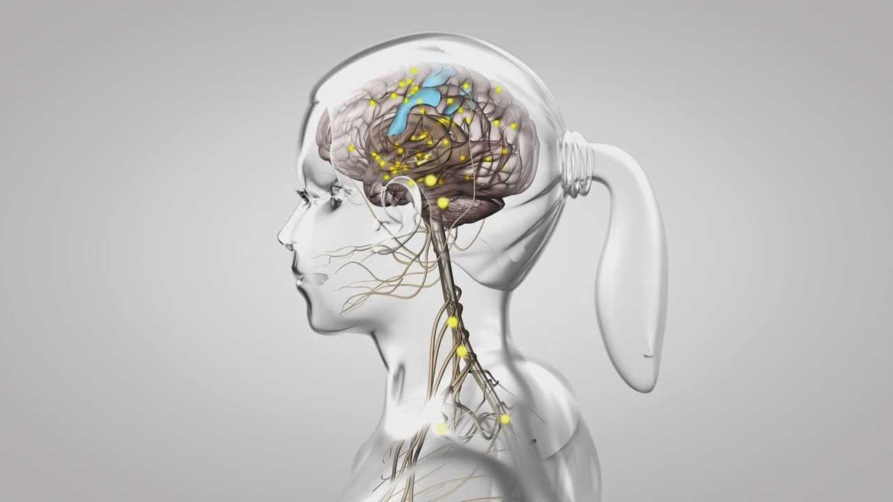


5.0 / 5 (0 votes)