Muscles of the face and scalp: Anatomy
Summary
TLDRThis video explores the muscles responsible for facial expressions, beginning with an overview of the scalp's structure and layers. It details key muscles like the occipitofrontalis, orbicularis oculi, and buccinator, explaining their origins, functions, and innervation by the facial nerve. Viewers learn how these muscles enable expressions like smiling, frowning, and eyebrow movement. The video also discusses muscles involved in lip and chin movements, emphasizing how each contributes to non-verbal communication. This informative guide aids in understanding facial anatomy, especially for students and clinicians.
Takeaways
- 😀 Facial expressions are an essential way of communication, revealing emotions like happiness or curiosity.
- 🧠 The scalp consists of five layers: Skin, Connective tissue, Aponeurosis, Loose connective tissue, and Pericranium.
- 🧍 Facial muscles originate from the mesoderm of the second pharyngeal arch and are innervated by branches of the facial nerve.
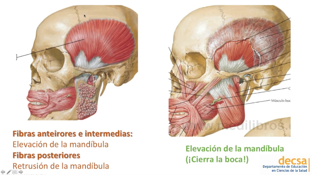
- 😲 The occipitofrontalis muscle, with its frontal and occipital bellies, elevates the eyebrows and wrinkles the forehead, showing surprise.
- 👁 The orbicularis oculi muscle has two parts: palpebral for gentle eyelid closure (blinking/sleeping) and orbital for tight closure (winking/squeezing).
- 🤔 The corrugator supercilii muscle draws the eyebrows downward and inward, creating vertical wrinkles during frowning.
- 👃 Muscles of the nose include the procerus, nasalis, and levator labii superioris alaeque nasi, which control nostril movements.
- 👄 The orbicularis oris encircles the mouth, helping close and protrude the lips (e.g., for kissing or whistling).
- 😁 Upper lip muscles like the zygomaticus major and minor elevate the lip to create a smile.
- 😐 Lower lip muscles, such as the depressor anguli oris, pull the mouth downward to express sadness or pout.
Q & A
What are facial expressions, and why are they important for communication?
-Facial expressions are movements of the facial muscles that convey emotions and reactions. They are essential for communication as they allow us to express feelings such as happiness, curiosity, or surprise without using words.
What are the five layers of the scalp, and how can they be remembered?
-The five layers of the scalp are Skin, Connective tissue, Aponeurosis (or epicranial aponeurosis), Loose connective tissue, and Pericranium. These layers can be remembered using the acronym 'SCALP,' which corresponds to the first letter of each layer.
Which cranial nerve innervates all facial muscles, and what are its branches?
-All facial muscles are innervated by the facial nerve, also known as Cranial Nerve VII. Its branches are the Posterior Auricular, Temporal, Zygomatic, Buccal, Marginal Mandibular, and Cervical nerves.
What is the function of the occipitofrontalis muscle, and how are its two bellies connected?
-The occipitofrontalis muscle functions to elevate the eyebrows and wrinkle the forehead, giving an expression of surprise. It has two bellies: the frontal and occipital bellies, which are connected by the epicranial aponeurosis.
What is the role of the orbicularis oculi muscle, and what are its two parts?
-The orbicularis oculi muscle is responsible for closing the eyelids. It has two parts: the palpebral part, which closes the eyelids gently (e.g., during blinking or sleeping), and the orbital part, which closes the eyelids tightly (e.g., when winking or squeezing the eyes shut).
Which muscle is responsible for frowning, and where is it located?
-The corrugator supercilii muscle is responsible for frowning. It is located deep in the eyebrow and pulls the eyebrows medially and inferiorly, creating vertical wrinkles at the root of the nose.
What are the three muscles of the nose, and what are their functions?
-The three muscles of the nose are the procerus, nasalis, and levator labii superioris alaeque nasi. The procerus depresses the medial ends of the eyebrows, creating transverse wrinkles on the nose. The nasalis has two parts: the transverse part narrows the nostrils, and the alar part flares the nostrils. The levator labii superioris alaeque nasi also flares the nostrils and raises the upper lip.
Which muscle encircles the mouth, and what are its functions?
-The orbicularis oris muscle encircles the mouth. It functions to close the mouth and protrude the lips, such as during kissing or whistling.
What are the muscles of the upper lip, and what are their general functions?
-The muscles of the upper lip include the risorius, levator anguli oris, zygomaticus major, zygomaticus minor, levator labii superioris, and levator labii superioris alaeque nasi. These muscles primarily elevate the upper lip and are involved in smiling, grinning, and other lip movements.
What is the function of the buccinator muscle, and where is it located?
-The buccinator muscle is located deep in the cheek and functions to press the cheek against the teeth, which is useful when drinking from a straw or during chewing. It helps maintain tension in the cheeks.
Outlines
😄 Introduction to Facial Expressions and the Scalp
This paragraph introduces the importance of facial expressions in human communication, illustrating examples like happiness and curiosity. It transitions into an anatomical discussion about the scalp, detailing its boundaries, layers, and the mnemonic to remember the five layers (SCALP: Skin, Connective tissue, Aponeurosis, Loose connective tissue, Pericranium). The first three layers form the scalp proper. The facial muscles, originating from the mesoderm of the second pharyngeal arch, are also briefly introduced.
🧠 Muscles of the Scalp and Face: Occipitofrontalis and Orbicularis Oculi
This paragraph discusses specific facial muscles, starting with the occipitofrontalis, which has two bellies (frontal and occipital). The frontal belly protracts the scalp, while the occipital belly retracts it, and together they elevate the eyebrows and wrinkle the forehead. The orbicularis oculi muscle is explained next, highlighting its two parts: the palpebral part, which gently closes the eyelids, and the orbital part, which tightly closes the eyelids.
🤨 Eyebrows and Nose Muscles: Corrugator Supercilii and Nasal Muscles
This section focuses on the muscles controlling eyebrow and nose movements. The corrugator supercilii, found beneath the eyebrow, contracts to create frowning expressions. The nose muscles—procerus, nasalis, and levator labii superioris alaeque nasi—are also described. These muscles depress the medial eyebrows and either narrow or flare the nostrils, depending on the specific muscle involved.
👄 Muscles of the Mouth and Lips
The focus here is on the muscles of the mouth, particularly the orbicularis oris, which encircles the mouth and controls lip movements like closing the mouth or protruding the lips for actions like kissing. Muscles of the upper lip include risorius, levator anguli oris, zygomaticus major and minor, and levator labii superioris, which are responsible for smiling and other lip movements. The muscles of the lower lip, such as depressor anguli oris and depressor labii inferioris, are also discussed, with their role in expressing sadness or pouting.
🙂 Chin, Neck, and Buccinator Muscles
This paragraph covers the mentalis muscle, which elevates and protrudes the lower lip, and the platysma, a broad neck muscle that depresses the mandible and tenses the skin of the neck. The buccinator muscle, found deep in the cheek, helps press the cheek against the teeth and is essential for actions like drinking through a straw. The buccinator also interacts with the orbicularis oris to perform its functions.
Mindmap
Keywords
💡Facial expressions
💡Scalp
💡Facial muscles
💡Occipitofrontalis muscle
💡Orbicularis oculi
💡Corrugator supercilii
💡Facial nerve
💡Orbicularis oris
💡Zygomaticus major
💡Buccinator
Highlights
Facial expressions are crucial for human communication, revealing emotions like happiness or curiosity.
The scalp consists of five layers: Skin, Connective Tissue, Aponeurosis, Loose Connective Tissue, and Pericranium, remembered by the mnemonic SCALP.
Muscles of facial expression are located in the skin of the scalp, face, and neck, attached to bones or fascia on one end and skin on the other.
Facial muscles develop from the mesoderm of the second pharyngeal arch, and are innervated by the facial nerve (cranial nerve 7).
The occipitofrontalis muscle has two bellies: the frontal belly and occipital belly, connected by the epicranial aponeurosis, helping elevate the eyebrows and wrinkle the forehead.
The orbicularis oculi muscle has two parts, the palpebral and orbital, and is responsible for closing the eyelids gently (blinking) or tightly (winking).
The corrugator supercilii muscle, innervated by the temporal nerve, pulls the eyebrows medially and inferiorly, creating frown lines.
The nasalis muscle, with its transverse and alar parts, controls the narrowing and flaring of the nostrils.
The levator labii superioris alaeque nasi elevates the upper lip and flares the nostrils.
The orbicularis oris muscle encircles the mouth, allowing the lips to close or protrude, like during kissing or whistling.
Muscles of the upper lip include the zygomaticus major and minor, levator labii superioris, and levator anguli oris, responsible for smiling and lifting the lip.
Muscles of the lower lip, including the depressor anguli oris and depressor labii inferioris, are responsible for expressing sadness and pulling the lip downward.
The mentalis muscle, located in the chin, elevates and protrudes the lower lip, contributing to pouting expressions.
The platysma muscle in the neck tenses the skin and depresses the mandible, expressing stress or tension.
The buccinator muscle, deep in the cheek, presses the cheek against the teeth, aiding in actions like drinking from a straw.
Transcripts
humans make many different facial
expressions and they are an extremely
important way that we communicate they
let people know when you happy like
after getting a good Mark on an anatomy
test or curious like when you learn
something new about the human body let's
learn about the muscles responsible for
our facial
expressions first let's start with the
scalp which is a layer of skin and
subcutaneous tissue that covers the
cranium it extends from the supraorbital
margins on the frontal bone to the
superior nucal line on the occipital
bone laterally it covers the temporal
fascia and extends to the zygomatic
arches on each
side the scalp is made of five layers
which can be remembered easily as the
first letter of each spells out the word
scalp going in the order from
superficial to deep s stands for skin C
stands for connective tissue a stands
for aerosis or epicranial
aerosis L stands for loose connective
tissue and P stands for parac Cranium
which is the periostium on the external
surface of the
cranium the first three layers skin
connective tissue and the aerosis are
connected tightly together forming a
single unit called the scalp
proper muscles of facial expression
simply known as the facial muscles are
found deep in the skin of the scalp face
and
neck most facial muscles are attached to
bones or fascia on one end and skin on
the other so that when they contract
they create facial
expressions
all facial muscles originate from the
mesoderm of the second Fingal Arch along
with the facial nerve during
embryological development a muscular
sheet forms and begins to spread over
the scalp face and neck which later
forms the facial
muscles this spreading carries the
branches of the nerve of the second arch
with it which is the facial nerve or
cranial nerve
7 so so all facial muscles are inated by
the branches of the facial nerve which
are the posterior aric temporal
zygomatic
Buckle marginal
mandibular and cervical nerves a way to
remember these last five branches that
emerge from the paraded gland is by
using the pneumonic to Zanzibar by
Motorcar now let's look at each of the
facial
muscles first up the occipital frontalis
muscle which has two
bellies the two bellies are connected by
the epicranial aerosis which is the
aerosis that makes the third layer of
the
scalp the frontal belly originates on
the epicranial aerosis and inserts on
the skin superior to the eyes and
nose it is innervated by the tempor
nerve the occipital belly originates on
the superior nucle line of the occipital
bone and inserts on the epicranial
aerosis the occipital belly is inated by
the posterior oric nerve now when the
frontal belly contracts it protracts the
scalp and when the occipital belly
contracts it draws the scalp back when
they work together they elevate the
eyebrows and wrinkle the forehead making
you look
surprised next up is the orbicularis
oculi muscle which has two main parts
the palpal part and the orbital part the
palpal part is found mostly within the
eyelid and it originates on the medial
palpal ligament and inserts on the
lateral palpebral
ligament the orbital part originates
from the medial orbital margin and
medial palpal ligament and inserts on
the skin around the orbit forming a
continuous ring that surrounds the
orbital orifice the Superior part of the
orbicularis oculi muscle is interated by
the temporal nerve while the zygomatic
nerve innervates the inferior part of
the
muscle the function of the orbicularis
oculi muscle is to close the eyelids
the palpal part closes the eyelids
gently like when you blink or sleep
while the orbital part closes the
eyelids tightly like when you wink or
squeeze the eyes
shut okay let's move on to the muscles
of the eyebrows and the nose the muscle
of the eyebrows is called the corrugator
super cyi which sounds like a spell
you'd learn at
Hogwarts but is actually a small muscle
found deep in the eyebrows and is
innervated by the temporal nerve
corrugator supercilii originates on the
medial part of the superciliary arch and
extends laterally to insert onto the
skin of the medial half of the eyebrow
when it contracts the corrugator
supercilii pulls the eyebrows medially
and inferiorly making vertical wrinkles
at the root of the nose which occurs
when
frowning now let's move on to the nose
there are three muscles of the
nose the procerus
nasalis and the levator labii superioris
alqua
Nai all three muscles of the nose are
innervated by the zygomatic
nerve the procerus originates on the
nasal bone and the lateral nasal
cartilage and inserts onto the skin in
the lower part of the forehead between
the
eyebrows when it contracts it depresses
the medial ends of the eyebrows and
creates transverse wrinkles on the
dorsum of the
nose the largest muscle of the nose is
the nasalis which is made of two parts a
transverse part and an AER part the
transverse part originates on the
maxilla on either side of the nose and
extends towards the dorsum of the nose
meeting in the midline to insert on an
aerosis the AER part also originates on
the maxela over the lateral inzer and
inserts on the AER
cartilage the transverse part Narrows
the nostrils and the AER part expands or
flares
them the levator labii superioris
alqui originates on the the frontal
process of the maxilla and inserts on
the upper lip and the AER cartilage of
the nose it flares the nostrils and
raises the upper
lip okay now let's take a short break
and try to remember which facial muscle
closes the
eyelids right it's the orbicularis oculi
muscle now let's tackle some muscles in
the oral region first up is the
orbicularis orus muscle which encircles
the mouth it originates on the maxilla
the mandible and on the angle of the
mouth called the
medis all of the fibers insert on the
mucus membrane of the lips the
orbicularis orus muscle is innervated by
the Buckle nerve and its function is to
close the mouth by bringing the lipss
together and protrude the lips like when
kissing or
whistling so next time you are whistling
a Happy tune remember it's the
orbicularis orus that does the
work as for the muscles of the lips
we'll divide them into the muscles of
the upper lip and the muscles of the
lower
lip the muscles of the upper lip are the
rosorius the levator anguli Ora
the zygomaticus major the zygomaticus
minor the levator labii superioris and
the levator labii superioris elake Nai
which we mentioned
earlier the rosorius originates on the
paraded fascia and the Buckle skin and
it inserts on the medis the rosorius
retracts the corner of the mouth the
levator anguli orus originates on the
infraorbital maxilla and also inserts on
the medis it raises the corner of the
mouth like when
grinning the zygomaticus major
originates on the zygomatic bone and
inserts on the medis it elevates the
corner of the mouth so we can smile the
zygomaticus minor originates on the
zygomatic bone and inserts into the skin
of the upper
lip
the levator Lei superioris originates on
the infraorbital margin of the maxela
and also inserts into the skin of the
upper
lip the zygomaticus minor levator labii
superioris and the levator Lei
superioris
elqui pull the upper lip
upward of course all muscles of the
upper lip are also dilators of the mouth
and they're all innervated by the
zygomatic and buckle
nerves now the muscles of the lower lip
are the depressor anguli orus and the
depressor labii
inferioris the depressor anguli orus
originates on the base of the mandible
and inserts on the
medis it pulls the corner of the mouth
downwards to express
sadness
the depressor labii inferioris
originates on the anterolateral body of
the mandible and the platisa muscle it
inserts into the skin of the lower lip
it pulls the lower lip downward when you
are sad or when you
pout just like the muscles of the upper
lip both muscles of the lower lip are
also dilators of the mouth the marginal
mandibular nerve innervates both the
depressor anguli orus and the oppressor
labii
inferioris okay now let's discuss a
muscle of the chin called
mentalis the mentalis originates on the
body of the mandible and inserts into
the skin of the chin it elevates and
protrudes the lower lip like when
pouting and is innervated by the
marginal mandibular
nerve lastly we have a broad muscle of
the neck NE called
plma it originates on the suprascapular
and the infraclavicular subcutaneous
tissue the platisa extends towards the
Mand and inserts on the base of the Mand
blending with the facial
muscles the
platismatia neck to express
stress since that was quite a bit of
information we can take a break here now
can you name all the muscles of the
upper lip in this
image before we wrap up let's look at
one final facial muscle called the buxin
nator the buxin nator is a rectangular
muscle found deep in the cheek it
originates on the alv processes of the
max and the mandible and the terago
mandibular
Ry now the terago mandibular Ry or the
terago mandibular ligament is a
thickening of the Buckle fenal fascia
the superior fenal constrictor
originates on the posterior side of the
terago mandibular
Rafi while the binat originates on the
anterior side the buxin nator extends
anteriorly where its fiber mingle with
the fibers of the orbicularis orus
muscle and inserts on the
medis the buxin nator is innervated by
the Buckle nerve and its main job is to
press the cheek against the teeth like
when you're drinking from a
straw all right as a quick recap the
scalp is a layer of skin and
subcutaneous tissue that covers the
cranium it is made of five layers skin
connective tissue aerosis loose
connective tissue and
pericranium all facial muscles originate
from the mesoderm of the second fenal
Arch which is why they are all
innervated by the branches of the facial
nerve the occipito frontalis muscle has
a frontal belly and an oxital belly
which are connected by the epicranial
aerosis its function is to elevate the
eyebrows and wrinkle the
forehead the orbicularis oculi muscle
surrounds the orbit its palpal part is
found within the eyelid and is used to
close the eyelids gently and its orbital
part is used to close the eyelids
tightly the muscle of the eyebrow is
called corrugator supercilii and it
draws the eyebrows downward and medially
which occurs when frowning the three mus
muscles of the nose are the
procerus
nasalis and the levator Lai superioris
alqua Nai the nasalis and the levator
Lai superioris alqua Nasi are used to
flare the
nostrils the orbicularis orus encircles
the mouth and its job is to close the
mouth or protrude the
lips the muscles of the upper lip are
the orus the levator anguli orus the
zygomaticus major the zygomaticus minor
the levator labii
superioris and the levator labii
superioris Al Nai all muscles are
involved in various lip
movements muscles of the lower lip are
the depressor anguli orus and the
depressor labii
inferioris both the muscles of the upper
and lower lip are dilators of the
mouth the mentalis is a muscle of the
chin and elevates and protrudes the
lower lip the platisa is found in the
neck and depresses the mandible draws
the corner of the mouth downwards and
tenses the skin of the lower face and
neck finally the binat is a rectangular
muscle found deep in the cheek and
presses the cheeks against the teeth
helping current and future clinicians
Focus learn retain and Thrive learn
more
5.0 / 5 (0 votes)