Cervical Spine Anatomy (eOrthopod)
Summary
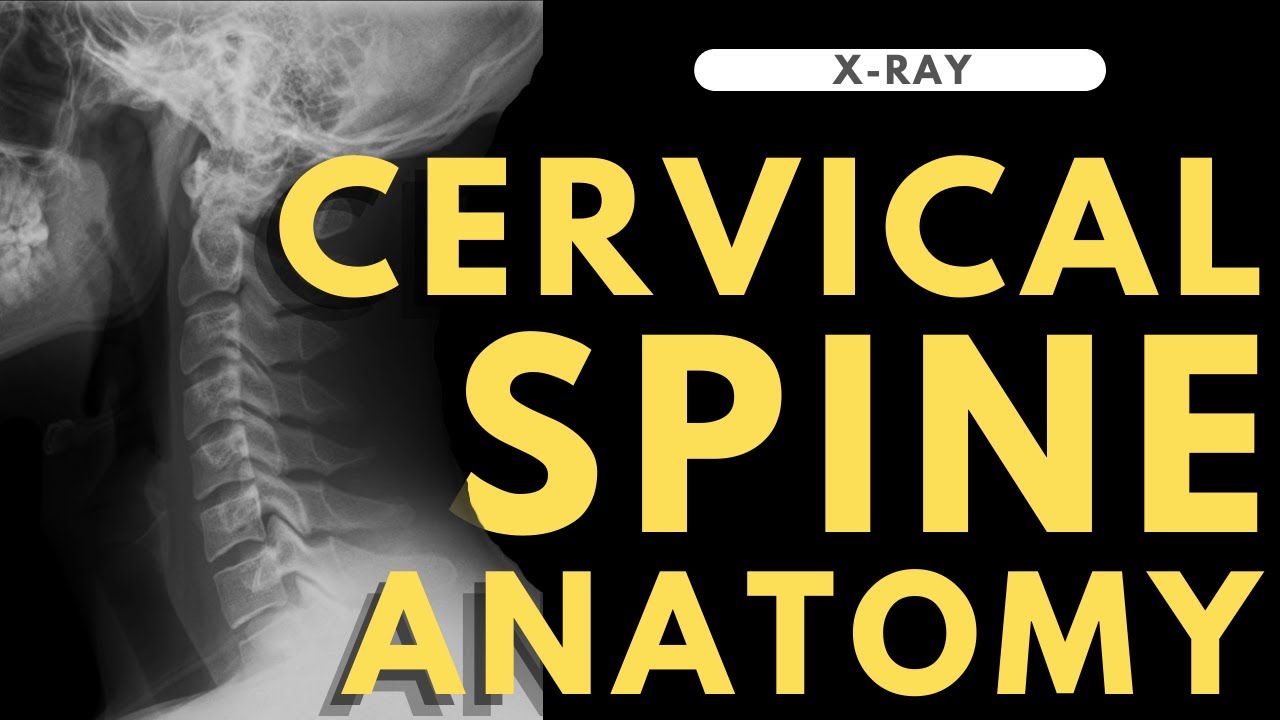
TLDRThe cervical spine, composed of seven vertebrae (C1-C7), is crucial for supporting the skull and facilitating head movement. It also protects the spinal cord, the brain's connection to the body. Key terms like 'anterior' and 'posterior' describe its parts, with C1, the atlas, and C2, the axis, being particularly significant for neck rotation. Each vertebra has a vertebral body and a bony ring that forms a protective spinal canal. The cervical spine features unique openings for blood vessels and facet joints that allow a wide range of motion. Understanding this complex structure can help manage neck pain and dysfunction.
Takeaways
- 🏌️ The cervical spine supports the skull and facilitates head movement for vision.
- 🛡️ It also protects the spinal cord, which is the link between the brain and the rest of the body.
- 🗣️ Key anatomic terms: 'anterior' for the front of the neck and 'posterior' for the back.
- 🦴 The human spine consists of 24 vertebrae, with 7 of them forming the cervical spine (C1 to C7).
- 🔝 The top cervical vertebra, C1, connects to the skull, and the cervical spine curves inward slightly.
- 🔄 The atlas (C1) and axis (C2) have a unique connection that allows for the neck's rotational movement.
- 💠 Each cervical vertebra from C2 to C7 has a vertebral body and a bony ring that forms a protective cover over the spinal cord.
- 🕳️ The spinal canal is the hollow tube inside the vertebral column that houses the spinal cord.
- 🔗 The cervical vertebrae have transverse foramina, which allow blood vessels to supply the brain.
- 🔄 Facet joints between vertebrae permit a wide range of neck movements.
- 🌿 The spinal cord is composed of nerve fibers, with nerve roots branching off at each vertebra level.
- 🔗 Ligaments and intervertebral discs provide stability and flexibility to the cervical spine.
- 💪 The cervical muscles, both anterior and posterior, play a crucial role in neck movement and support.
Q & A
What is the primary function of the cervical spine?
-The cervical spine supports the skull and allows us to move our head to direct our vision. It also protects the spinal cord, which is the connection between our brain and the rest of our body.
What do the terms 'anterior' and 'posterior' refer to in the context of the cervical spine?
-In the context of the cervical spine, 'anterior' refers to the front of the neck, while 'posterior' refers to the back of the neck.
How many vertebrae make up the cervical spine and what are they commonly referred to as?
-The cervical spine is made up of seven vertebrae, which are commonly referred to as C1 to C7.
What is unique about the connection between the atlas (C1) and the axis (C2)?
-The atlas (C1) sits on top of the axis (C2), which has a large bony knob on top called the dens. The dens points up and fits through a hole in the atlas, allowing the neck most of its ability to turn left and right.
What is the main section of each cervical vertebra from C2 to C7 called?
-The main section of each cervical vertebra from C2 to C7 is called the vertebral body, which is a round block of bone.
What is the function of the spinal canal within the cervical spine?
-The spinal canal is a hollow tube formed by the stacked vertebrae that surrounds and protects the spinal cord.
What are the spinous processes and where are they located?
-The spinous processes are bony projections that project posteriorly at the point where the two lamina bones join together at the back of the spine. They can be felt as you rub your fingers up and down the back of your neck.
What is the purpose of the transverse foramen in the cervical vertebrae?
-The transverse foramen is an opening that passes through each transverse process in the cervical vertebrae, providing a passageway for arteries that supply blood to the back of the brain.
How do facet joints contribute to the movement of the cervical spine?
-Facet joints connect the vertebrae together and slide against one another, allowing the neck to move in many directions. They are covered by articular cartilage, which allows for smooth and frictionless movement.
What is the structure of an intervertebral disc and what is its main function?
-An intervertebral disc is made of two parts: the nucleus pulposus, which is a spongy material that provides most of the shock absorption in the spine, and the annulus, a series of strong ligamentous rings that attach to the vertebrae above and below the disc.
How do the muscles in the anterior and posterior cervical areas contribute to the function of the cervical spine?
-The anterior cervical muscles run from the rib cage and collar bone to the cervical vertebrae, jaw, and skull, while the posterior cervical muscles cover the bones along the back of the spine. These muscles help in the movement and stabilization of the cervical spine.
Outlines

This section is available to paid users only. Please upgrade to access this part.
Upgrade NowMindmap

This section is available to paid users only. Please upgrade to access this part.
Upgrade NowKeywords

This section is available to paid users only. Please upgrade to access this part.
Upgrade NowHighlights

This section is available to paid users only. Please upgrade to access this part.
Upgrade NowTranscripts

This section is available to paid users only. Please upgrade to access this part.
Upgrade NowBrowse More Related Video

VERTEBRAL COLUMN ANATOMY (1/2)
Cervical spine anatomy | Radiology anatomy part 1 prep | C-spine X-ray interpretation

Atlas & Axis Cervical Vertebrae (C1-C2) Anatomy

Vertebral landmarks

How To Do The Head Tilt-Chin Lift and Jaw Thrust Maneuvers | Merck Manual Professional Version
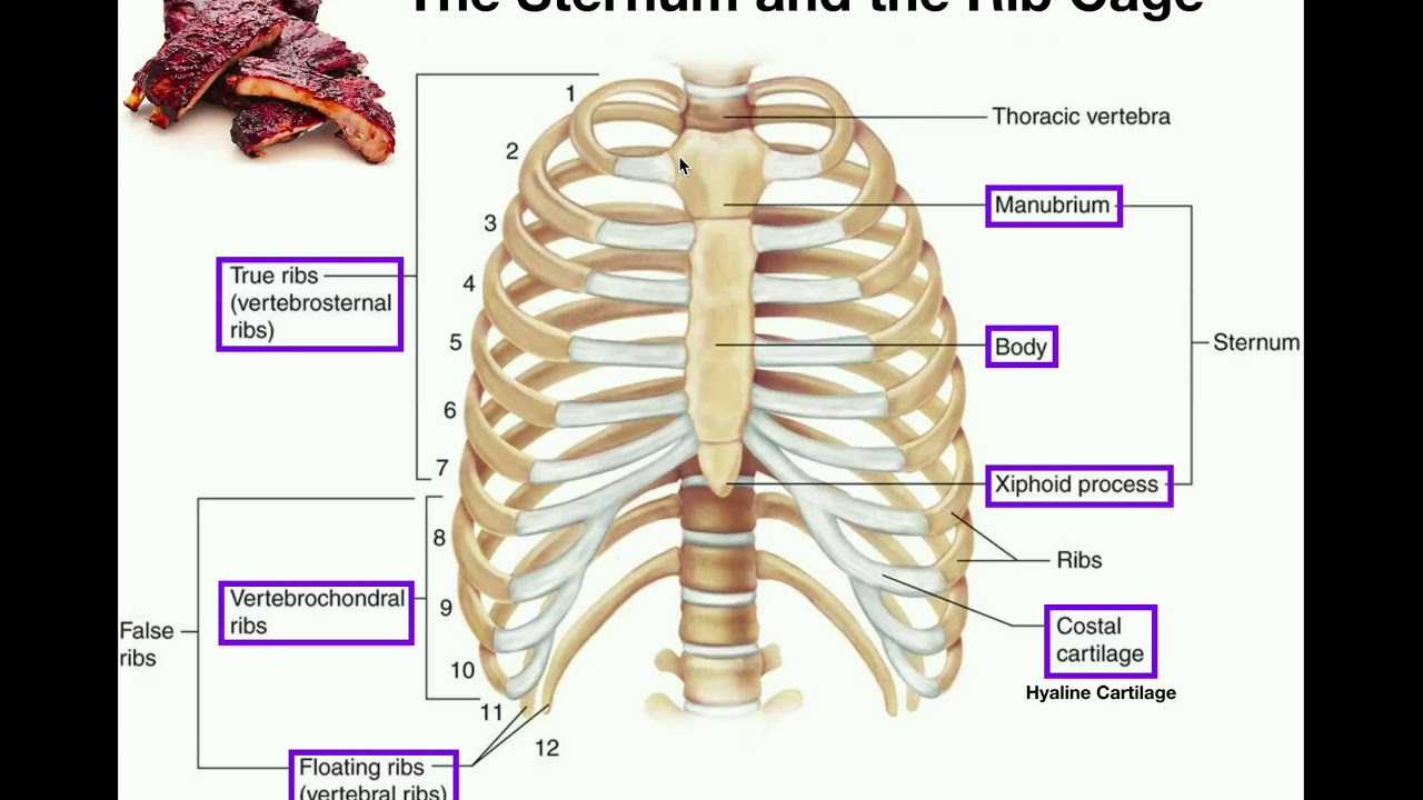
Anatomy | The Sternum, Rib Cage, & Vertebrae
5.0 / 5 (0 votes)