Blood Flow Through the Heart [Made Easy] - Cardiac Circulation Animation

EZmed
5 Mar 202108:50

Summary

TLDRThis Easy Med video simplifies the complex blood flow through the heart, using a 12-step process divided into the right and left sides. It starts with deoxygenated blood entering the right atrium via the superior and inferior vena cava, then moves through the tricuspid valve into the right ventricle, and is pumped to the lungs via the pulmonary valve. Oxygenated blood returns from the lungs through the pulmonary veins to the left atrium, passes through the mitral valve into the left ventricle, and is finally ejected into the aorta for systemic circulation. The video employs diagrams and a 2x2 table to clarify these steps, making it easier to understand and remember the heart's function.

Takeaways

  • πŸ“š The video is part of the Easy Med channel, which simplifies medical and science topics.
  • πŸ”— The channel provides free notes and an Easy Med blog that correlate with the videos, found on easymedlearning.com.
  • 🧠 The video's focus is on the blood flow through the heart, aiming to help viewers label a detailed diagram by the end.
  • πŸ’‘ The video uses simple tricks and a step-by-step approach to explain the blood flow, making it easier to remember.
  • 🟦 The right side of the heart handles deoxygenated blood, which is pumped to the lungs for oxygenation.
  • πŸŸ₯ The left side of the heart receives oxygenated blood from the lungs and pumps it to the rest of the body.
  • πŸ”„ The blood flow through the heart is divided into 12 steps, with six steps on each side, following a similar pattern.
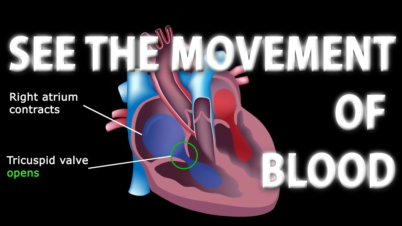
  • 🩸 The right atrium receives deoxygenated blood from the superior and inferior vena cava, then pumps it to the right ventricle.
  • πŸ«€ The left atrium collects oxygenated blood from the pulmonary veins, then sends it to the left ventricle.
  • πŸ” The video emphasizes the importance of understanding the valves (tricuspid, pulmonary, mitral, aortic) in the blood flow process.
  • πŸ“ˆ The video concludes with a realistic animation of blood flow, reinforcing the learned concepts.

Q & A

  • What is the primary purpose of the Easy Med channel?

    -The primary purpose of the Easy Med channel is to make medical and science topics easy to understand by providing educational videos, notes, and a blog that correlate with the video content.

  • How can viewers access additional materials related to the videos?

    -Viewers can access additional materials such as notes and an easy med blog by visiting the Easy Med website, easymedlearning.com, which is linked in the video description.

  • What is the focus of the video script provided?

    -The focus of the video script is to explain the blood flow through the heart, using simple tricks and a step-by-step approach to help viewers understand and label a diagram of the heart's blood flow.

  • How is the heart divided for the purpose of understanding blood flow?

    -The heart is divided into two sides for understanding blood flow: the right side, shown in blue, and the left side, shown in red. Each side has six main steps or structures through which blood flows.

  • What are the two main blood vessels that bring deoxygenated blood to the right side of the heart?

    -The two main blood vessels that bring deoxygenated blood to the right side of the heart are the superior vena cava and the inferior vena cava.

  • What is the role of the tricuspid valve in the blood flow through the heart?

    -The tricuspid valve allows blood to flow from the right atrium to the right ventricle, preventing backflow during the process.

  • What is the function of the main pulmonary artery in the context of blood flow?

    -The main pulmonary artery carries deoxygenated blood from the right side of the heart to the lungs to become oxygenated.

  • How does the left side of the heart contribute to the overall blood flow?

    -The left side of the heart pumps oxygenated blood it receives from the lungs to the rest of the body through the pulmonary veins, left atrium, left ventricle, and aorta.

  • What is the significance of the mitral and aortic valves in the blood flow through the heart?

    -The mitral valve allows blood to flow from the left atrium to the left ventricle, while the aortic valve allows blood to flow from the left ventricle into the aorta, ensuring unidirectional flow.

  • What is the main goal of the right side of the heart in terms of blood flow?

    -The main goal of the right side of the heart is to pump deoxygenated blood it receives from the rest of the body to the lungs to become oxygenated.

  • What is the main goal of the left side of the heart in terms of blood flow?

    -The main goal of the left side of the heart is to pump oxygenated blood it receives from the lungs to the rest of the body.

Outlines

00:00

🩺 Introduction to Blood Flow Through the Heart

This paragraph introduces the topic of the video, which is the blood flow through the heart. It emphasizes the educational resources available on the Easy Med channel, such as video notes and an accompanying blog on the website easymedlearning.com. The video promises to teach viewers how to label a diagram of the heart's blood flow by the end, using simple tricks for memorization. The host also references a previous video that covered the anatomy of the heart, suggesting that viewers watch it for a comprehensive understanding. The heart is divided into two sides for easier understanding: the right side, shown in blue, and the left side, shown in red, each with six main steps or structures involved in blood flow.

05:00

πŸ” Detailed Explanation of Blood Flow in the Heart

This paragraph delves into the specifics of blood flow through the heart, starting with the right side. It describes the role of the superior and inferior vena cava in carrying deoxygenated blood to the right atrium. The blood then moves through the tricuspid valve into the right ventricle and is pumped into the pulmonary artery through the pulmonary valve during systole. The function of the right side of the heart is to send deoxygenated blood to the lungs for oxygenation. The paragraph then transitions to the left side of the heart, explaining how oxygenated blood from the lungs enters through the pulmonary veins into the left atrium, moves through the mitral valve into the left ventricle, and is then pumped into the aorta through the aortic valve during systole. The left side's function is to distribute oxygenated blood to the rest of the body. The paragraph concludes with a recap using a 2x2 table from a previous video to visualize the blood flow and a transition to a more realistic anatomical image for further explanation.

Mindmap

Keywords

πŸ’‘Blood flow

Blood flow refers to the circulation of blood through the heart and blood vessels. In the video, understanding blood flow is central to grasping the heart's function. The script describes the step-by-step journey of blood through the heart's chambers and valves, highlighting the process for both oxygenated and deoxygenated blood.

πŸ’‘Heart

The heart is a muscular organ that pumps blood throughout the body via the circulatory system. The video script uses the heart as the primary focus, explaining the blood flow through its various chambers and the role of different valves in ensuring one-way flow, which is crucial for maintaining circulation.

πŸ’‘Right side of the heart

The right side of the heart, as mentioned in the script, is responsible for receiving deoxygenated blood from the body and pumping it to the lungs for oxygenation. The script outlines the specific steps and structures involved, such as the right atrium and ventricle, and the tricuspid and pulmonary valves.

πŸ’‘Left side of the heart

The left side of the heart receives oxygenated blood from the lungs and pumps it to the rest of the body. The video script emphasizes the parallel process on the left side, involving the left atrium and ventricle, and the mitral and aortic valves, which are essential for efficient blood circulation.

πŸ’‘Superior vena cava

The superior vena cava is a large vein that carries deoxygenated blood from the upper body to the right atrium of the heart. In the context of the video, it is the first step in the blood flow through the right side of the heart, illustrating the initial entry point of blood returning from the body.

πŸ’‘Inferior vena cava

The inferior vena cava is another large vein that carries deoxygenated blood from the lower body to the right atrium. The script uses this term to explain the dual entry points of deoxygenated blood into the heart, complementing the superior vena cava.

πŸ’‘Pulmonary veins

Pulmonary veins transport oxygenated blood from the lungs to the left atrium of the heart. The video script highlights the role of pulmonary veins as the return pathway for oxygen-rich blood, which is then distributed to the body by the left side of the heart.

πŸ’‘Valves

Valves in the heart, such as the tricuspid, mitral, pulmonary, and aortic valves, ensure that blood flows in one direction through the heart. The script explains how these valves open and close at specific times during the cardiac cycle to facilitate the correct flow of blood.

πŸ’‘Cardiac cycle

The cardiac cycle refers to the series of events that occur in the heart as it contracts and relaxes. The video script mentions diastole and systole, which are the relaxation and contraction phases of the heart, respectively. Understanding the cardiac cycle is key to comprehending how blood moves through the heart.

πŸ’‘Diastole

Diastole is the phase of the cardiac cycle when the heart is at rest and filling with blood. The script uses diastole to explain the period during which blood flows from the atria to the ventricles, preparing the heart for the next contraction.

πŸ’‘Systole

Systole is the phase of the cardiac cycle when the heart contracts and pumps blood. The video script describes systole as the time when the ventricles squeeze to push blood into the pulmonary artery and aorta, distributing it to the lungs and the rest of the body.

Highlights

Introduction to the Easy Med channel and its resources for learning medical and science topics.

The video's focus on explaining blood flow through the heart with simple tricks for memorization.

The use of a diagram to label and understand the entire cardiovascular system by the end of the video.

Division of the heart into two sides, right and left, each with six main steps or structures for blood flow.

The similarity in pattern between the right and left sides of the heart in terms of blood flow.

Detailed explanation of the first step involving the superior and inferior vena cava.

Description of the right atrium as the entry point for deoxygenated blood from the body.

Explanation of the tricuspid valve's role in blood flow from the right atrium to the right ventricle.

The function of the pulmonary valve and main pulmonary artery in sending deoxygenated blood to the lungs.

The role of the left side of the heart in pumping oxygenated blood to the rest of the body.

Introduction of the pulmonary veins as carriers of oxygenated blood from the lungs to the left atrium.

The mitral valve's function in allowing blood to flow from the left atrium to the left ventricle.

Description of the aortic valve and aorta's role in distributing oxygenated blood to the body.

The use of a 2x2 table to visualize blood flow through the heart's main structures.

Application of learned concepts to a realistic heart diagram for better understanding.

Animation summarizing the blood flow through the heart, highlighting deoxygenated and oxygenated blood paths.

Encouragement for viewers to engage with the video by liking, commenting, and subscribing for more educational content.

Transcripts

play00:02

[Music]

play00:14

welcome back to the easy med channel

play00:15

where medical and science topics are

play00:17

made easy

play00:18

every easy med video comes with notes

play00:20

and an easy med blog that correlates

play00:21

with the video

play00:22

all found on the easymed website

play00:24

easymedlearning.com linked down below in

play00:26

the description

play00:27

so if you don't want to miss out on all

play00:28

this free material that will save you

play00:30

time and studying

play00:31

and help you throughout your career make

play00:33

sure to subscribe to the channel

play00:35

in this video today we're going to be

play00:36

talking about the blood flow through the

play00:37

heart

play00:38

we've got some simple tricks that will

play00:39

help you remember the material

play00:41

by the end of this video you'll be able

play00:42

to label this entire diagram

play00:44

we're also going to be using this

play00:46

diagram today to walk through the blood

play00:47

flow of the heart step by step

play00:49

you might remember in the last easy med

play00:51

video we used the same diagram to learn

play00:53

the anatomy of the heart

play00:54

by the end of that video we were able to

play00:56

label all the main cardiac structures

play00:57

shown here

play00:59

we simplified the anatomy of the heart

play01:00

using this cartoon image and 2x2 table

play01:03

that video is full of tricks and ways to

play01:05

remember the names and anatomical

play01:06

features of the main cardiac structures

play01:09

so i highly encourage you check out the

play01:10

video it'll be linked down below in the

play01:12

description

play01:12

that will teach you the anatomy of the

play01:14

heart and now we're going to walk

play01:15

through the blood flow through those

play01:16

structures

play01:17

the easiest way to go about this is to

play01:19

divide the heart into two sides

play01:21

first we have the right side shown in

play01:22

blue there are six main steps or six

play01:25

main structures

play01:26

in which blood flows through the right

play01:27

side of the heart then we have the left

play01:29

side shown in red

play01:30

same thing here there are six main steps

play01:32

or six main structures

play01:33

in which blood flows through the left

play01:35

side of the heart we're going to walk

play01:36

through these 12 steps one by one

play01:38

and just like we did in the last video

play01:40

about the anatomy of the heart we're

play01:41

going to first start off with this

play01:42

cartoon image

play01:43

and then we'll go back to the original

play01:45

diagrams to bring it all together

play01:47

one of the first things you'll notice if

play01:48

you take a look at the 12 steps as a

play01:50

whole

play01:50

is that the pattern between the right

play01:52

and left side of the heart is similar

play01:54

steps one and six involve a blood vessel

play01:56

and this makes sense because that's how

play01:58

blood enters and exits that side of the

play02:00

heart

play02:00

steps two through five go chamber valve

play02:03

chamber valve

play02:04

so if you remember this general pattern

play02:06

this will help you remember the order in

play02:07

which blood

play02:08

flows through each side of the heart

play02:10

let's now walk through these 12 steps

play02:11

starting with the right side of the

play02:12

heart

play02:13

the first step is the superior vena cava

play02:15

and inferior vena cava

play02:16

these are the main blood vessels that

play02:18

carry the deoxygenated venous blood from

play02:20

the rest of the body

play02:21

to the right side of the heart

play02:22

specifically the right atrium

play02:24

the superior vena cava is located

play02:26

superiorly and it carries the

play02:27

deoxygenated venous blood from the upper

play02:29

body to the right atrium

play02:31

the inferior vena cava is located

play02:33

inferiorly and it carries the

play02:34

deoxygenated venous blood from the lower

play02:36

body to the right atrium

play02:38

and this brings us to step two which is

play02:40

the right atrium

play02:41

the right atrium is the first chamber of

play02:43

the heart in which the deoxygenated

play02:45

blood from the rest of the body

play02:46

enters blood will then flow from the

play02:48

right atrium to

play02:50

another chamber known as the right

play02:51

ventricle this occurs during diastole

play02:53

when the heart is at rest and filling

play02:55

with blood

play02:56

there's a previous easy med video where

play02:58

we talk about the cardiac cycle along

play03:00

with diastole and systole

play03:01

and how the pressures in the heart

play03:03

change that video will be linked down

play03:05

below in the description so feel free to

play03:06

go check it out

play03:08

now going back to those steps you may

play03:09

have noticed that we skipped the third

play03:11

one

play03:11

as blood flows from the right atrium to

play03:13

the right ventricle it travels through a

play03:15

valve known as the tricuspid valve

play03:17

so let's recap this so far step one was

play03:20

the superior vena cava and inferior vena

play03:22

cava

play03:22

they were the blood vessels that carried

play03:24

the deoxygenated blood from the rest of

play03:25

the body to the right atrium which is

play03:27

step number two

play03:28

blood then flows from the right atrium

play03:31

through the tricuspid

play03:32

valve which is step number three and

play03:34

enters another chamber of the heart

play03:35

known as the right ventricle

play03:36

which is step four during systole when

play03:39

the heart is squeezing and contracting

play03:41

it pumps blood forward from the right

play03:43

ventricle to the main pulmonary artery

play03:45

or pulmonary trunk

play03:46

as blood flows from the right ventricle

play03:48

to the main pulmonary artery it travels

play03:50

through another valve known as the

play03:51

pulmonary valve

play03:53

so the pulmonary valve is step number

play03:55

five and the main pulmonary artery is

play03:57

step number six

play03:58

the function of the main pulmonary

play03:59

artery is to then carry the deoxygenated

play04:01

blood from the right side of the heart

play04:03

to the lungs to become oxygenated it

play04:05

does so using the right and left

play04:07

pulmonary arteries

play04:08

which we'll see some better pictures of

play04:09

coming up so the main goal of the right

play04:12

side of the heart

play04:12

is to pump the deoxygenated blood it

play04:14

receives from the rest of the body

play04:16

to the lungs to become oxygenated an

play04:18

easy way you can remember the function

play04:20

of the right side of the heart is that

play04:21

it pumps blood right to the lungs

play04:24

so now that the deoxygenated blood has

play04:25

become oxygenated we need to pump this

play04:27

blood to the rest of the body and that's

play04:29

where the left side of the heart comes

play04:30

into play

play04:31

starting with the first step we again

play04:33

have blood vessels involved they're the

play04:35

pulmonary veins and their function is to

play04:36

carry oxygenated blood from the lungs to

play04:38

the left side of the heart

play04:40

specifically the left atrium we'll see

play04:42

some better pictures of the pulmonary

play04:44

veins coming up

play04:45

the second step is the left atrium the

play04:47

left atrium is the chamber of the heart

play04:49

that receives oxygenated blood from the

play04:51

lungs

play04:52

blood will then flow from the left

play04:53

atrium to the left ventricle during

play04:55

diastole

play04:56

as blood flows from the left atrium to

play04:58

the left ventricle it travels through

play05:00

another valve known as the mitral valve

play05:02

so you should be noticing a similar

play05:04

pattern on the left side as you did on

play05:06

the right side

play05:07

first we have blood vessels that allow

play05:08

the blood to enter that side of the

play05:10

heart

play05:11

then we entered an atrium then we

play05:13

traveled through a valve

play05:14

and now we've entered another ventricle

play05:16

during systole when that heart is

play05:18

squeezing and contracting

play05:19

it's going to pump blood forward from

play05:21

the left ventricle into the aorta

play05:23

as blood flows from the left ventricle

play05:25

into the aorta it passes through another

play05:27

valve known as the aortic valve

play05:29

so you should be seeing a similar

play05:30

pattern here again between that left and

play05:32

right side

play05:33

we've got the aortic valve which is step

play05:35

number five we have the aorta which is

play05:36

step number six

play05:38

the function of the aorta is to carry

play05:40

oxygenated blood from the left side of

play05:41

the heart to the rest of the body

play05:43

so the main goal of the left side of the

play05:45

heart is to pump oxygenated blood it

play05:47

receives from the lungs to the rest of

play05:48

the body

play05:49

an easy way to think about the function

play05:51

of the left side of the heart is that it

play05:52

pumps blood that has left the lungs

play05:55

let's now use the two by two table we

play05:57

made in the last video about the anatomy

play05:59

of the heart

play06:00

and this will give us another way to

play06:01

visualize the blood flow through the

play06:02

heart

play06:03

so first we have the superior vena cava

play06:05

inferior vena cava that carry

play06:06

deoxygenated blood from the body to the

play06:08

right atrium

play06:10

blood will then flow from the right

play06:11

atrium through the tricuspid valve

play06:13

and enter the right ventricle blood will

play06:16

then exit the right ventricle

play06:17

travel through the pulmonary valve and

play06:19

enter the main pulmonary artery

play06:20

to be delivered to the lungs to become

play06:22

oxygenated the oxygenated blood will

play06:24

then travel from the lungs

play06:26

through the pulmonary veins and enter

play06:27

the left atrium

play06:29

blood will then flow from the left

play06:30

atrium through the mitral valve and

play06:32

enter the left ventricle

play06:34

blood exits the left ventricle travels

play06:36

through the aortic valve

play06:38

and enters the aorta to be delivered to

play06:39

the rest of the body

play06:41

now that we have a good understanding of

play06:42

the blood flow through the heart using

play06:44

cartoon diagrams

play06:45

let's apply this to a more realistic

play06:47

image you can see the blue arrows

play06:49

represent the flow of deoxygenated blood

play06:51

through the right side of the heart

play06:52

and the red arrows represent the flow of

play06:54

oxygenated blood through the left side

play06:56

of the heart

play06:57

we're now going to remove the arrows and

play06:58

labels so if you need more time to look

play07:00

at this diagram hit pause in the video

play07:02

and we're going to bring all this

play07:03

together with a quick animation

play07:05

first we have all the deoxygenated

play07:07

venous blood from the rest of the body

play07:09

that needs to go to the right side of

play07:10

the heart to be pumped to the lungs

play07:12

so the superior vena cava will carry the

play07:14

deoxygenated blood from the upper body

play07:16

and the inferior vena cava will carry

play07:18

the deoxygenated blood from the lower

play07:19

body

play07:20

the deoxygenated blood will first enter

play07:22

the right atrium

play07:24

during diastole when the heart is

play07:25

relaxed the blood will flow from the

play07:27

right atrium through the tricuspid valve

play07:30

and enter the right ventricle then

play07:32

during systole when that ventricle is

play07:34

squeezing and contracting

play07:35

it will pump the deoxygenated blood from

play07:37

the right ventricle through the

play07:38

pulmonary valve and enter the main

play07:40

pulmonary artery

play07:41

from there the deoxygenated blood will

play07:43

travel through the right and left

play07:44

pulmonary arteries and enter the lungs

play07:46

to become

play07:47

oxygenated oxygenated blood will then

play07:49

travel from the lungs through the

play07:51

pulmonary veins and enter the left

play07:52

atrium

play07:54

during diastole when that heart is

play07:55

relaxed oxygenated blood will travel

play07:57

from the left atrium through the mitral

play07:59

valve and enter the left ventricle

play08:01

then during systole when the ventricle

play08:03

is contracting and squeezing

play08:04

oxygenated blood will be pumped forward

play08:06

through the aortic valve and into the

play08:08

aorta to be delivered to the rest of the

play08:10

body

play08:11

so now if we go back to the original

play08:12

diagram we started the video with you

play08:14

should be able to label and understand

play08:16

the entire image

play08:17

and hopefully this video made it easier

play08:19

for you if you found the content useful

play08:21

please give the video a quick like or

play08:22

drop a comment down below before you go

play08:24

and if you haven't already make sure to

play08:26

hit that subscribe button so you don't

play08:27

miss out on future videos that make

play08:29

medical and science topics easy

play08:31

as well as all the free notes and blogs

play08:33

that correlate with the videos

play08:34

all of this will help save you time and

play08:36

studying and help you throughout your

play08:37

career

play08:38

you can find the easymed blog and notes

play08:40

for this video linked down below in the

play08:41

description

play08:42

or you can go to easymedlearning.com

play08:44

thanks for watching and i hope you check

play08:46

out future videos

Rate This
β˜…
β˜…
β˜…
β˜…
β˜…

5.0 / 5 (0 votes)

Related Tags
Blood FlowHeart AnatomyMedical EducationCardiac CycleDeoxygenated BloodOxygenated BloodTricuspid ValvePulmonary ValveMitral ValveAortic Valve