04 History of Ultrasound: Lecture by Dr. Eric Blackwell on History and Physics
Summary
TLDRThis script recounts the evolution of ultrasound technology from its inception in 1949 for battlefield use to its modern applications in medical imaging. It highlights the transition from rudimentary oscilloscope images to advanced 3D imaging, showcasing remarkable advancements in resolution and portability. The speaker reflects on the pioneers who shaped the field and the current capabilities of ultrasound, including its use in detecting fetal abnormalities and monitoring patient conditions in real-time.
Takeaways
- 🕰️ The first known article on the use of ultrasound in humans was published in 1949 by Dr. George Ludwig, who was exploring its potential to detect foreign bodies in battlefield injuries.
- 🔍 Early ultrasound technology was primarily used to measure distances to structures within the body, with images appearing as spikes on an oscilloscope rather than the detailed images we have today.
- 🌐 The evolution of ultrasound has been remarkable, from bistable black and white displays to current high-resolution 3D capabilities that allow for detailed examination of soft tissues and bone.
- 👶 Ultrasound technology has advanced to the point where it can capture detailed images of early pregnancies, including the ability to see the yolk sac, amniotic fluid, and placental tissue.
- ❤️ The resolution of modern ultrasound equipment is so high that it can detect sub-millimeter defects, such as a ventricular septal defect in a fetal heart.
- 😀 The speaker humorously notes the ability to see fetal emotions in 3D ultrasound, showcasing the depth of detail possible with current technology.
- 🙏 The speaker acknowledges the contributions of pioneers in the field, such as Dr. Bill McKinney, who was instrumental in advancing the use of ultrasound in medicine.
- 🎥 The script mentions the transition from military surplus sonar equipment to medical ultrasound devices, highlighting the repurposed technology's role in early ultrasound development.
- 📺 The evolution from contact B scanners to real-time moving pictures represents a significant technological leap in ultrasound imaging.
- 💻 The development of portable and handheld ultrasound devices, resembling laptop computers, has made ultrasound more accessible and practical for point-of-care use.
- 🔌 The latest advancements include cordless ultrasound transducers, which offer greater flexibility and convenience, especially in sterile environments like surgery.
Q & A
What was the first known application of ultrasound in humans?
-The first known application of ultrasound in humans was in 1949 by Dr. George Ludwig from the Department of Defense, who used it to look for foreign bodies like glass or wood in battlefield injuries that didn't show up on X-rays.
Why was the early ultrasound not effective for detecting foreign bodies in the battlefield?
-The early ultrasound was not effective for detecting foreign bodies because ultrasound waves cannot penetrate certain materials like glass or wood, which are not radio opaque.
How did the ultrasound technology evolve from its first use to the 1970s?
-Ultrasound technology evolved from the initial use in 1949 to the 1970s with the development of images that were displayed on oscilloscopes, which were used to measure distances to structures within the body.
What was the limitation of early ultrasound images in the 1970s?
-The limitation of early ultrasound images in the 1970s was that they were displayed on oscilloscopes with bistable images, either on or off, with no shades of gray, and required a Polaroid camera to capture a permanent picture.
How has the ultrasound technology advanced in terms of image quality and capabilities?
-Ultrasound technology has advanced significantly with the introduction of 3D imaging, color Doppler for blood flow visualization, and high-resolution images that can detect minute details such as sub-millimeter defects in a fetal heart.
What is the significance of the 'point of care' concept in the context of early ultrasound applications?
-The 'point of care' concept refers to the ability to perform medical examinations at the patient's bedside or in the field. The first application of ultrasound was essentially a point of care tool, although it required plugging into an AC power line and was not portable.
What was the role of Dr. Bill McKinney in the development of ultrasound technology?
-Dr. Bill McKinney was a neurologist who became the president of the American Institute of Ultrasound in Medicine. He was instrumental in promoting the use of ultrasound and brought many people into the field, including the speaker of the script.
How did the surplus of sonar equipment from World War II contribute to the development of ultrasound technology?
-The surplus of sonar equipment from World War II provided a foundation for researchers to experiment with ultrasound technology. The equipment was repurposed for medical use, leading to the development of early ultrasound machines.
What was the 'water path' concept in early ultrasound scans?
-The 'water path' concept involved using a plastic bag filled with water to create a medium for the ultrasound waves to travel through. This was necessary because the technology required a water-based environment for the ultrasound waves to be effective.
How did the transition from B-mode scanners to contact B scanners impact the practicality of ultrasound technology?
-The transition from B-mode scanners, which required a water path, to contact B scanners that made direct contact with the skin, made the technology more practical for day-to-day patient care by eliminating the need for a water-filled medium and improving image quality.
What are the benefits of the recent advancements in portable ultrasound devices?
-Recent advancements in portable ultrasound devices, such as the development of handheld units and wireless transducers, have made ultrasound technology more accessible and convenient for various medical applications, including point-of-care diagnostics and surgical assistance.
Outlines

This section is available to paid users only. Please upgrade to access this part.
Upgrade NowMindmap

This section is available to paid users only. Please upgrade to access this part.
Upgrade NowKeywords

This section is available to paid users only. Please upgrade to access this part.
Upgrade NowHighlights

This section is available to paid users only. Please upgrade to access this part.
Upgrade NowTranscripts

This section is available to paid users only. Please upgrade to access this part.
Upgrade Now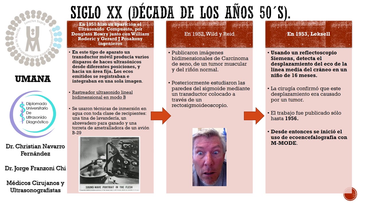


5.0 / 5 (0 votes)