Fetal Circulation | Cardiovascular system | Step 1 Simplified
Summary
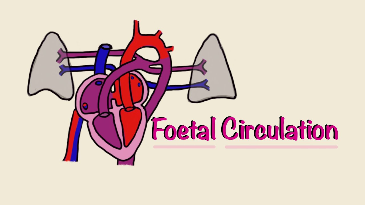
TLDRThis script explores fetal circulation, highlighting the role of the placenta for gas exchange instead of the lungs. It details key shunts like the ductus arteriosus, umbilical arteries, and the foramen ovale, which ensure efficient oxygen delivery to vital organs. Post-birth, the script explains how changes in pulmonary vascular resistance, atrial pressures, and prostaglandin levels lead to the closure of these shunts, transitioning the baby to adult circulation. It also briefly touches on congenital heart diseases.
Takeaways
- 🌟 The placenta is the organ for gas exchange in fetal circulation, not the lungs.
- 🚫 Fetal lungs have high pulmonary vascular resistance, preventing blood flow through them efficiently.
- 🔄 Deoxygenated blood from the veins is diverted from the pulmonary artery to the aorta through the ductus arteriosus.
- 🔄 Umbilical arteries carry deoxygenated blood to the placenta for oxygenation.
- 🌀 The umbilical vein returns oxygenated blood to the fetus, contrary to typical arterial function.
- 🚸 The ductus venosus shunts blood from the umbilical veins, bypassing the liver to increase oxygen delivery to the heart.
- 🕳️ The foramen ovale allows oxygenated blood to bypass the lungs by moving from the right to the left atrium.
- 🔄 The ductus arteriosus shunts deoxygenated blood from the superior vena cava to the aorta, bypassing the lungs and directing it to the placenta.
- 🌬️ At birth, the first breath decreases pulmonary vascular resistance, allowing blood to flow through the lungs.
- 🔧 After birth, the closure of the foramen ovale and ductus arteriosus occurs due to increased oxygen levels and decreased prostaglandins.
Q & A
What is the primary organ for gas exchange in fetal circulation?
-The primary organ for gas exchange in fetal circulation is the placenta, not the lungs.
Why are the fetal lungs not capable of respiration until week 25?
-The fetal lungs are not capable of respiration until week 25 because they are still developing and have very high pulmonary vascular resistance, making it difficult for blood to flow through them.
What is the role of the ductus arteriosus in fetal circulation?
-The ductus arteriosus diverts deoxygenated blood from the pulmonary artery to the aorta, bypassing the lungs and directing it towards the placenta for gas exchange.
How do umbilical arteries and veins function differently in fetal circulation compared to adult circulation?
-In fetal circulation, umbilical arteries carry deoxygenated blood to the placenta, while umbilical veins return oxygenated blood to the fetus. In adult circulation, arteries carry oxygenated blood and veins carry deoxygenated blood.
What is the purpose of the ductus venosus in fetal circulation?
-The ductus venosus bypasses the hepatic circulation, allowing oxygenated blood from the umbilical veins to bypass the liver and directly enter the inferior vena cava, ensuring efficient delivery of oxygen to the heart.
Why does the foramen ovale allow blood to flow from the right atrium to the left atrium in fetal circulation?
-The foramen ovale allows oxygenated blood from the right atrium to flow directly to the left atrium, ensuring that blood rich in oxygen is directed to vital organs like the brain and heart.
What changes occur in the circulatory system at birth to transition from fetal to adult circulation?
-At birth, the baby takes its first breath, which increases lung volume and decreases pulmonary vascular resistance. This leads to closure of the foramen ovale and ductus arteriosus, and a decrease in prostaglandins causes closure of the ductus venosus.
How does the closure of the foramen ovale occur after birth?
-The closure of the foramen ovale occurs after birth as the left atrial pressure exceeds the right atrial pressure due to increased blood flow to the lungs, causing the flap to close like a trapdoor.
What is the role of prostaglandins in maintaining the patency of the ductus arteriosus during fetal life?
-Prostaglandins, specifically prostaglandin E1 and E2, keep the ductus arteriosus open during fetal life by preventing it from closing.
How can a patent ductus arteriosus be treated after birth if it fails to close naturally?
-A patent ductus arteriosus can be treated with indomethacin, a non-steroidal anti-inflammatory drug that blocks prostaglandin production, leading to closure of the ductus arteriosus.
Outlines
👶 Fetal Circulation and Its Unique Characteristics
This paragraph explains the distinct features of fetal circulation compared to adult circulation. The placenta plays a crucial role as the organ for gas exchange since the fetal lungs are not yet functional for respiration. The fetal lungs have high pulmonary vascular resistance, causing deoxygenated blood to be diverted from the pulmonary artery to the aorta via the ductus arteriosus. This blood then flows through the umbilical arteries to the placenta for oxygenation. Unlike in adults, umbilical arteries carry deoxygenated blood, while umbilical veins carry oxygenated blood back to the fetus. The oxygenated blood bypasses the liver through the ductus venosus, which connects the umbilical veins to the inferior vena cava, avoiding the congested portal vein system. Another important shunt is the foramen ovale, which allows oxygenated blood to pass from the right atrium to the left atrium and then be pumped out through the aorta, ensuring oxygenated blood reaches the brain and heart. Lastly, the ductus arteriosus ensures deoxygenated blood from the superior vena cava is directed away from the brain and towards the placenta.
🌟 Transition from Fetal to Neonatal Circulation
The second paragraph discusses the changes that occur in circulation at birth. The baby's first breath leads to a decrease in pulmonary vascular resistance as the lungs expand and blood vessels stretch out. This allows blood to flow more easily through the lungs. With the lungs now responsible for oxygenation, blood from the right side of the heart goes to the lungs instead of the placenta. The increased left atrial pressure compared to the right atrial pressure causes the foramen ovale to close. Additionally, the decrease in prostaglandins due to placental separation and the increase in oxygenation lead to the closure of the ductus arteriosus and ductus venosus. The paragraph also mentions the use of indomethacin, a non-steroidal anti-inflammatory drug, to treat patent ductus arteriosus by blocking prostaglandin production, which is necessary to keep the ductus arteriosus open. The summary highlights the three main circulatory changes at birth: decreased pulmonary vascular resistance, closure of the foramen ovale, and closure of the ductus arteriosus and ductus venosus, marking the transition from fetal to neonatal life.
Mindmap
Keywords
💡Fetal Circulation
💡Placenta
💡Ductus Arteriosus
💡Umbilical Arteries
💡Umbilical Vein
💡Ductus Venosus
💡Foramen Ovale
💡Pulmonary Vascular Resistance
💡Prostaglandins
💡Indomethacin
💡Congenital Heart Diseases
Highlights
Fetal circulation is the direct opposite of adult circulation.
The placenta is the organ for gas exchange in fetal circulation, not the lungs.
Fetal lungs are not capable of respiration until week 25 due to high pulmonary vascular resistance.
Deoxygenated blood is diverted from the pulmonary artery to the aorta through the ductus arteriosus.
Umbilical arteries carry deoxygenated blood to the placenta for gas exchange.
Umbilical vein returns oxygenated blood to the fetus.
The ductus venosus shunt bypasses the hepatic circulation to deliver oxygenated blood to the heart.
The foramen ovale shunt allows oxygenated blood to bypass the lungs and go directly to the left atrium.
The ductus arteriosus shunts deoxygenated blood from the SVC to the aorta to bypass the lungs.
At birth, the baby's first breath decreases pulmonary vascular resistance.
Blood from the right side of the heart goes to the lungs for oxygenation after birth.
The foramen ovale closes due to increased left atrial pressure after birth.
Decreased prostaglandins and increased oxygenation lead to closure of the ductus arteriosus and ductus venosus.
Indomethacin, a non-steroidal anti-inflammatory drug, can be used to decrease prostaglandins and close a patent ductus arteriosus.
Three main circulatory changes at birth: decreased pulmonary vascular resistance, closure of foramen ovale, and closure of ductus arteriosus and ductus venosus.
Congenital heart diseases will be discussed after understanding fetal circulation.
Transcripts
so let's turn our attention to the fetal
circulation which in some ways is the
direct opposite of the actual adult
circulation so in the fetal circulation
the key thing is the placenta this is
where this is the organ for gas exchange
for fetuses not the lungs and this is
the mother's placenta note this is a
mother's placenta and this is the
fetuses circulation okay and the reason
why the placenta is the location of gas
exchanges not the lungs is first the
fetal lungs are not capable of
respiration until week 25 they're still
developing and even after that the
fetuses have a very high pulmonary
vascular resistance there are other long
other blood vessels and the lungs are
all scrunched up and so it's very hard
for blood to flow through them so
deoxygenated blood coming from the veins
will be diverted from the poner artery
remember to go from the veins to the
right side of the heart to the pulmonary
artery they're gonna be diverted to the
aorta through the ductus arteriosus so
that's ductus arteriosus it makes total
sense cuz it sorry I'm gonna go back
because it connects the pulmonary artery
and the aorta then it's gonna go down
the aorta as you can see here so this is
our ductus arteriosus take it from the
pulmonary artery today order it's gonna
go down the aorta it's gonna split and
eventually it's gonna go to the
umbilical veins so that's two umbilical
I'm sorry umbilical arteries and there's
two umbilical arteries as you can see
because they come from the branches of
the aorta and the two umbilical arteries
will go into the placenta for gas
exchange to retrieve oxygen so umbilical
arteries I want to point out one key
thing is that in the fetus much the
mythical arteries carry a lot of low
oxygenated blood okay it's not super low
but it's not super high like you would
normally expect in humans where humans
have oxygenated blood in the arteries in
contrast at least the umbilical arteries
the oxygenated blood and umbilical veins
had the oxygenated blood because the
arteries to go to the placenta to get
get oxygen and see this big fat red
thing this is the vein umbilical vein
which is confusing because usually when
you see red a red vessel you think it's
an artery but this umbilical vein
returns it's oxygenated blood to the
fetus via the IVC it's gonna connect to
the IVC so there's three shunts to know
about on the fetal circulation the first
one is from the umbilical veins and it's
gonna bypass the hepatic circulation and
that is through what shunt us through
the ductus venosus again that make sense
because doctors venosus connects the
veins in the inferior vena cava and the
reason why we have this shunt is because
the liver the hepatic circulation with a
portal vein portal vein system that's
like it's like traffic is like sitting
in traffic is very congested and we want
to get as much higher oxygenated blood
to the circuit system as much as
possible and we want a fast pass so this
ductus venosus is our fast fast
it's our carpool lane that's gonna let
us skip the portal venous circulation
and go directly to the heart now what's
gonna happen when this oxygenated blood
goes to the right atrium is it's
actually going to go through another
shunt and this shot is called the
foramen ovale and this foramen connects
the left atrium and the right atrium and
the left atrium so as you can see here
this this one you can imagine that it
shoots through it it's gonna keep going
and visit hole there it's gonna go from
the right atrium to the left atrium okay
that's what it's gonna do it's gonna go
to the left side of the heart now and
it's go today or do-- and the reason why
we want to do this is because then it's
gonna shoot out the aorta as you wanna
see here now and then it can go up all
these branches and these branches are
gonna take your nice oxygenated blood up
into the brain as well as to the heart
your heart will also get this blood so
you wanted to get this oxygenated blood
to the brain and to the heart so that's
why we have this foramen ovale
finally we have the ductus arteriosus
which
we already talked about and the reason
why we need this because we have
deoxygenated blood coming from the SVC
okay deoxygenated blood coming from the
SVC and this is gonna go down and it's
gonna follow gravity it's gonna follow
the way that makes the most sense it's
not going to go through that formula
valley because that doesn't make sense
the how is it gonna just suddenly shift
direction it's gonna go down it's the
right ventricle it's gonna go through
the right ventricle into the pulmonary
artery but remember you can't get that
blood into the lungs because of that
high resistance so this deoxygenated
blood is gonna get shunted and it's
gonna get shunted you want to shunt it
to the placenta so how are you gonna
shine it you have to shine it remember
to do it to get to the person you have
to go to lay order so you're gonna shine
it through this ductus arteriosus goes
from the pulmonary arteries to through
the ductus arteriosus into the order so
you're getting this deoxygenated blood
to really order and you're also note
that you're gonna skip all these
branches and ruin these branches you
don't wanna go you don't want this
deoxygenated blood going up to the brain
so you're gonna skip all these branches
because this shine is gonna make you
skip it all and it's gonna go straight
down the descending order and again
remember through the umbilical arteries
to the placenta so this is a beautiful
thing
the shunts all make complete sense why
we need them and just fascinating how we
have these shunts that do exactly what
we need them to do so at birth we're
gonna have circulatory changes to make
us more normal so the first change
that's gonna happen is the baby's gonna
take a first breath and when it takes a
breath the oxygens gonna fill up the
lungs like a balloon everything's gonna
stretch out including all the blood
vessels of the lungs and what happens to
pulmonary vascular resistance if your
blood vessels stretch out well your
radius is going to increase and when
your radius increases your vascular
resistance will decrease it's gonna be a
lot easier for blood to go through those
vessels the second thing that happens is
blood from the right side right side
heart of the heart will go to the lungs
for oxygenation rather than the placenta
and because we have blood flowing
through the lungs and then the lungs is
then you're gonna drain into the left
atrium the left atrial pressure will now
exceed the right atrial pressure and now
another thing is the right atrial
pressure will decrease
museum decreased umbilical vein flow all
that all that Venus flows knowledge
diverted to the lungs instead of the
placenta so because of this you have
closure of the foramen ovale I'm gonna
show this to you okay
this is prenatal when there still have a
fetus when they still have that foramen
ovale
and because the right atrial pressure is
greater than the left atrial pressure so
blood wants to go this way blood wants
to go down and pressure goes down the
pressure gradient so it's gonna open and
this like this septum primum
opens like I don't know like a trapdoor
and that's blood go through but at birth
we talked about all these changes that
are gonna make the left atrium greater
than the right atrium and because of
that blood is gonna want to go the other
way it's gonna go down the pressure
gradient and when it does this it kind
of closes that trapdoor as we see in
this picture and that's how you're
framing ovalle closes finally the last
thing we're gonna have increases in
oxygenation through your lungs and
you're gonna have decreased
prostaglandins from the placenta and
this was caught this will cause closure
of the ductus arteriosus and the ductus
venosus and the reason why is i focus on
the prostaglandins because
prostaglandins will normally make a
prostaglandin e1 and e2 keep the ductus
arteriosus alive that's what they do
they keep it patent they keep it open
that's this what that's what this is
it's the key that stays open so if your
prostaglandins decreased due to that
placental separation then your ductus
arteriosus will die it will close off
now the opposite will hold true let's
say you have a you have someone not the
opposite but let's say you do have
someone now for some reason their
prostaglandins don't go down and they
have a patent ductus arteriosus which is
a problem we'll talk about in a second
we have a treatment for that all you
have to do is decrease prostaglandins
somehow and the way we do that is with
indomethacin
do you know what type of drug and what
mechanism of action this is this is the
end set it's a non-steroidal
anti-inflammatory drug what what this
does is it blocks prostaglandin
production because that's an
inflammatory molecule so you brach plus
design and production you decreased
prostaglandins and now you have nothing
keeping that ductus arteriosus alive so
you can close that patent ductus
arteriosus so again let me just
summarize three circuits on circulatory
changes at birth three main ones when
you think of a deep breath you get
decreased pulmonary vascular resistance
this in turn will lead to your closure
of the formation for a Manoa Valley by
changing the pressure in the left atrium
in the right atrium and finally because
of that decreased prostaglandin and that
increase in oxygenation your closure or
your ductus arteriosus and your ductus
venosus so now you see that all three of
our fronts all three of our fetal shunts
have now closed so again another very
beautiful thing of how we transition
from the fetal life to the non funeral
life now we're going to talk about
congenital heart diseases
Посмотреть больше похожих видео
5.0 / 5 (0 votes)