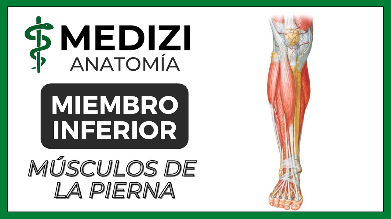
🥇 MÚSCULOS DE LA PIERNA. Compartimientos (Anteriores, Laterales y Posteriores

Anatomía Fácil por Juan José Sánchez
13 Feb 202027:49

Summary

TLDREste video educativo, presentado por Juan José Sánchez, ofrece una visión detallada de la miología del miembro inferior, centrando la atención en los músculos de la pierna. Se explica la importancia de comprender la anatomía de los huesos tibia y fibia para entender la función de los músculos. Se detallan los compartimentos de la pierna y se describen los músculos del compartimento anterior, incluyendo el tibialis anterior, extensor hallucis longus y otros, señalando sus orígenes, inserciones y funciones. Además, se menciona la innervation de los músculos y se invita a los espectadores a suscribirse y seguir el canal para más contenido anatómico.

Takeaways

  • 🦿 El vídeo de Juan José Sánchez se centra en la miología del miembro inferior, específicamente en los músculos de la pierna.
  • 📚 Se menciona que los músculos de la región del tobillo se discuten en un vídeo anterior, mientras que los del pie se tratarán en un próximo video.
  • 🗺 Se describe la fascia del tobillo, también conocida como aponeurosis, que envuelve los músculos de la región y es una continuación de la fascia lata del muslo.
  • 🔍 Se enfatiza la importancia de comprender la anatomía ósea del tobillo y del pie para entender la inserción y función de los músculos.
  • 🏋️‍♂️ Se explica que los músculos del compartimento anterior de la pierna realizan movimientos de dorsiflexión e inversión del pie.
  • 🦾 Se describen cuatro músculos en el compartimento anterior: tibialis anterior, extensor hallucis longus, extensor común de los dedos y el músculo peronéo anterior.
  • 🤸‍♀️ Los músculos del compartimento lateral, compuestos por los peronéos largo y corto, son responsables de la eversión del pie y, en el caso del peronéo largo, también de la plantarflexión.
  • 🥾 En el compartimento posterior se encuentran los músculos que realizan movimientos de plantarflexión e inversión del pie, formando el tríceps sural.
  • 🧠 La innervación de los músculos de la pierna se basa en compartimentos, siendo la nervadura del compartimento anterior por la nervio tibial anterior, la del lateral por el nervio peronéo superficial y la del posterior por el nervio tibial.

Q & A

  • ¿Cuál es la región del muslo conocida en anatomía?

    -La región del muslo también se conoce como la región sural.

  • ¿Qué es la aponeurosis o fascia del muslo y de qué es responsable?

    -La aponeurosis o fascia del muslo es una continuación de la fascia lata, la cual se encarga de envolver todos los músculos que se encuentran en esta región del muslo.

  • ¿Cuál es la importancia de estudiar los huesos del muslo para comprender sus músculos?

    -Es importante estudiar los huesos del muslo, como el tibia y el fibula, ya que son los lugares donde se insertan estos músculos y son esenciales para entender su función y movilidad.

  • ¿Cuál es la función del tibialis anterior y en qué compartimiento del muslo se encuentra?

    -El tibialis anterior es el músculo más anterior del muslo y su función es realizar la dorsiflexión e inversión del pie. Se encuentra en el compartimiento anterior del muslo.

  • ¿Cómo se inserta el extensor de los dedos comunes y qué función cumple?

    -El extensor de los dedos comunes se inserta en los tendones que van hacia los dedos laterales, es decir, el segundo, tercero, cuarto y quinto dedo, y su función es realizar la extensión de estos dedos.

  • ¿Cuál es la diferencia entre la inversión y la eversión del pie y cuáles músculos están involucrados?

    -La inversión del pie es un movimiento que permite ver el solado del pie hacia el medio, mientras que la eversión es el opuesto. Los músculos que están involucrados en estos movimientos son principalmente los músculos que se insertan medialmente (inversores) y lateralmente (evertores) del pie.

  • ¿Qué músculo se encuentra en el compartimiento posterior del muslo y tiene la función de invertir el pie?

    -El compartimiento posterior del muslo contiene el tibialis posterior, el cual tiene la función de invertir el pie.

  • ¿Cuál es la función del tríceps sureo y qué músculos lo componen?

    -El tríceps sureo es un grupo de músculos que incluye los dos gemelos o gastrocnemios y el sóleus. Su función es realizar la plantarflexión del pie y ayudar a mantener la postura erguida, siendo también importantes para actividades como bailar, saltar y correr.

  • ¿Cómo se llama el músculo que se encuentra en el compartimiento lateral del muslo y tiene una función incierta?

    -El músculo ubicado en el compartimiento lateral del muslo con una función poco definida es el músculo peronéo anterior o también conocido como el tercer peronéo.

  • ¿Cuál es la innervation de los músculos del compartimiento anterior del muslo?

    -Los músculos del compartimiento anterior del muslo son innervados por el nervio tibial anterior, que es una rama del nervio isquiático popliteo externo, también conocido como nervio peronéo profundo.

Outlines

00:00

🦿 Introducción a la Miología del Miembro Inferior

El vídeo comienza con Juan José Sánchez saludando y presentando un nuevo video sobre anatomía, específicamente la miología del miembro inferior, con enfoque en los músculos de la pierna. Se menciona que en videos anteriores se habló de los músculos de la región de la cadera y del muslo, y se anticipa que en futuras sesiones se abordarán los músculos del pie. Se presenta una imagen de una pierna con vistas anterior y posterior, envuelta en una aponeurosi conocida como la fásica de la pierna, que es una continuación de la fásica lata. Se destaca la importancia de comprender las estructuras óseas como el tibia y el fibula, así como las del pie, para entender la inserción de los músculos. Se describe la división de la pierna en compartimentos por medio de septos y se menciona que se explorarán los músculos del compartimento anterior, incluyendo su origen, inserción, función e innervación.

05:03

🏃‍♂️ Músculos del Compartmento Anterior de la Pierna

Se detalla la función y características de los cuatro músculos del compartimento anterior de la pierna: el tibialis anterior, el extensor de hallux (dedo gordo), el extensor común de los dedos y el peronéo anterior. Se explica que el tibialis anterior se origina en el cóndilo lateral del tibia y se inserta en el primer cunéiforme y el primer metatarsiano, realizando la dorsiflexión e inversión del pie. El extensor de hallux se origina en el fibula y el espacio interósseo, y se inserta en la falange distal del dedo gordo, siendo responsable de su extensión. El extensor común de los dedos tiene una amplia inserción y se divide en cuatro tendones que se dirigen a los dedos laterales, siendo responsables de su extensión. El peronéo anterior es un músculo pequeño que se origina en la superficie anterior del fibula y se inserta en la base de los metatarsianos cuarto y quinto, con una función poco definida en la dorsiflexión del pie.

10:05

🚶‍♂️ Músculos del Compartmento Lateral de la Pierna

Se describen los dos músculos del compartimento lateral de la pierna: el peronéo largo y el peronéo corto. Ambos se originan en el muslo y pasan por el malleolo lateral, pero se diferencian por su inserción y función. El peronéo largo se inserta en el primer cunéiforme y el primer metatarsiano, similar al tibialis anterior, pero su función es opuesta, realizando la eversión y la plantarflexión del pie. El peronéo corto solo realiza la eversión del pie. Se menciona la importancia de la distinción entre estos músculos y se destaca su papel en la estabilidad y movimiento del pie.

15:13

💪 Músculos del Compartmento Posterior de la Pierna: Superficiales

Se exploran los tres músculos superficiales del compartimento posterior de la pierna, conocidos colectivamente como el tríceps sureno. Se describe el gastoceso, compuesto por el bíceps femoral externo y el bíceps femoral interno, y el sóleo, un músculo poderoso que se origina en la cabeza peronéa y el diafisis peronéa. Estos músculos se insertan a través del tendon común calcáneo, también conocido como el tendon de Aquiles, en el calcáneo. El tríceps sureno es esencial para la plantarflexión y la inversión del pie, y juega un papel crucial en la postura y el movimiento, como caminar, saltar y correr.

20:13

🧘 Músculos del Compartmento Posterior de la Pierna: Profundos

Se examina la función y origen de los cuatro músculos profundos del compartimento posterior de la pierna: el poplíteo, el flexor largo del hallux, el tibialis posterior y el flexor común de los dedos. El poplíteo se origina en el cóndilo femoral lateral y se inserta en la tibia, realizando la rotación medial de la tibia o la rotación lateral del fémur. El flexor largo del hallux se origina en el diafisis peronéa y se inserta en la falange distal del dedo gordo, siendo responsable de su flexión. El tibialis posterior, con una amplia inserción en el tibia, el fibula y el espacio interósseo, se inserta en la tuberosidad del escafóideo y tiene una función de inversión del pie. El flexor común de los dedos se origina en la línea soleal y se inserta en las falanges distales de los dedos del pie, realizando su flexión.

25:14

🔋 Innervación de los Músculos de la Pierna

Se explica la innervación de los músculos de la pierna por compartimentos. Los músculos del compartimento anterior son innervados por el nervio tibial anterior, un ramal del nervio isquiático popliteo externo. Los músculos del compartimento lateral son innervados por el nervio cutáneo musculocutáneo, también conocido como el nervio peronéo superficial. Los músculos del compartimento posterior, tanto superficiales como profundos, son innervados por el nervio tibial, un ramal del nervio isquiático popliteo interno. Se resalta la simplicidad de la innervación de los músculos de la pierna y se invita al espectador a suscribirse al canal y seguir al presentador en Instagram para más contenido sobre anatomía.

Mindmap

Keywords

💡Miología

Miología se refiere al estudio de los músculos en la anatomía humana. En el video, el presentador explora la miología del miembro inferior, específicamente los músculos de la pierna, que son fundamentales para el movimiento y la postura. Esta palabra es clave ya que establece el tema central del video y guía a los espectadores en el aprendizaje de la anatomía muscular de la región.

💡Región sural

La región sural es el término anatómico utilizado para referirse a la parte de la pierna. En el guion, se menciona que la fascia o aponeurosis que cubre la pierna también se conoce como la región sural, lo que indica su importancia en la estructura y el estudio de la pierna. Este término ayuda a los espectadores a entender la terminología utilizada en la anatomía y cómo se relaciona con la parte específica del cuerpo que se está examinando.

💡Fascia de la pierna

La fascia de la pierna es una aponeurosis que envuelve los músculos de la pierna, continuando desde la fascia lata del muslo. Se destaca en el video como un elemento crucial que sostiene y separa los compartimentos musculares de la pierna. Esta estructura es esencial para comprender cómo se organizan los músculos y cómo interactúan entre sí dentro de la pierna.

💡Tibia y Fibula

Tibia y fibula son los huesos del antebrazo en la pierna humana. En el video, se enfatiza la importancia de conocer estos huesos para entender la inserción y la función de los músculos de la pierna. Los huesos proporcionan el marco sobre el cual se insertan los músculos y son esenciales para la movilidad y la estabilidad del miembro inferior.

💡Músculos del compartimiento anterior

El video describe los músculos ubicados en el compartimiento anterior de la pierna, incluyendo el tibialis anterior, el extensor de hallux y el músculo peronéo anterior. Estos músculos son importantes para las funciones de dorsiflexión e inversión del pie. El conocimiento de estos músculos es fundamental para comprender los movimientos del pie y su papel en la movilidad y la postura.

💡Dorsiflexión

La dorsiflexión es un movimiento del pie en el que se elevan los dedos hacia arriba, implicando una extensión de la articulación del tobillo. El video explica cómo ciertos músculos, como el tibialis anterior, participan en esta acción. Este concepto es crucial para entender cómo los músculos controlan los movimientos del pie y cómo estos afectan la marcha y la postura.

💡Inversión y Eversión

La inversión y la eversión son movimientos específicos del pie que implican la rotación del pie hacia adentro (inversión) o hacia afuera (eversión). En el video, se discuten los músculos que participan en estos movimientos, como el tibialis anterior para la inversión y los músculos peronéos para la eversión. Estos términos son importantes para comprender la estabilidad y la movilidad del pie durante las actividades diarias y deportivas.

💡Músculos peronéos

Los músculos peronéos son un par de músculos ubicados en el compartimiento lateral de la pierna, incluyendo el peronéo largo y corto. Estos músculos son esenciales para la eversión del pie y la pronación del pie. El video los describe en detalle, destacando su origen, inserción y función, lo que ayuda a los espectadores a entender su papel en la movilidad y la estabilidad del pie.

💡Triceps Sure

El triceps sure, también conocido como tríceps de la pierna, es un grupo muscular formado por el gastrocnemio y el sóleo. En el video, se explica cómo estos músculos trabajan juntos para realizar la plantarflexión y la inversión del pie. El triceps sure es un músculo poderoso que juega un papel crucial en la postura y en actividades como caminar, correr y saltar.

💡Nervios motores

Los nervios motores son conductos neurológicos que envían señales desde el cerebro y la médula espinal a los músculos, provocando su收缩. En el video, se menciona la innervation de los músculos de la pierna por diferentes nervios como el nervio tibial, el nervio peronéo superficial y el nervio peronéo profundo. La comprensión de la innervation es crucial para entender cómo se controlan los movimientos musculares y cómo se pueden afectar en condiciones neurológicas o lesiones.

Highlights

Introducción al estudio de la miología del miembro inferior, específicamente los músculos de la pierna.

Importancia de comprender la anatomía de la región del tobillo antes de estudiar los músculos de la pierna.

Descripción de la fascia o aponeurosis de la pierna y su función en envolver los músculos de la región.

Explicación de la división de la pierna en compartimentos para facilitar el estudio de sus músculos.

Identificación de los compartimentos anteriores, laterales y posteriores de la pierna y sus músculos correspondientes.

Funciones de los cuatro músculos del compartimento anterior de la pierna: tibialis anterior, extensor hallucis longus, extensor de los dedos y peronéus anterior.

Movimientos básicos del pie y su relación con los músculos: dorsiflexión, plantarflexión, inversión y eversión.

Descripción detallada del origen, inserción y función del músculo tibialis anterior.

Importancia del músculo extensor hallucis longus en la extensión del dedo gordo y su contribución a la dorsiflexión del pie.

Función del músculo extensor común de los dedos en la extensión de los dedos del pie y su relación con la eversión.

Características y función del músculo peronéus anterior, incluyendo su origen y inserción.

Introducción a los músculos del compartimento lateral de la pierna: peronéus longus y peronéus brevis.

Descripción del origen y función del músculo peronéus longus y su papel en la eversión y plantarflexión del pie.

Funciones del músculo peronéus brevis y su contribución única a la eversión del pie.

Detalles sobre el tríceps sureo, compuesto por los músculos gastrocnemio y soleo, y su función en la plantarflexión y inversión del pie.

Descripción de los músculos del compartimento posterior profundo de la pierna: poplíteo, flexor hallucis longus, tibialis posterior y flexor común de los dedos.

Función del músculo poplíteo en la rotación media de la tibia y su importancia en la estabilidad del pie.

Importancia del músculo flexor hallucis longus en la flexión de la falange distal del dedo gordo del pie.

Descripción del origen y función del músculo tibialis posterior, incluyendo su inserción en los metatarsales y su papel en la inversión del pie.

Función del músculo flexor común de los dedos en la flexión de las falanges distales de los dedos del pie.

Revisión de la innervation de los músculos de la pierna por compartimentos y sus nervios correspondientes.

Conclusión del video con una invitación a suscribirse al canal y seguir al presentador en Instagram.

Transcripts

play00:00

Hello, how are you, a pleasure to greet you, welcome to a new anatomical video of easy anatomy by

play00:06

Juan José Sánchez, today I bring you a video of lower limb myology, we will talk about the

play00:12

muscles of the leg, in previous videos we talked about the muscles of the hip region

play00:17

, the muscles of the thigh region and well now we will talk about the muscles of the leg and

play00:22

in a next video we will finish the myology of the lower limb talking about the muscles of the foot,

play00:29

so this image that I bring you below is a anterior and posterior view of the leg

play00:36

wrapped by a fascia or aponeurosis that is called, aponeurosis or fascia of the leg,

play00:41

it is a continuation of the fascia lata which is the aponeurosis that covers my thigh and is the one that is

play00:46

responsible for wrapping everything that is the muscles that are found in this region of the leg,

play00:53

this region of the leg is also known in anatomy with the name of sural region, just

play00:59

as the region of the thigh was known as crural, this is known by the name of the sural region,

play01:04

so every time you hear the word sural or suro it refers to the leg, in this section that we

play01:10

are going to make axial, transverse at the level of what the leg is, we are going to see this image, so that you can

play01:17

locate this It would be anterior, this would be posterior, this would be lateral because I see the fibula and this

play01:24

would be medial because I see the tibia, it is important that for you to understand these

play01:30

leg muscles you first study what the tibia and fibula are, videos that are going to find in my videos,

play01:36

if you dominate the tibia and fibula you will not be able to understand these muscles of the leg, you also

play01:41

have to understand the bones of the foot, because they are the places where these muscles are inserted,

play01:47

so in this cross section of the leg, remember that this is lateral and this is medial

play01:53

we are going to find this large aponeurosis that is the fascia of the leg, see that it originates at the

play01:58

level of what is the anterior edge of the tibia, it envelops the entire leg and reaches the medial edge

play02:05

of the same, this aponeurosis or fascia of the leg emits deep septa that is what

play02:12

will divide my leg into compartments, it is important that they study the leg by

play02:17

compartments so that they can understand it, it is the easiest way to study it, Now

play02:22

this membrane that you see here, which I have indicated in blue, is the one that joins the lateral edge of what

play02:29

is the tibia with the medial edge of the fibula, that membrane that joins it is the interosseous membrane,

play02:38

now of that aponeurosis or fascia of the leg, two septa are derived towards the deep part,

play02:46

this would be the anterior intermuscular septum which then goes from the fascia of the leg

play02:52

to what is the anterior edge of the fibula and then there would be this which would be the

play03:00

posterior intermuscular septum which also goes from that fascia of the leg to the posterior edge of it,

play03:06

then between the interosseous membrane and these two intermuscular partitions three compartments will form

play03:12

, an anterior compartment, a then lateral compartment and a

play03:18

posterior compartment, now that posterior compartment to its time it is separated by this structure

play03:28

that goes from the medial edge of the tibia to the posterior edge of the fibula, that is the transversal fascia

play03:35

of the leg and that transverse fascia of the leg then divides that posterior compartment

play03:41

in two, in a posterior compartment deep compartment that is more anterior and a

play03:47

superficial posterior compartment that is much more posterior, then we are going to see that in the anterior compartment

play03:54

we find four muscles, in the lateral compartment we are going to find two muscles and in the

play04:00

posterior compartment we find seven muscles, which are divided in three

play04:06

superficial muscles and four deep muscles, we are then going to elucidate each of these

play04:11

muscles, its origin, its insertion, its function and its innervation. Do not take off from the video

play04:19

[Music] I invite you to subscribe here the lower right corner to success [ Music]

play04:34

and don't forget to like it, it's very important that you subscribe to the channel so that you

play04:42

can help me grow and have full access to my videos here in the lower right corner,

play04:47

click on it, let's start first with these muscles that I shaded them here in red, which

play04:52

are the muscles of the anterior compartment, but before you know these muscles you have to

play04:58

know the basic movements of the foot, in the foot we can do dorsiflexion

play05:03

and plant flexion movements, dorsiflexion would be like the extension of the foot which is simply standing with

play05:12

your heels, that is, raising the toes upwards, that is called dorsiflexion

play05:17

, which would be more or less like extension, but in feet it is called dorsiflexion, while the

play05:22

opposite movement, which is when I stand on the tip of my feet, is plantar flexion, which

play05:28

in standing would basically be like the flexion movement, the The foot makes two special movements,

play05:36

it makes an inversion and eversion movement, we should not confuse it with abduction and adduction,

play05:41

the foot cannot abduct and adduct, simply that movement of the complete leg,

play05:46

of the complete lower limb of abduction and adduction is up on the hip, the foot

play05:52

can make me in an inversion movement which is I try to see the sole of the foot medially

play05:57

and it can do the opposite which would be the eversion movement, then we are going to see some muscles

play06:02

that are inverters and other muscles that are going to be evertors, let's start then with the

play06:07

previous compartment, which I remind you were four muscles, we will talk first about the

play06:12

tibialis anterior muscle, then the extensor hallucis longus, the big toe, which is the first toe

play06:18

as well . It is known anatomically by the name of hallux, so I will sometimes find it

play06:24

with the name of hallux and sometimes with the name of big toe, then we will talk about the

play06:30

common extensor of the fingers and finally we will talk about the anterior peroneus muscle, which In this image

play06:35

it does not look good, later I will explain what it is, then this tibialis anterior muscle is the most

play06:41

anterior that is found at the level of the leg, it will originate in the external condyle of the tibia

play06:47

and at the level of what is the interosseous membrane, also has its origin at the level of what is the

play06:53

tibial diaphysis, specifically the upper two thirds of the tibial diaphysis, now this

play06:59

muscle is directed downwards and we are going to see that all these muscles of the anterior compartment

play07:03

pass through this retinaculum, this is called the famous extensor retinaculum, because all the muscles that

play07:11

are going to pass there have an extensor function or they also call it the anterior retinaculum of the foot,

play07:17

so this muscle, tibialis anterior passes deep to this extensor retinaculum, it goes

play07:24

downwards here we can see it cut away , here we can see its origin in the lateral condyle of the

play07:30

tibia and its origin at the level of the tibial shaft, it then heads downward to insert itself at the

play07:35

level of what is the first cuneiform and the first metatarsal, but specifically it

play07:41

does so in the medial part of these two bones, its function is going to be then if it contracts it makes my

play07:48

foot go up and I stand on my heels that is why it does dorsiflexion and once again I say

play07:54

to this anatomical pearl that every muscle that is insert medially into the foot it makes inversion, because

play08:00

by raising the medial part it makes me see the sole of the foot medially, I remind you

play08:05

that it is the inversion movement, then this tibialis anterior muscle does both dorsiflexion

play08:10

and inversion of the foot, At the end of the video I am going to tell you what the innervation of each one is,

play08:16

you will see that the innervation of the muscles of the leg is extremely easy, so far

play08:21

the easiest innervation I have done of all the muscles in the body, Now we go from medial

play08:27

to lateral, we already talked about the tibialis anterior, we are now going to talk about the extensor hallucis longus

play08:33

, which you already know is the same extensor hallux longus, this muscle originates at the level

play08:38

of the fibula and at the level of the interosseous membrane. If we see this image here, what you see here in

play08:44

red would be the origin of the tibialis anterior, we go towards the lateral and this one that comes here shaded

play08:51

in green would now be the origin of the extensor tibialis longus, its origin then at the level

play08:56

of the interosseous membrane and at the level of what is the peroneal diaphysis,

play09:02

this muscle, which would be this one, is then directed downwards, I remind you that it is the tibialis anterior, so we are talking now

play09:07

about this one, it is directed downwards, it passes through the extensor retinaculum to be inserted into the which is

play09:14

the distal phalanx of the first finger, it is said to be at the base of the distal phalanx on its dorsal side,

play09:18

So its function is going to be to extend that first toe, but since it also passes under

play09:27

the retinaculum when it contracts, in addition to extending that first toe, it also helps me

play09:32

extend my foot and remember, this movement of extension of the foot is called dorsiflexion of the

play09:37

foot. , now very lateral to this muscle that is the long extensor of the hallux we have the

play09:45

common extensor of the fingers muscle, this common extensor of the fingers muscle has a wide origin,

play09:51

it originates at the level of what is the external tibial condyle, at the level of the peroneal diaphysis,

play09:57

it is said that at the level of three quarters of the tibial diaphysis and it originates at the level of the

play10:05

interosseous membrane as you can see here, then this muscle is directed downwards, see it here,

play10:11

it would be this one that you see here , it passes deep into what is the extensor retinaculum and is divided

play10:18

into four tendons, these four tendons are going to go towards the most lateral fingers, that is, the second,

play10:26

third, fourth and fifth fingers, now these four tendons are inserted, see clearly through of an

play10:32

aponeurosis and metatarsophalangeal joints and they will be divided distally into three, let's say,

play10:41

mini tendons, a central tendon that reaches the middle phalanx and two lateral tendons that reach

play10:47

the distal phalanx, this common extensor of the fingers its function will then be make me extend my

play10:53

fingers, but to do this I extend those four of them, which you already know are from the second to the fifth,

play10:57

nothing more, notice that the common one does not touch the big toe, I am also going to do dorsal flexion and also,

play11:04

as it is being inserted, it is more lateral in the leg, remember that everything that is inserted

play11:08

laterally makes eversion, while I told you that everything that is inserted medially makes inversion,

play11:15

so the fourth and last muscle of this group is a small muscle that is the

play11:21

anterior peroneus muscle, it is also called the third peroneus muscle, some authors say that this

play11:27

anterior peroneus muscle is part of the lateral muscles but they mostly group it within

play11:32

the muscles of the anterior compartment, then this rather small muscle that would be the

play11:38

anterior peroneus originates on the anterior surface of the fibula of the fibular diaphysis and at the level of the

play11:43

interosseous membrane, it heads downwards, joining by a serous bursa with the common extensor of the fingers,

play11:52

then they separate and it inserts at the base of the fourth and fifth metatarsals, despite the fact that

play11:59

you See that nothing has an insertion in the fifth, in the theoretical books they say that

play12:04

it is both the fourth and the fifth metatarsal, the function of this muscle is quite uncertain,

play12:09

it is said that it has a minor function in what is the dorsiflexion of the foot,

play12:15

then these would be the four muscles of the anterior compartment, let's now move on

play12:22

to the muscles of the lateral compartment, which are only two muscles, which

play12:26

are the famous peroneal muscles, these peroneal muscles are two, see that we are looking at the foot,

play12:33

the leg sorry, from a lateral view to remind you this would be the tibialis anterior,

play12:38

this one that makes me see if it is small here would be the hallux extensor, this would be the common extensor

play12:47

of the fingers and this small one that is half seen here would be the third fibula or

play12:53

peroneus anterior, now we are going to talk about these from the lateral compartment, we would have

play12:58

one that is more posterior and more lateral or more superficial, which would be the peroneus longus and we will see this

play13:06

muscle that is more medial and its tendon is more anterior, which would be the peroneus lateralis. short,

play13:12

the way to recognize them is that the tendon that is anterior is from the peroneus brevis muscle,

play13:19

as you see here, while the tendon that is posterior is from the peroneus longus muscle,

play13:24

let's start with the peroneus longus muscle that is going to have origin in what is the peroneal head

play13:30

and at the level of what is the external condyle of the tibia, although here I am shading it

play13:36

in black and you do not see that it has origins in theory, they insist that it also has an origin

play13:41

at the level of the external condyle of the tibia, then it also originates at the level of the peroneal head

play13:46

as I told you and at the level of the peroneal diaphysis, now the muscle that is most medial would be the

play13:52

peroneus brevis, which only originates at the level of the peroneal shaft, both

play13:57

muscles remember that the anterior was the peroneus longus and the posterior was the peroneus lateralis

play14:02

shortl they pass under the external malleolus of the lateral malleolus now both muscles although

play14:11

they are united in a long distance by what is a serous bag, then they separate and pass through

play14:17

this retinaculum which is called the lateral retinaculum of the leg, of the foot, lateral retinaculum of the foot,

play14:24

is one upper and one lower, they pass through there and the one that is inserted first, which is the one that seems to

play14:32

be long, is the peroneus brevis, which you can see is the anterior tendon, while the one that

play14:36

is disappears in the lateral peroneus longus, you will say well because of the long one it looks like the short one, I'm

play14:40

going to explain why this one that disappeared first, which is the posterior one,

play14:44

it is the long lateral peroneus muscle, see here this is lateral because I see the fifth and this is medial

play14:50

because I see the big toe as the fattest one than the others, so this tendon that is

play14:55

inserting into the styloid process of the fifth toe is the peroneus brevis, which

play15:01

was the most anterior of the two, I remember here, the most anterior of the two,

play15:04

see the styloid process of the fifth metatarsal, while the one you see that does not reach but

play15:12

disappeared, which was the most posterior of the two, see how it crosses the entire foot obliquely,

play15:17

from lateral to medial. , that would be the peroneus longus, this peroneus

play15:25

longus muscle is inserted at the level of the first cuneiform and the first metatarsal, what does

play15:32

this remind you of? It is reminiscent of the tibialis anterior muscle, but I told them that the tibialis anterior,

play15:38

which is cut here, was inserted into the same bones, that is, the first uniform

play15:42

and the first metatarsal but in the medial part while the peroneus longus lateralis. They make

play15:48

the same bones but on the lateral side, so I'm sorry that their function is totally

play15:55

opposite. When we talk about the tibialis anterior, we talked about it being a foot inverter and that

play16:01

apart from being a foot inverter, it also made me dorsiflex my foot, while this one, which is

play16:09

the peroneus longus, is quite the opposite, it makes my foot evert and makes me plantarflex,

play16:16

that is, it makes me able to stand with the tips of my fingers, while the peroneus

play16:22

brevis, for its part, This one can only make my foot evert, it cannot make me the

play16:28

plantar flexion movement. These would then be the muscles of the lateral compartment.

play16:33

We then quickly move on to the muscles of the posterior compartment, reminding them

play16:39

that they are divided into two groups, a superficial group made up of three muscles and a deep group

play16:44

made up of four muscles, we are going to start first with the three of the superficial group,

play16:50

something important is that these muscles of the superficial group together form the famous triceps

play16:58

surae, but not the three and I will explain well how This happens, then we are going to talk about the

play17:05

most posterior, which would be the most superficial, from a posterior view of the leg to the deepest,

play17:12

which would be the most anterior, first we start with the famous calves of the leg,

play17:16

the gastrocnemius, these muscles are made up of two bellies completely

play17:20

independent of each other, we would then have the twins of the leg, we would have an

play17:26

external twin or external gastrocnemius that originates at the level of the external femoral condyle and we would have

play17:34

an internal gastrocnemius or an internal twin that originates in the popliteal face femoral, we are going to

play17:41

see it in a much better cut, these muscles are part of the limits of the famous popliteal fossa,

play17:48

fossa quite important in anatomy that I invite you to look for in my popliteal fossa video

play17:53

on the channel, so here in This cut we then see how the lateral or external gastrocnemius

play17:59

originates at the level of the femoral condyle, while this would be the internal gastrocnemius originates

play18:04

at the level of the femoral political face, both muscles, both calves are going to be inserted at the level

play18:12

of This tendon, which is the common calcaneal tendon, or rather they will form the common

play18:18

calcaneal tendon when they join the tendon of the muscle that is anterior to them, that is, deep to them,

play18:24

which is the soleus muscle, then we are going to talk about that muscle. soleus, this soleus muscle has a

play18:30

wide origin, it is a quite powerful muscle, which forms the largest mass at the level of what it is

play18:36

the calf, then that soleus originates at the level of what is the peroneal head at the level of

play18:43

the peroneal diaphysis but it also has an arch that surrounds what are the popliteal vessels and

play18:49

this nerve which is the internal popliteal sciatic and is through This fibrous arch, which is called

play18:55

the soleus fibrous arch, is inserted into the line that is posterior to the tibia, which is the famous

play19:02

oblique line, the soleus line. This muscle will then be inserted together with the two

play19:09

gastrocnemius, forming the calcaneal tendon. which is the famous Achilles tendon, it inserts into the

play19:16

back of the calcaneus bone, so if this powerful muscle called the triceps surae,

play19:23

I repeat triceps surae, means that it has three bellies, what are the three bellies going to be?

play19:30

The two twins, which are the gastrocnemius, and the one anterior to them, which is the soleus,

play19:34

all three make up the triceps of the leg or triceps surae, then this triceps surae is inserted through

play19:41

the Achilles tendon or calcaneus at the level of what It is the back part of the calcaneal bone,

play19:46

its function will be to make my leg plantarflex, that is to say that you can stand on tiptoe

play19:52

and invert my foot, it is a very important postural muscle, it is the one that prevents you from

play19:57

staggering towards forward when you walk, so that when you walk you don't go forward,

play20:01

it is a muscle that we use to dance, to jump, to be able to run, it is a fairly powerful muscle,

play20:08

so the third muscle of this group is the superficial thin plantar muscle,

play20:13

a very unimportant that has its origin at the level of the lateral femoral supracondylar crest

play20:19

and at the level of the popliteal surface, it forms a long tendon that is found between the gastrocnemius

play20:25

and the soleus, passes lateral to the gastrocnemius and is inserted into the same calcaneal tendon for its

play20:31

part lateral, its function is said to be very little understood, it is the name that the authors give

play20:37

to this function, finally we will talk then about the deep muscles of this

play20:45

posterior compartment, which I remind you there were four, then we are going to first cut the gastrocnemius,

play20:51

we see them like this, now we cut to what is the soleus and we are like this to be able to see the

play20:57

deep compartment of the posterior part, we are going to talk first about the popliteus muscle,

play21:02

then we will find the long flexor of the big toe or hallux muscle, then we will talk about the

play21:09

tibialis posterior muscle and finally we will talk about the common flexor digitorum longus muscle,

play21:15

which would then be in order from lateral to medial, let's start first with the

play21:22

popliteus muscle which would be the one you see here, this is a posterior view This popliteus muscle

play21:28

originates at the level of the lateral femoral condyle at the level of what is the fibula through this

play21:35

reflex tendon and at the level of what is the external meniscus, then this popliteus muscle is going to

play21:44

insert, obliquely crosses the public fossa and is inserted at the level of the tibia,

play21:49

in an area that is above this line that you see here, which is the famous soleus line,

play21:54

the function of this popliteus muscle will be to rotate the tibia medially, it does medial rotation

play22:02

of the tibia because it is where they are inserted or if I have a fixed leg he is going to make me

play22:09

lateral rotation of the femur, then it is a muscle that can do both functions, one of the few

play22:14

muscles in the body that can do it, then well now We said then the popliteus muscle,

play22:19

we are going to talk about the flexor hallucis longus or flexor hallux longus, this muscle

play22:26

originates at the level of the peroneal diaphysis, it goes downwards, becoming medial

play22:35

to what is the calcaneus and Passing below what is the internal malleolus, see that the three

play22:41

deep ones cross the leg obliquely, from lateral to medial, it then becomes posterior

play22:47

to the internal malleolus, it passes through this retinaculum, which would be the retinaculum of the flexors, these muscles

play22:54

of The back part of the leg are more of a flexor, remember that the previous ones were more

play22:58

of an extensor, so once we see the sole of the foot here we section several muscles

play23:04

we can find the flexor longus tendon of the big toe, then this muscle goes on to through

play23:12

what is the sole of the foot to reach what is the distal phalanx of the first toe,

play23:17

Its function then is going to be to flex the distal phalanx of the first finger, that is,

play23:23

to bring that finger downwards, now we are going to talk about the tibialis posterior muscle, this has

play23:28

a wide origin at the level of what the tibia is at the level of what It is the fibula and at the level of what

play23:34

is the interosseous membrane, as you see here, this muscle also goes downwards,

play23:39

its tendon crosses posteriorly to the common flexor digitorum longus, which is this one, and becomes inferior

play23:46

to the internal malleolus and also passes deeply to this which would be the retinaculum of the flexors, this

play23:53

tibialis posterior as you can see here what would be this one that remember that it is made deep

play23:57

to this one, that this would be the common flexor digitorum longus and this would be the tibialis posterior,

play24:02

this which See back it would be the flexor longus muscle of the big toe, see it as it follows towards the

play24:08

big toe, towards the hallux, then this this tendon that would be that of the tibialis posterior is inserted at the

play24:15

level of the tuberosity of the scaphoid but in its insertion, see it here, In addition to inserting

play24:21

into the tuberosity of the scaphoid, it is said that through aponeurotic expansions

play24:26

it is inserted into all the metatarsals, except the talus. Remember

play24:32

that the talus is the only bone that does not have direct muscular insertions. Now,

play24:38

which one? Is it going to be the function of the tibialis posterior? Its function is to invert the foot, the last

play24:45

muscle that we will talk about is the flexor as a length of the fingers, which would be the most medial posteromuscle,

play24:50

this muscle has a wide origin at the level of the tibia below the line of the soleus,

play24:56

then what originates on the line of the sun is the soleus, who inserts above it is the

play25:01

poplithium and who originates below it is the common flexor digitorum longus, this muscle

play25:07

is directed downwards by the tendon It is placed behind what would be the posterior tibialis,

play25:14

it also passes through the retinaculum of the flexors and once we see it here, which is located

play25:21

at the level of the leg or foot, rather, the sole of the foot, it is divided in four tendons,

play25:28

that these four tendons are going to go to the four most lateral toes, now he is going to pass through a

play25:32

channel that is going to make this muscle that we are not going to see because it is a muscle of the foot, which would be

play25:37

the flexor short of the fingers, then the superior passage, that is, deeper, remember that

play25:44

in anatomical position it would be superior to this, which would be the flexor brevis of the fingers to

play25:48

be inserted into the distal phalanx of the second to fifth fingers and its function will then be

play25:52

flexion of those distal phalanges of the second to the fifth finger, now the innervation

play25:59

of these muscles I told you that it is very easy because it is an innervation by compartments,

play26:03

all the muscles of the anterior compartment are innervated by the anterior tibial nerve,

play26:08

this anterior tibial nerve It is a branch of the external sciaticopopliteal nerve, it is also known

play26:16

as the deep peroneal nerve, I am referring to this anterior tibial nerve,

play26:20

sometimes the tibialis anterior muscle itself and the common extensor of the fingers are

play26:28

directly innervated by the external sciaticopopliteus but the Most of the time, it is through one of

play26:33

the branches of the external popliteal sciatic, which is the anterior tibial nerve or deep peroneal nerve

play26:38

. The muscles of the lateral compartment are then innervated by the second branch of the

play26:44

external popliteal sciatic, which is the musculocutaneous nerve. Also known as the superficial peroneal nerve

play26:49

, here many times the lateral peroneus longus is innervated directly

play26:56

by the external popliteal sciatic but most of the time, it is then innervated by a

play27:02

branch of the lateral sciatic nerve in particular and the compartment posterior without exception,

play27:07

both the superficial and the deep ones, are innervated by the internal popliteal sciatic nerve,

play27:14

which is known as the tibial nerve, well only the tibial nerve, so this has been the entire

play27:20

video of leg muscles friends, I hope If you liked it, I remind you to subscribe to my

play27:25

channel in the little circle that appears here, like the very important video and you can

play27:29

follow me on Instagram @juan_sanchez1315 here I leave you then the videos of thigh muscles,

play27:37

of leg muscles and bones, it is important that you look for all those muscles on my

play27:42

channel so that you can understand the lower limb very well, thank you very much for watching the video

Rate This

5.0 / 5 (0 votes)

Связанные теги
MiologíaMiembro InferiorMusculosAnatomíaVideo TutorialSaludEducativoCienciaMédicoFisiología
Вам нужно краткое изложение на английском?