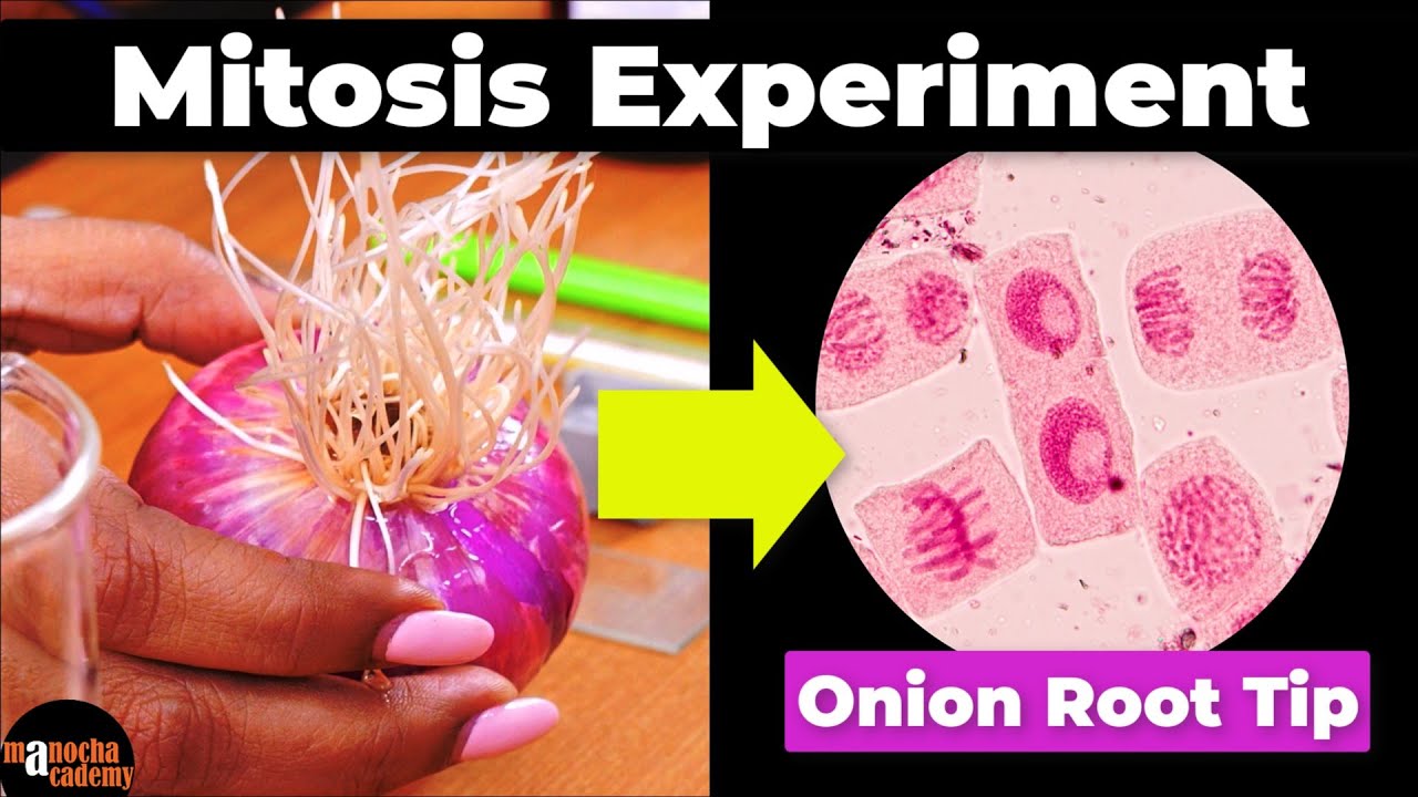
Mitosis in Onion Root tip Experiment
Summary
TLDRThis video guides viewers through the process of preparing root tips of Allium species to observe mitosis under a microscope. Mitosis is a cell cycle stage where chromosomes replicate and separate to form two identical cells. The experiment requires an onion or garlic bulb, various chemicals, and lab equipment. After growing root tips, they are fixed, preserved, and stained to highlight chromosomes. The stained root tips are then squashed on a slide for microscopic observation. The video demonstrates steps to prepare, squash, and observe the cells under different magnifications, capturing various stages of mitotic division.
Takeaways
- 🧬 Mitosis is a cell cycle stage where chromosomes replicate and separate to form two genetically identical cells.
- 🧅 The experiment requires an onion or garlic bulb, beakers, toothpicks, and various chemicals and tools.
- 🌱 Allow roots to grow by submerging the base of the bulb in water for a few days.
- 🔪 Cut out root tips and place them in Carnoy fixative fluid to preserve the DNA.
- ⏳ Leave the root tips in the fixative for 48 hours for proper fixation.
- 💧 If storing for future use, transfer fixed root tips to 70% ethanol for dehydration and preservation.
- 🔬 Use 1N hydrochloric acid to soften cell walls and facilitate squashing of root material.
- 🌈 Stain the root tips with acetyl carbine or acido-orcin stain to color the nuclear material for observation.
- 📚 Create a slide by squashing the root tips between a slide and a coverslip, avoiding air bubbles.
- 🔍 Observe the slide under a compound light microscope, using different magnifications to view mitotic stages.
- 📸 Capture images of the cells using a mobile phone and a tripod for documentation.
Q & A
What is the purpose of preparing root tips of Allium species?
-The purpose is to observe cells in various stages of mitotic division, where chromosomes replicate and separate to form two genetically identical cells.
What are the differences between mitosis and meiosis?
-In mitosis, the chromosome number is maintained in both daughter cells, whereas in meiosis, the chromosome number is halved.
What materials are needed for the experiment described in the script?
-Materials needed include an onion or garlic bulb, beakers, toothpicks, Carnoy's fixative fluid, 70% ethanol, one normal hydrochloric acid, acetyl carbine or aceto-orcein stain, glass slides and cover slips, a blade or scalpel, watch glasses or petri dishes, spirit lamp or Bunsen burner, blotting paper, droppers, thumb forceps, scissors, a compound light microscope, and immersion oil.
Why is it necessary to keep the base of the bulb in contact with water?
-The base of the bulb needs to touch the water level to facilitate root growth for the experiment.
How long should the root tips be left in Carnoy's fixative fluid?
-The root tips should be left in Carnoy's fixative fluid for about 48 hours.
What is the purpose of using 70% ethanol after fixing the root tips?
-70% ethanol is used to dehydrate the root tissue, which helps preserve the DNA for potential future genetic studies.
Why is the root material softened with one normal hydrochloric acid?
-Hydrochloric acid softens the cell walls and weakens cellular connections, making it easier to squash the root material for slide preparation.
What is the role of acetyl carbine or aceto-orcein stain in the experiment?
-These stains are used to color the nuclear material of the root cells, making it easier to observe the cells under a microscope.
How long should the root tips be left in the stain before microscopic observation?
-The root tips should be left in the stain for about 5 to 10 minutes.
What is the significance of squashing the root tips between the slide and the coverslip?
-Squashing the root tips helps to spread out the cells evenly, allowing for clear observation of individual cells without overlapping.
Why is immersion oil used during high-power microscopic observation?
-Immersion oil is used to reduce light refraction and improve the resolution of the image when observing under high magnification, such as with a 100x oil immersion lens.
Outlines
🔬 Preparing Allium Root Tips for Mitosis Observation
This video tutorial guides viewers through the process of preparing root tips from Allium species (onion or garlic) for observing cells in various stages of mitotic division. Mitosis is a critical stage in the cell cycle where chromosomes replicate and separate to form two genetically identical daughter cells. The experiment requires materials such as an Allium bulb, beakers, toothpicks, tap water, Carnoy's fixative fluid, 70% ethanol, one normal hydrochloric acid, acetic orcein stain, glass slides, cover slips, a scalpel, watch glasses, blotting paper, droppers, thumb forceps, scissors, a compound light microscope, and immersion oil. The process begins with growing roots in water, fixing them in Carnoy's fluid, dehydrating in ethanol, softening in hydrochloric acid, staining with acetic orcein, and finally squashing the root tips on a slide for microscopic examination. The video also covers the use of a microscope, including low and high-power observation, and the use of immersion oil for high-resolution viewing. Additionally, it suggests a method for capturing micro photographs of the cells.
Mindmap
Keywords
💡Mitosis
💡Allium species
💡Chromosomes
💡Carnoy’s fluid
💡Hydrochloric acid
💡Acetocarmine or Aceto-orcein stain
💡Root tips
💡Squashing
💡Microscopic observation
💡Immersion oil
Highlights
Preparing root tips of Allium species to observe cells in various stages of mitotic division
Mitosis is a stage in the cell cycle where chromosomes replicate and separate
Chromosome number is maintained in both daughter cells after mitosis, unlike meiosis
Required materials for the experiment include onion or garlic bulb, beakers, toothpicks, and various chemicals
Fixing the onion or garlic bulb in water to encourage root growth
Cutting out root tips and transferring them into Carnoy's fixative fluid
Carnoy's fluid fixes the DNA of the root tip cells
Preserving root tips in 70% ethanol for long-term storage
Using 1N hydrochloric acid to soften cell walls and weaken cellular connections
Warming the root tips in acid to prepare for staining
Rinsing root tips in distilled water after acid treatment
Staining root tips with acetyl carbine or acido-orcin stain for chromosomal visualization
Transferring stained root tips onto a glass slide for microscopic observation
Squashing the root tips between a slide and a coverslip to spread out the cells
Observing the spread-out root tip cells under a compound light microscope
Using a 40x or 100x oil immersion lens for high magnification observation
Adding immersion oil and using fine adjustment for focusing under high magnification
Taking micro photographs of mitotic stages using a mobile phone and tripod
Slideshow of various mitotic stages of cell division in Allium species
Transcripts
in this video we'll be preparing root
tips of allium species to observe cells
in various stages of mitotic division
mitosis is one of the stages in the cell
cycle where the chromosome of a cell
replicate and separate to eventually
form two genetically identical cells
unlike in meiosis chromosome number is
maintained in both daughter cells for
the experiment you will need onion or
garlic bulb
beakers toothpicks car noise fluid 70
ethanol one normal hydrochloric acid
acetyl carbine or acetyl or seen stain
glass lights and cover slips
blade or scalpel
watch glasses or petri dishes tiny vials
or containers spirit lamp or bunsen
burner or any flame source
blotting paper droppers
thumb forceps and a pair of scissor
compound light microscope and immersion
oil
to begin the experiment take a onion or
garlic bulb and fix it on a beaker
containing tap water using toothpicks
like you see here
make sure the base of the bulb touches
the water level
keep this in a safe corner for a couple
of days
once the roots have grown about two to
three centimeters cut out one centimeter
of root tips and transfer them into a
tiny vial containing carnous fixative
fluid
carnose fluid fixes the dna of the root
tip cells
leave the roots in the fixative for
about 48 hours
if you are planning on storing the root
tips for a longer time for use in future
you may transfer the fixed root tips in
a second vial containing 70 ethanol
ethanol dehydrates the root tissue and
thereby preserves dna
root tips preserved in ethanol and
preferably refrigerated will keep well
for a couple of years for genetic
studies
either ways take a few root tips from
carnous fluid or ethanol and transfer
them onto a watch glass containing one
normal hydrochloric acid
acid will soften the cell walls and
weaken cellular connections so it
becomes easy to squash the root material
later
gently warm the watch glass on a flame
for about 5 seconds
expose the root tips in the acid for
about 2 minutes
give the root tips a couple of washings
in distilled water
now transfer the roots onto a watch
glass containing acetyl carbine or
acido-orcin stain
these are excellent chromosomal stains
and will impart a deep red coloration to
the nuclear material of the root cells
warm the stain on the flame for about 5
seconds and leave the root tips in the
stain for about 5 to 10 minutes
now transfer the root tips onto a clean
glass light containing a drop of water
using a sharp blade or scalpel remove
about a millimeter of the root tip and
discard the rest
the very tips of the roots are the
regions with active cell division and
they are all that we require for mitotic
experiments
gently lower a cover slip on the root
tips making sure to avoid any air
bubbles
using the blunt end of a forcep or a
pencil gently tap the coverslip a few
times until the root tips are uniformly
squashed in between the slide and the
coverslip
a properly squashed slide will appear
faint cloudy pink to almost colorless
the root tip slide is now ready for
microscopic observation
this is how a finely squashed root tip
appears under 10x of the microscope
as you can see the cells of the root
tips are nicely spread out with almost
no overlappings
if root tips are not squashed enough you
may repeat the squashing process until
you get well spread root cells
scan the slide for cells showing mitotic
division stages under low power and then
proceed to observation under high power
such as 40x or 100x oil immersion lens
for observations under 100x objective
you will need to add a drop of immersion
oil on top of the coverslip and gently
lower the objective lens until it
touches the oil surface
from there use the fine adjustment knob
for focusing
you may take micro photographs of the
rare stages by employing a simple
technique using a mobile phone and a
tripod like so
and here's a quick slideshow of the
various mitotic stages of cell division
in root tip cells of allium species
you
5.0 / 5 (0 votes)