Type II hypersensitivity (cytotoxic hypersensitivity) - causes, symptoms, & pathology
Summary
TLDRThe video script delves into type II hypersensitivity, a condition where the immune system mistakenly attacks healthy cells. It explains how antibodies, particularly IgG and IgM, can lead to tissue damage through mechanisms like complement system activation, cell lysis, and antibody-dependent cell-mediated cytotoxicity. The script also touches on non-cytotoxic effects and diagnostics like the Coomb’s test, highlighting the specificity and potential severity of this immune response.
Takeaways
- 🚨 Hypersensitivity refers to the immune system's overreaction to substances, causing harm instead of protection.
- 🔍 There are four types of hypersensitivities, with Type II, or cytotoxic hypersensitivity, involving antibody-mediated destruction of healthy cells.
- 🏠 Type II hypersensitivity disorders are often tissue-specific, with antibodies targeting a specific tissue or organ.
- 🌐 Type III hypersensitivities are systemic and involve antibody-antigen complexes causing widespread inflammation.
- 🛡️ Central tolerance is a process that eliminates self-reactive immune cells during their development in primary lymphoid organs.
- 🚫 Some self-reactive B and T cells escape central tolerance and can lead to autoimmune diseases.
- 🏥 In Type II hypersensitivity, self-reactive B cells produce antibodies that attach to antigens on host cells, which can be intrinsic or extrinsic.
- 💊 Medications like penicillin can act as extrinsic antigens, binding to host cells and triggering an immune response.
- 🩸 The complement system is activated in Type II hypersensitivity, leading to the destruction of cells bound to the antigen-antibody complex.
- 🧬 The direct Coomb’s test is used to detect antibodies on red blood cells in conditions like autoimmune hemolytic anemia.
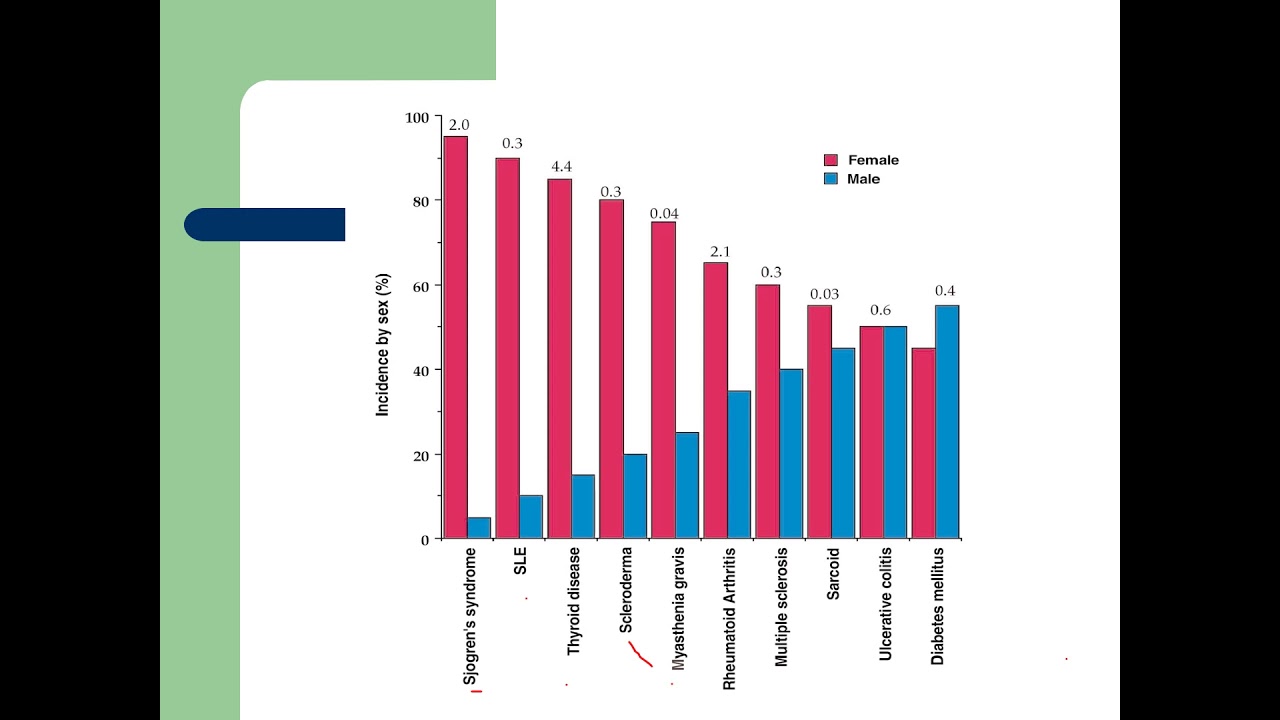
- 🔄 Non-cytotoxic Type II hypersensitivities can disrupt cell function rather than causing cell death, as seen in diseases like Myasthenia gravis and Grave’s disease.
Q & A
What is hypersensitivity and how does it differ from a normal immune response?
-Hypersensitivity refers to an immune system reaction that damages the body instead of protecting it. Unlike a normal immune response that targets 'non-self' entities, hypersensitivity involves the immune system mistakenly reacting to substances that are not harmful, leading to adverse effects on the body's own tissues.
How many types of hypersensitivities are there, and what is Type II hypersensitivity?
-There are four types of hypersensitivities. Type II hypersensitivity, also known as cytotoxic hypersensitivity, involves the destruction of healthy cells by antibodies, often targeting specific tissues or organs.
What is the role of central tolerance in preventing autoimmune diseases?
-Central tolerance is a process during which self-reactive immune cells are destroyed or inactivated while they are still in the primary lymphoid organs, such as the thymus for T cells and bone marrow for B cells. This helps to prevent autoimmune diseases by eliminating cells that could potentially attack the body's own tissues.
How do self-reactive B cells contribute to Type II hypersensitivity?
-In Type II hypersensitivity, self-reactive B cells that have escaped central tolerance become activated and produce antibodies like IgM or IgG, which attach to antigens on host cells, leading to tissue damage or autoimmune disease.
What are the two types of antigens involved in Type II hypersensitivity?
-The two types of antigens involved in Type II hypersensitivity are intrinsic antigens, which are normally produced by the host cell, and extrinsic antigens, which come from external sources like infections or medications, such as penicillin.
Can you explain the cytotoxic mechanism involving the complement system in Type II hypersensitivity?
-The complement system is activated by IgG or IgM antibodies binding to antigens on host cells. This activation leads to a cascade of events involving complement proteins, ultimately resulting in the destruction of the host cell, such as a red blood cell bound to penicillin.
What is the difference between the direct and indirect Coomb’s test?
-The direct Coomb’s test detects antibodies on the surface of red blood cells, indicating an autoimmune response. The indirect Coomb’s test checks for the presence of antibodies or complement in the serum that could cause agglutination with known antigens on laboratory red blood cells, and is used to determine pre-existing antibodies, such as in blood group incompatibility.
How does the membrane attack complex (MAC) contribute to cell lysis in Type II hypersensitivity?
-The MAC, formed by complement proteins C5b, C6-C8, and multiple C9 molecules, inserts itself into the cell membrane, creating a channel that allows fluid and molecules to flow in and out. This leads to cell swelling and eventual lysis, causing cell death.
What is opsonization and how does it relate to Type II hypersensitivity?
-Opsonization is the process where cells are marked for phagocytosis by having antibodies or complement proteins bound to their surface. In Type II hypersensitivity, opsonized cells are targeted and destroyed by phagocytes like macrophages and neutrophils.
Can you describe antibody-dependent cell-mediated cytotoxicity (ADCC) in the context of Type II hypersensitivity?
-ADCC is a mechanism where natural killer cells recognize the Fc tail of antibodies bound to antigens on host cells and release toxic granules containing perforins and granzymes. These substances induce cell death through apoptosis, a process that does not cause inflammation.
What are non-cytotoxic Type II hypersensitivities and how do they differ from cytotoxic ones?
-Non-cytotoxic Type II hypersensitivities involve antibodies binding to antigens in a way that disrupts cell function rather than causing cell death. Examples include Myasthenia gravis, where antibodies block the acetylcholine receptor, and Grave’s disease, where antibodies activate receptors, leading to cellular dysfunction.
Outlines
🛡️ Type II Hypersensitivity and Its Cytotoxic Mechanisms
This paragraph delves into the concept of hypersensitivity, specifically focusing on type II, also known as cytotoxic hypersensitivity. It explains how the immune system's overreaction to certain antigens can lead to the destruction of healthy cells. The paragraph outlines the role of self-reactive B cells, the production of IgM and IgG antibodies, and the involvement of extrinsic and intrinsic antigens. It further details the cytotoxic mechanisms at play, including the activation of the complement system, the formation of the membrane attack complex (MAC), and the process of cell lysis. Additionally, it touches on non-cytotoxic effects of type II hypersensitivity, such as antibody-mediated cellular dysfunction, and provides examples of diseases associated with this type of immune response, like autoimmune hemolytic anemia and Goodpasture’s syndrome.
🧬 Diagnostic Tests and Additional Cytotoxic Mechanisms in Type II Hypersensitivity
The second paragraph continues the discussion on type II hypersensitivity by focusing on diagnostic approaches and additional cytotoxic mechanisms. It introduces the direct and indirect Coomb’s tests used to detect the presence of antibodies on red blood cells, which can indicate conditions like hemolytic anemia or blood group incompatibility. The paragraph also describes opsonization, where IgG antibodies and complement proteins target cells for phagocytosis by macrophages and neutrophils. Furthermore, it explains antibody-dependent cell-mediated cytotoxicity (ADCC), where natural killer cells recognize antibody-coated cells and induce apoptosis. Lastly, it addresses non-cytotoxic effects where antibodies disrupt cellular function, as seen in Myasthenia gravis and Grave’s disease, emphasizing the tissue-specific nature and cytotoxic potential of type II hypersensitivity.
Mindmap
Keywords
💡Hypersensitivity
💡Type II Hypersensitivity
💡Cytotoxic Hypersensitivity
💡Antibody-Mediated
💡Central Tolerance
💡Autoimmune Disease
💡Intrinsic Antigens
💡Extrinsic Antigens
💡Complement System
💡Cytotoxic
💡Direct Coomb’s Test
💡Antibody-Dependent Cell-Mediated Cytotoxicity (ADCC)
💡Cellular Dysfunction
Highlights
Hypersensitivity is an immune system reaction that damages the body instead of protecting it.
Type II hypersensitivity, also known as cytotoxic hypersensitivity, involves antibody-mediated destruction of healthy cells.
Type II disorders are tissue-specific, with antibodies targeting a specific tissue or organ.
Type III hypersensitivities are systemic and involve antibody complexes that can cause inflammation.
Central tolerance is a process that eliminates self-reactive immune cells during their development in the thymus and bone marrow.
Escaped self-reactive B and T cells can cause autoimmune diseases.
In type II hypersensitivity, self-reactive B cells produce antibodies that attach to host cells.
Type II hypersensitivity involves intrinsic and extrinsic antigens on host cells.
Antigen-antibody complexes can be problematic when bound to host tissue.
The complement system is activated in type II hypersensitivity, leading to cell death.
Cytotoxic mechanisms in type II hypersensitivity include the formation of the membrane attack complex (MAC).
Type II hypersensitivity can result in hemolytic anemia, thrombocytopenia, or neutropenia.
Goodpasture’s syndrome is an example of type II hypersensitivity involving intrinsic antigens.
The direct Coomb’s test is used to detect antibodies on red blood cells in autoimmune hemolytic anemia.
The indirect Coomb’s test checks for blood group incompatibility and antibodies in the serum.
Opsonization targets cells for phagocytosis by macrophages and neutrophils in type II hypersensitivity.
Antibody-dependent cell-mediated cytotoxicity (ADCC) involves natural killer cells and causes cell death.
Non-cytotoxic type II hypersensitivities can disrupt cell function without causing cell death.
Myasthenia gravis and Grave’s disease are examples of non-cytotoxic type II hypersensitivity affecting cellular function.
Type II hypersensitivity is characterized by antibody mediation, cytotoxicity, and tissue specificity.
Transcripts
Having a hypersensitivity means that the immune system is reacting to something in a way that
damages the body rather than protecting it.
There are four different types of hypersensitivities, and the second type or type II hypersensitivity
is sometimes called cytotoxic hypersensitivity because a lot of disorders caused by this
hypersensitivity involve antibody mediated destruction of healthy cells.
These disorders tend to be tissue specific meaning that the antibodies are generally
specific to one type of tissue or organ.
There are other antibody-mediated hypersensitivities that are systemic, and these are generally
Type III hypersensitivities.
Our immune system is setup to fight anything that is considered “non-self” right?
Anything that’s not “self”, or you.
This works in large part because of a process called central tolerance which is when developing
immune cells that are self-reactive get destroyed or inactivated, whereas immune cells that
aren’t are allowed to survive.
This happens while they are still in their primary lymphoid organs, which is the thymus
for T cells and the bone marrow for B cells.
This process, though, is not perfect and some self-reactive B and T cells will escape.
These escaped self-reactive cells can then attack healthy tissue and result in autoimmune
disease.
In type II hypersensitivity these escaped self-reactive B cells become activated and
produce IgM or, with the help of CD4 positive T helper cells, IgG antibodies that attach
to antigens on host cells.
There are two type of antigens involved with type II hypersensitivity: intrinsic meaning
an antigen the host cell normally makes or extrinsic which is an antigen from an infection
or even some medications, like penicillin that gets attached to the host cell.
Alright so let’s say a drug, like penicillin, binds to a red blood cell - well it becomes
an extrinsic antigen.
An IgG or more rarely an IgM antibody that is penicillin specific might bind to the penicillin
molecule, creating an antigen-antibody complex.
Now it’s worth mentioning that antigen-antibody complexes can happen in the course of a normal
infection, but its when an antibody is complexed to host tissue, that things start to become
a problem.
The first cytotoxic mechanism of type II hypersensitivity is activation of the complement system.
The complement system is a family of small proteins that work in an enzymatic cascade
to fight off bacterial infections using a variety of mechanisms.
In this case, the IgG or IgM antibodies activate complement proteins which ultimately will
kill the red blood cell bound to penicillin which is complexed now with IgG or IgM.
The process gets started when C1, the first of the complement proteins, which binds the
Fc portion of the antibody.
C1 then engages other members of the complement family - C2 through C9, some of which are
activated by being cleaved or chopped by an enzyme.
The cleaved fragments C3a, C4a, and C5a act as chemotactic factors, meaning they attract
certain cells, in this case neutrophils.
Once neutrophils join the party, they degranulate or dump a bunch of enzymes like peroxidase,
myeloperoxidase, and proteinase 3 which all help generate little oxygen radicals that
are highly cytotoxic to cells and can cause tissue damage.
When it comes to drug reactions, like penicillin, Type II hypersensitivity can result in hemolytic
anemia (sometimes called autoimmune hemolytic anemia), as well as thrombocytopenia, or neutropenia,
since these are the blood cell types that are often affected.
This mechanism is also involved in diseases like Goodpasture’s syndrome, where antibodies
bind to intrinsic antigens on collagen of the basement membrane in their glomeruli in
the kidney or their alveoli in the lungs, rather than extrinsic antigens in the penicillin
example.
The second cytotoxic mechanism requires us to follow the complement system through to
the end, that said, C5b, and C6-C8, and a bunch of C9 come together to form the membrane
attack complex, or MAC.
The MAC “attacks” the cell by inserting itself into the cell membrane, punching a
hole or creating a channel that allows fluid and molecules to flow in and out of the cell,
and this is not good for the cell’s overall health, right?
Because due to the osmotic difference, fluid rushes into the cell, and the cell swells
and eventually bursts, called cell lysis, and it dies.
And that’s the second mechanism, and this is where the name cytotoxic comes from.
In our example if you suspect autoimmune hemolytic anemia, the antibody in question can be detected
using a direct Coomb’s test.
In the direct Coomb’s test, the person’s RBCs are separated from the plasma, and mixed
with Coombs reagent which is anti-human globulin, an antibody against human antibodies.
If the red blood cells agglutinate or clump up, that means that they probably had antibodies
on the surface.
Besides a direct test, there is also an indirect Coomb’s test which is usually done to check
for blood group incompatibility.
For an indirect Coomb’s test the patient’s serum is mixed with laboratory red blood cells
that have known antigens on their surface, and then once again mixed with Coombs reagent.
If there is red blood cells agglutination, that indicates the presence of antibodies
or complement in the serum.
The indirect Coomb’s test is done to determine if you have antibodies before you are exposed
to an antigen.
For example, this could be like a mismatched blood transfusion or a second pregnancy with
a mismatched Rh factor between mother and child.
Alright, so the third cytotoxic mechanism of type II hypersensitivity happens when IgG
antibodies coat a blood cell and are bound by C3b, another one of those complement protein
fragments.
At this point, we would say that the cell has been opsonized, which means it’s targeted
for phagocytosis which is where they get engulfed and destroyed by phagocytes like macrophages
and neutrophils.
Once opsonized, the antigen-antibody complex and the cell it's attached to encounters a
phagocyte in the body’s blood filtration organ—the spleen, and the phagocytes target
cells by binding to the Fc tail of the antibody, or the C3b bound to the IgG, then engulfs
and destroys the cell.
Alright, so the last three mechanisms involve the complement system, in one way or another,
the last couple mechanisms are a little different, so the fourth mechanism is called antibody-dependent
cell-mediated cytotoxicity or ADCC.
In this case, the bound antigen-antibody complex gets recognized by immune cells called natural
killer cells, yeah.
The natural killer cell recognizes the Fc tail of the antibody and releases toxic granules.
These granules contain perforins which just like the MAC, form pores in the cell, except
this time the pore also allows entry of enzymes that are like silent assassins called granzymes
as well as granulysin which work together to cause cell death in an apoptotic or “quiet
death” sort of way, such that there’s no surrounding inflammation.
Okay so far all of the mechanisms have lead to cell death or cytotoxicity, right?
There are though non-cytotoxic Type II Hypersensitivities as well, where it just sort of disrupts function,
called antibody-mediated cellular dysfunction.
Sometimes when an antibody binds to its antigen, it just, sort of gets in the way.
When this happens it can change the way the cell is supposed to function.
This is the case in the autoimmune disease Myasthenia gravis where antibodies specific
for the acetylcholine receptor in muscles simply blocks the binding of acetylcholine
which causes the muscles to not get stimulated and progressively weaken over time.
This mechanism is also involved in Grave’s disease, where antibodies target receptors
that stimulate thyroid hormone production, but in this case they not only get in the
way, they actually activate the receptors, causing overproduction of thyroid hormone,
or hyperthyroidism.
There you have it - a handful of different ways that antibody binding to cells can cause
them to get destroyed or become less effective.
The important things to remember about Type II Hypersensitivity is that they are antibody
mediated, they generally lead to cytotoxicity, and they are tissue specific.
5.0 / 5 (0 votes)