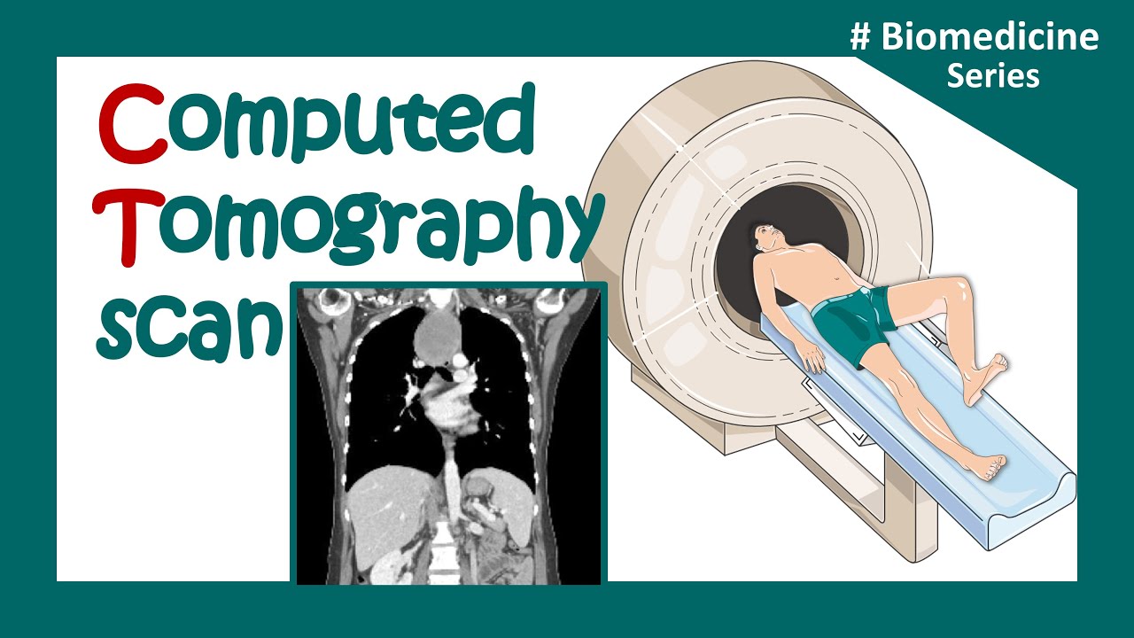
CT Artifacts
Summary
TLDRThis podcast episode delves into physics-based artifacts in CT scans, which can degrade image quality and mimic clinical lesions. It categorizes artifacts into physics-based, patient-based, scanner-based, and technique-based, focusing on beam hardening, partial volume, and under sampling artifacts. The discussion covers their causes and solutions, such as calibration corrections and beam hardening software, emphasizing the importance of proper patient positioning and scan parameter selection.
Takeaways
- 🛠️ Physics-based artifacts in CT imaging result from physical processes during data acquisition, and they can seriously degrade image quality.
- 📉 Artifacts can mimic clinical lesions, leading to diagnostic errors for physicians interpreting CT images.
- 🧠 Artifacts are categorized into four types: physics-based, patient-based (due to movement or metallic objects), scanner-based (imperfections in the scanner), and those from image reconstruction techniques (helical or multi-section).
- 🔬 Beam hardening is a key physics-based artifact that occurs when lower-energy x-rays are absorbed as they pass through tissue, leading to image distortions such as cupping and streaking artifacts.
- 🛡️ To minimize beam hardening, CT scanners use filtration, calibration correction, and beam hardening correction software.
- 🌀 Cupping artifacts arise when x-rays passing through the center of an object harden more than those passing through its edges, causing distorted image intensities.
- 🎯 Streaks and dark bands form between dense objects in the body due to differential x-ray absorption, commonly seen when contrast media is used.
- 🔧 Partial volume artifacts occur when dense objects extend into the x-ray beam’s path. These are mitigated by using thinner image slices.
- 🔍 Photon starvation appears in obese patients or highly attenuating regions like the shoulders, creating noisy streak artifacts, reduced by automatic tube current modulation.
- 📏 Under-sampling artifacts, like view aliasing, happen when there are too few projections per rotation, and are reduced by increasing the number of projections.
Q & A
What are artifacts in CT imaging?
-Artifacts in CT imaging are errors in the perception or representation of information introduced by imaging techniques. They can degrade the quality of images and, in some cases, mimic clinical lesions, which can confuse the diagnosis.
What are the main categories of artifacts in CT imaging?
-The main categories of artifacts in CT imaging are physics-based artifacts, patient-based artifacts, scanner-based artifacts, and helical or multi-section technique artifacts.
What causes physics-based artifacts in CT imaging?
-Physics-based artifacts are caused by physical processes involved in the acquisition of CT data. These include beam hardening, partial volume effects, and under-sampling during data collection.
How can patient-based artifacts occur?
-Patient-based artifacts can occur due to patient movement or the presence of metallic materials either inside or on the patient, which interferes with image quality.
What is beam hardening in CT imaging, and why is it important?
-Beam hardening occurs when lower-energy X-ray photons are absorbed as the X-ray passes through denser materials, causing a shift in the X-ray spectrum toward higher energy. Beam hardening is necessary to remove soft X-rays that would otherwise be absorbed by the skin and not contribute to image formation.
What are cupping artifacts, and how do they occur?
-Cupping artifacts occur when X-rays passing through the center of a uniform object are more hardened than those passing through the edges, causing the CT number to appear higher at the edges and lower in the center. This results in a 'cupping' appearance in the image.
What are streaks and dark bands in CT imaging?
-Streaks and dark bands are beam hardening artifacts that occur between two dense objects in the image. These streaks can be caused by highly attenuating structures like contrast media in vessels, leading to darker areas or streaking in adjacent regions.
How can beam hardening artifacts be minimized in CT imaging?
-Beam hardening artifacts can be minimized using filtration, calibration correction, and beam hardening correction software. Filtration removes soft X-rays, calibration correction adjusts for cupping effects, and beam hardening correction software iteratively minimizes artifacts.
What is partial volume artifact, and how is it corrected?
-Partial volume artifact occurs when dense objects are partially in the path of the X-ray beam, causing blurring or streaking. It can be corrected by using thinner slices during image acquisition to capture finer details.
What is photon starvation, and how is it addressed in CT scans?
-Photon starvation occurs when insufficient X-rays penetrate dense regions, such as in obese patients or areas like the shoulders. It can lead to noisy images with streaks. This can be addressed by using automatic tube current modulation to increase the X-ray intensity in these regions.
Outlines

このセクションは有料ユーザー限定です。 アクセスするには、アップグレードをお願いします。
今すぐアップグレードMindmap

このセクションは有料ユーザー限定です。 アクセスするには、アップグレードをお願いします。
今すぐアップグレードKeywords

このセクションは有料ユーザー限定です。 アクセスするには、アップグレードをお願いします。
今すぐアップグレードHighlights

このセクションは有料ユーザー限定です。 アクセスするには、アップグレードをお願いします。
今すぐアップグレードTranscripts

このセクションは有料ユーザー限定です。 アクセスするには、アップグレードをお願いします。
今すぐアップグレード関連動画をさらに表示

CT Artifacts Patient-based Artifacts

CT (Computed Tomography) Scans - A Level Physics

Nuclear Chemistry Medical Applications
CT scan | computerized tomography (CT) scan |What is a CT scan used for? | Clinical application
PENGOLAHAN CITRA DIGITAL, Materi #1 (Video 4)

UQx Bioimg101x 3.2.4 CT Reconstruction & Back Projection
5.0 / 5 (0 votes)
