SDS PAGE : How does it works?

Animated biology With arpan
16 Jun 201906:16

Summary

TLDRIn this video, the host explains SDS-PAGE (SDS polyacrylamide gel electrophoresis) in under five minutes. SDS-PAGE is a technique used to separate proteins based on their molecular weight. The video covers the difference between stacking and resolving gels, the role of SDS and beta-mercaptoethanol in denaturing proteins, and how proteins are separated using electrophoresis. The process involves various components like chloride and glycine ions. Finally, the host mentions protein staining methods and points viewers to a related video on Western blotting for further analysis.

Takeaways

  • 🧬 SDS-PAGE (Sodium Dodecyl Sulfate-Polyacrylamide Gel Electrophoresis) is a technique used to separate proteins based on their molecular weight.
  • 🔬 The gel used in SDS-PAGE consists of a stacking gel and a resolving gel, each with different pH levels and pore sizes to facilitate protein separation.
  • 🌡️ The stacking gel has a pH of 6.8 and larger pores with lower ionic strength, while the resolving gel has a pH of 8.8 with smaller pores and higher ionic strength.
  • 🧪 Polyacrylamide gels are made from acrylamide and bisacrylamide, and their ratio can be adjusted to create different pore sizes for protein separation.
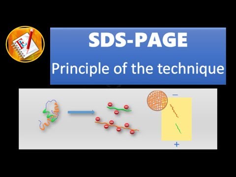
  • 🔄 Proteins are denatured with SDS and beta-mercaptoethanol to break disulfide bonds and mask charges, resulting in uniform negatively charged proteins.
  • 🚫 The electrophoresis process involves the movement of proteins from the negative to the positive terminal due to their negative charge from SDS coating.
  • 🏃‍♂️ Small ions like chloride move quickly towards the positive terminal, followed by proteins, with larger, slower glycine ions lagging behind.
  • 🏁 Proteins are 'stacked' at the interface of the stacking and resolving gels before being separated in the resolving gel based on their molecular weight.
  • 🖼️ After electrophoresis, gels can be stained with Coomassie Brilliant Blue or silver stain to visualize protein bands for analysis.
  • 🔍 Western blotting is a further technique used to confirm the presence of a protein of interest in a sample after SDS-PAGE separation.

Q & A

  • What is SDS-PAGE?

    -SDS-PAGE, or SDS polyacrylamide gel electrophoresis, is a technique used to separate proteins based on their molecular weight by utilizing a gel system.

  • What are the two types of gels used in SDS-PAGE?

    -The two types of gels used in SDS-PAGE are the stacking gel, with a pH of 6.8, and the resolving gel, with a pH of 8.8.

  • Why is there a pH difference between the stacking gel and the resolving gel?

    -The pH difference ensures proper separation of proteins. The stacking gel's lower pH helps to 'stack' the proteins at the boundary, while the resolving gel's higher pH facilitates their separation based on molecular weight.

  • What role does pore size play in protein separation in SDS-PAGE?

    -The pore size, which depends on the concentration of acrylamide and bis-acrylamide, affects the separation of proteins. Smaller proteins require higher concentrations of gel to be properly separated.

  • Why is SDS and beta-mercaptoethanol added to the protein solution?

    -SDS denatures proteins and masks their charges with a uniform negative charge, while beta-mercaptoethanol breaks disulfide bonds, ensuring proteins are fully denatured and linearized.

  • How do proteins move during electrophoresis in SDS-PAGE?

    -Since SDS coats proteins with a negative charge, they move towards the positive terminal during electrophoresis, allowing separation based on their molecular weight.

  • What is the role of chloride ions and glycine in the running buffer?

    -Chloride ions, being small and fast, lead the proteins in the electrophoretic process, while glycine ions, which move slowly at pH 6.8, lag behind, aiding in the stacking of proteins in the stacking gel.

  • How are proteins separated in the resolving gel?

    -Once proteins reach the resolving gel, they are separated based on their molecular weight, with larger proteins moving slower than smaller ones.

  • What staining techniques can be used to visualize proteins after SDS-PAGE?

    -Proteins can be stained using techniques like Coomassie Brilliant Blue staining or silver staining to visualize the separated proteins on the gel.

  • What additional step is required to confirm the presence of a specific protein in the sample?

    -To confirm the presence of a specific protein, a Western blot is performed after SDS-PAGE, where the protein of interest is detected using specific antibodies.

Outlines

00:00

🧬 Understanding SDS-PAGE: Protein Separation Technique

This paragraph introduces the concept of SDS-polyacrylamide gel electrophoresis (SDS-PAGE), a technique used to separate proteins based on their molecular weight. The video explains the gel-based separation process involving two types of gels: the stacking gel and the resolving gel. The stacking gel has a pH of 6.8 and larger pores with lower ionic strength, while the resolving gel has a pH of 8.8, smaller pores, and higher ionic strength. The percentage of polyacrylamide and bisacrylamide in the gels can be varied to create different pore sizes, which is crucial for separating proteins of different sizes. The paragraph also discusses the use of SDS and beta-mercaptoethanol to denature proteins, remove disulfide bonds, and mask charges, resulting in a uniform negative charge. This preparation allows proteins to be separated based on their size during electrophoresis.

05:01

🔬 SDS-PAGE Electrophoresis Process and Visualization

This paragraph delves into the electrophoresis process of SDS-PAGE, where negatively charged proteins move towards the positive terminal. The movement of chloride ions, which are small and move quickly, is contrasted with the bulkier proteins that move more slowly. The paragraph explains the role of glycine ions at pH 6.8, which behave as cations and move slowly, contributing to the stacking of proteins at the top of the resolving gel. This stacking effect aligns proteins at the starting line, allowing them to be separated based on their molecular weight. The video concludes with a discussion on visualizing the results of the electrophoresis through staining methods like Coomassie brilliant blue or silver staining. It also mentions the use of Western blot for further analysis of specific proteins of interest, with a call to action for viewers to watch a related video on Western blotting.

Mindmap

Keywords

💡SDS-PAGE

SDS-PAGE stands for Sodium Dodecyl Sulfate-Polyacrylamide Gel Electrophoresis. It is a widely used technique in biochemistry and molecular biology for the separation of proteins based on their molecular weight. In the script, SDS-PAGE is the main subject of the video, explaining how proteins are separated using this method. The video describes how proteins are denatured, coated with SDS, and then loaded onto a gel to be separated.

💡Polyacrylamide Gel

Polyacrylamide gel is a gel made from polyacrylamide, a synthetic polymer, and is used in gel electrophoresis to separate biomolecules like proteins and nucleic acids. The video script explains that the gel is composed of two components, acrylamide and bisacrylamide, which polymerize to form a matrix with different pore sizes, allowing for the separation of proteins based on their size.

💡Stacking Gel

The stacking gel is the top layer of the gel in SDS-PAGE, where samples are loaded. It has a lower concentration of polyacrylamide and larger pores compared to the resolving gel, which allows for the stacking of proteins before they enter the resolving gel. The video script mentions that the stacking gel has a pH of 6.8, which is different from the resolving gel, contributing to the separation process.

💡Resolving Gel

The resolving gel is the lower layer of the gel in SDS-PAGE, where the actual separation of proteins occurs. It has a higher concentration of polyacrylamide and smaller pores, which allows for the separation of proteins based on their molecular weight. The script explains that the percentage of the resolving gel can vary, affecting the pore size and thus the resolution of the separation.

💡Molecular Weight

Molecular weight refers to the mass of a molecule, which in the context of the video, is used to describe the size of proteins. The video script discusses how proteins are separated in the gel based on their molecular weight, with smaller proteins moving faster through the gel matrix than larger ones.

💡SDS

SDS, or Sodium Dodecyl Sulfate, is a detergent that denatures proteins, causing them to unfold and coat the proteins with a uniform negative charge. This ensures that all proteins move at the same rate in the electric field during electrophoresis, allowing for separation based on size rather than charge. The script describes how SDS treatment prepares the proteins for loading onto the gel.

💡Beta Mercaptoethanol

Beta mercaptoethanol is a reducing agent used in conjunction with SDS to break disulfide bonds within proteins, further aiding in their denaturation. The script mentions that beta mercaptoethanol is added to the protein solution along with SDS to ensure complete denaturation before electrophoresis.

💡Running Buffer

The running buffer is the liquid medium in which the gel electrophoresis takes place. It contains ions that facilitate the movement of charged particles in an electric field. The script explains that one component of the running buffer is chloride ion, which moves quickly towards the positive terminal, helping to drive the proteins through the gel.

💡Glycine

Glycine is an amino acid that, at the pH of the running buffer, behaves as a cation and moves slowly in the electric field. The script describes how glycine ions lag behind the proteins, contributing to the stacking effect at the start of the resolving gel and aiding in the separation process.

💡Western Blot

Western blot is a technique used to detect specific proteins in a sample after they have been separated by SDS-PAGE. It involves transferring the proteins from the gel to a membrane and probing them with specific antibodies. The video script suggests that viewers watch another video for a detailed explanation of Western blot, indicating its importance in protein analysis.

Highlights

Introduction to HTS-PAGE, a technique for separating proteins by molecular weight.

Explanation of the gel-based separation system using a combination of running and resolving gels.

Difference in pH between stacking gel (6.8) and resolving gel (8.8) and its significance.

Varying percentages of resolving gel to achieve different pore sizes for protein separation.

Composition of polyacrylamide gel made from acrylamide and bisacrylamide.

Adjustable ratio of acrylamide to bisacrylamide to create different pore sizes in the gel.

Use of HTS and beta mercaptoethanol to denature proteins and mask charges.

Impact of SDS treatment on protein structure, leading to uniform charge and linearization.

Loading of protein samples onto the gel for electrophoresis.

Movement of negatively charged proteins from negative to positive terminal during electrophoresis.

Role of chloride ions as leaders in the electrophoretic process due to their small size.

Glycine ions' behavior at pH 6.8 and their slow movement in the gel.

Protein stacking at the resolving gel interface, preparing for molecular weight-based separation.

Proteins' movement and separation in the resolving gel based on their molecular weight.

Completion of gel running, visualization of the unstained gel, and subsequent staining methods.

Use of Coomassie Brilliant Blue or silver staining for visualizing protein bands on the gel.

Necessity of Western blot for identifying a protein of interest in the sample.

Encouragement to watch the Western blot video for further understanding.

Closing remarks, appreciation for watching, and call to action for likes, shares, and subscriptions.

Transcripts

play00:00

hi and welcome to my video series of

play00:03

piyo techniques explained in five

play00:05

minutes where I explained the concept in

play00:06

biology in less than 5 minutes or so so

play00:08

if you haven't yet subscribed to my

play00:10

channel

play00:10

hit that subscribe button right now so

play00:13

today's video is about HTS page so SDS

play00:18

page or SDS polyacrylamide gel

play00:20

electrophoresis it's a technique by

play00:22

which we can separate proteins according

play00:24

to their molecular weight now there are

play00:27

this system is based on gel based

play00:30

separation and the jail is a complex of

play00:33

two type of jails are running jail and

play00:35

resolving jail and using a combination

play00:37

of running gel and the resolving gel

play00:39

which we would be discussing in this

play00:40

video one can finally resolve the

play00:44

proteins according to their molecular

play00:45

weight so let's go step by step and

play00:48

understand this process process in

play00:50

details so here imagine this is the jail

play00:54

that you need to run for the

play00:56

polyacrylamide gel electrophoresis it

play00:58

has two basic region one is Technogel

play01:01

that is on the top where you load the

play01:03

samples and here is something called

play01:05

resolving gel so what is different

play01:08

between these two region the stacking

play01:10

gel has a pH of 6.8 and the loading J

play01:13

and the resolving gel has a pH of 8.8

play01:17

and why there is a pH difference we

play01:19

would get to know very soon also the

play01:21

percentage of these gels are very

play01:23

different the percentage of resolving

play01:25

gel varies across the usage of the user

play01:29

they can use different percentage of

play01:32

resolving gel and different percent of

play01:34

resolving gel has different pore size

play01:36

which would allow them to separate the

play01:38

samples properly so generally the

play01:41

stacking gel has larger pore and lower

play01:44

ionic strength whereas the resolving gel

play01:46

has smaller pores and higher ionic

play01:48

strength why is so we would get to know

play01:50

soon so the poly acrylamide which is

play01:54

used the poly acrylamide gel which is

play01:57

used to separate proteins is basically

play01:59

made up of two components

play02:00

acrylamide and miss acrylamide so they

play02:03

polymerize to form this whole shell-like

play02:06

structure and the percentage of these

play02:09

poly acrylamide and miss acrylamide can

play02:12

be changed and their ratio could be

play02:13

varied

play02:14

to get different type of pore size and

play02:16

this pore size difference is important

play02:20

because let's say we need to separate a

play02:22

protein which is very small in size so

play02:25

we would prefer using a higher

play02:27

concentration of jail right because

play02:31

protein size could be different and in

play02:33

order to resolve them we need proper

play02:34

size now let's just try to start this

play02:38

process by let's say we have a protein

play02:41

solution then we add into the protein

play02:44

solution

play02:44

HTS and Peter mercaptoethanol and why an

play02:47

HTS because SDS and beta mercaptoethanol

play02:50

would first of all denature the protein

play02:53

get rid of all the disulfide bonds and

play02:56

kind of mask all of different charges on

play02:59

the protein by negative charge so after

play03:02

that you put it in incubator to boil it

play03:05

for some time so this is the situation

play03:07

of the protein before a seediest

play03:09

treatment after SDS treatment you can

play03:11

see the charge are uniformly Mark's mask

play03:14

by the SDS and everything is like

play03:17

uniform and linearized so all the

play03:20

secondary structures are broken after

play03:21

this treatment now you would load this

play03:24

gel and start the gel for and try to

play03:29

separate the proteins according to the

play03:32

electro phoretic principle now the

play03:35

proteins are now negatively charged

play03:37

because their SDS coated so they would

play03:40

move from negative to the positive

play03:42

terminal but there are multiple things

play03:45

in the solution then use running buffer

play03:47

one of the component of the running

play03:49

buffer is chloride ion so chloride ion

play03:52

is a small ion it would quickly go to

play03:54

the positive and so it's the leader so

play03:57

it runs very fast after that the protein

play04:01

results pretty much slowly because the

play04:04

protein is big bulkier but it has also

play04:06

negative charge so it would be attracted

play04:07

to the positive terminal it would move

play04:09

towards the resolving gel side and at

play04:13

the end there is a glycine ion now at pH

play04:16

6.8 glycine and behave like a cation and

play04:19

since it's a cation

play04:21

it's mobility is ultra slow and it is

play04:26

it is lagged behind in this whole

play04:28

process so they move very slowly now

play04:31

let's say they're moving and at the end

play04:36

of this process what would be happening

play04:38

is the proteins would be stacked on top

play04:41

of the resolving gel as if they are like

play04:46

making their position in the starting

play04:50

line and from that they would be

play04:52

resolved according to the function of

play04:54

their molecular weight right so the pH

play04:57

of the running gel is closer to the pKa

play05:00

of glycine that is why it becomes also

play05:04

negatively charged and it quickly moves

play05:07

just like the chlorine ion and now the

play05:09

protein is free and it would move like

play05:11

its own pace and it would be separated

play05:13

by its own molecular weight so after

play05:17

that let's say we stop the timer when

play05:19

the gel running is complete because we

play05:21

can understand by the die front and then

play05:24

this is how our unstained gel look like

play05:26

we can stain the gel by different method

play05:29

one such method is using Kumasi

play05:32

brilliant blue or sometimes silver

play05:34

staining or anything so you can stain

play05:36

the gel and you know the expected band

play05:39

size so you can ultimately check the

play05:42

stain gel and check the known protein

play05:44

markers to understand that whether your

play05:48

protein of interest is there whether the

play05:50

resolving is happening but in order to

play05:51

understand a particular protein of

play05:53

interest is present in your protein

play05:55

sample or not you have to do Western

play05:57

blot and if you haven't yet washed my

play05:59

Western blot video quickly watch the

play06:01

Western blot and the link is in the

play06:03

description and thanks for listening I

play06:06

hope you enjoyed this video if you liked

play06:08

this video give it a quick thumbs up and

play06:10

don't forget to Like share and subscribe

play06:12

thank you

Rate This

5.0 / 5 (0 votes)

関連タグ
HTS-PAGEProtein SeparationBiologyMolecular WeightGel ElectrophoresisSDS-PAGEPolyacrylamide GelBiotechnologyLab TechniquesEducational Video
英語で要約が必要ですか?