General Embryology - Detailed Animation On Second Week Of Development
Summary
TLDRThe script details the early stages of human development post-fertilization, focusing on the implantation of the blastocyst into the uterine wall. It describes the differentiation of the trophoblast and embryoblast, the formation of the amniotic cavity, and the establishment of uteroplacental circulation. By the end of the second week, the bilaminar embryonic disc is connected to the trophoblast, laying the groundwork for the future umbilical cord.
Takeaways
- 🌱 Implantation of the blastocyst typically happens 6 to 8 days post-fertilization.
- 🌼 By day 8, the blastocyst has implanted into the uterine endometrium, consisting of the Trophoblast and the Inner Cell Mass (Embryoblast).
- 🔍 The Trophoblast differentiates into the cytotrophoblast and syncytiotrophoblast upon contact with the endometrium.
- 🌿 The Embryoblast forms a bilaminar embryonic disc made up of the hypoblast and epiblast.
- 🌌 An amniotic cavity starts to form between the epiblast and cytotrophoblast.
- 🕸️ The hypoblast cells migrate to form the exosomic membrane, contributing to the primitive yolk sac's walls.
- 📅 By day 12, the syncytiotrophoblast forms lacunae which later merge into a lacuna network for maternal blood flow.
- 🌐 Maternal sinusoids dilate, and the syncytiotrophoblast's expansion allows for uteroplacental circulation.
- 🔬 Extraembryonic mesoderm appears, leading to the formation of the chorionic cavity.
- 🔗 Around day 13, the secondary yolk sac forms, and by the end of the second week, the connecting stalk (future umbilical cord) links the bilaminar embryonic disc to the Trophoblast.
Q & A
When does the implantation of the blastocyst typically occur after fertilization?
-Implantation of the blastocyst usually occurs 6 to 8 days after fertilization.
What are the two main components of the blastocyst at the time of implantation?
-At the time of implantation, the blastocyst is composed of the outer cell mass, the trophoblast, and the inner cell mass, the embryoblast.
What differentiates from the trophoblast when it makes contact with the endometrium?
-When the trophoblast makes contact with the endometrium, it differentiates into two layers: an inner cytotrophoblast and an outer syncytiotrophoblast.
What is the amniotic cavity and how does it form?
-The amniotic cavity is a cavity that begins to appear between the epiblast and the cytotrophoblast soon after the embryonic disc has formed.
What is the exosomic membrane and how is it formed?
-The exosomic membrane is a thin membrane that covers the inner surface of the cytotrophoblast and is formed by cells originating from the hypoblast.
What is the primitive yolk sac and how is it formed?
-The primitive yolk sac is formed by the exosomic membrane and cells of the hypoblast together, which form its walls.
What are lacunae and how do they relate to the developing embryo?
-Lacunae are small holes that begin to form in the syncytiotrophoblast as it continues to expand. They later fuse to form large interconnecting spaces called lacuna networks.
How is uteroplacental circulation established during early development?
-Uteroplacental circulation is established as the syncytiotrophoblast expands, eroding the lining of the sinusoids and uterine glands, allowing maternal blood and uterine secretions to flow into the lacunar networks.
What is the extraembryonic mesoderm and when does it appear?
-The extraembryonic mesoderm is a new population of cells that appear between the inner surface of the cytotrophoblast and the outer surface of the primitive yolk sac.
What is the chorionic cavity and how does it form?
-The chorionic cavity is a single cavity that forms in the extraembryonic mesoderm as large cavities begin to appear and gradually fuse.
What is the secondary yolk sac and how is it formed?
-The secondary yolk sac is a smaller cavity that forms when a large portion of the exocoelomic cavity is pinched off around 13 days after fertilization.
What is the connecting stalk and its role in development?
-The connecting stalk is a band of extraembryonic mesoderm that joins the bilaminar embryonic disc to the trophoblast by the end of the second week of development, which will become the future umbilical cord.
Outlines

このセクションは有料ユーザー限定です。 アクセスするには、アップグレードをお願いします。
今すぐアップグレードMindmap

このセクションは有料ユーザー限定です。 アクセスするには、アップグレードをお願いします。
今すぐアップグレードKeywords

このセクションは有料ユーザー限定です。 アクセスするには、アップグレードをお願いします。
今すぐアップグレードHighlights

このセクションは有料ユーザー限定です。 アクセスするには、アップグレードをお願いします。
今すぐアップグレードTranscripts

このセクションは有料ユーザー限定です。 アクセスするには、アップグレードをお願いします。
今すぐアップグレード関連動画をさらに表示
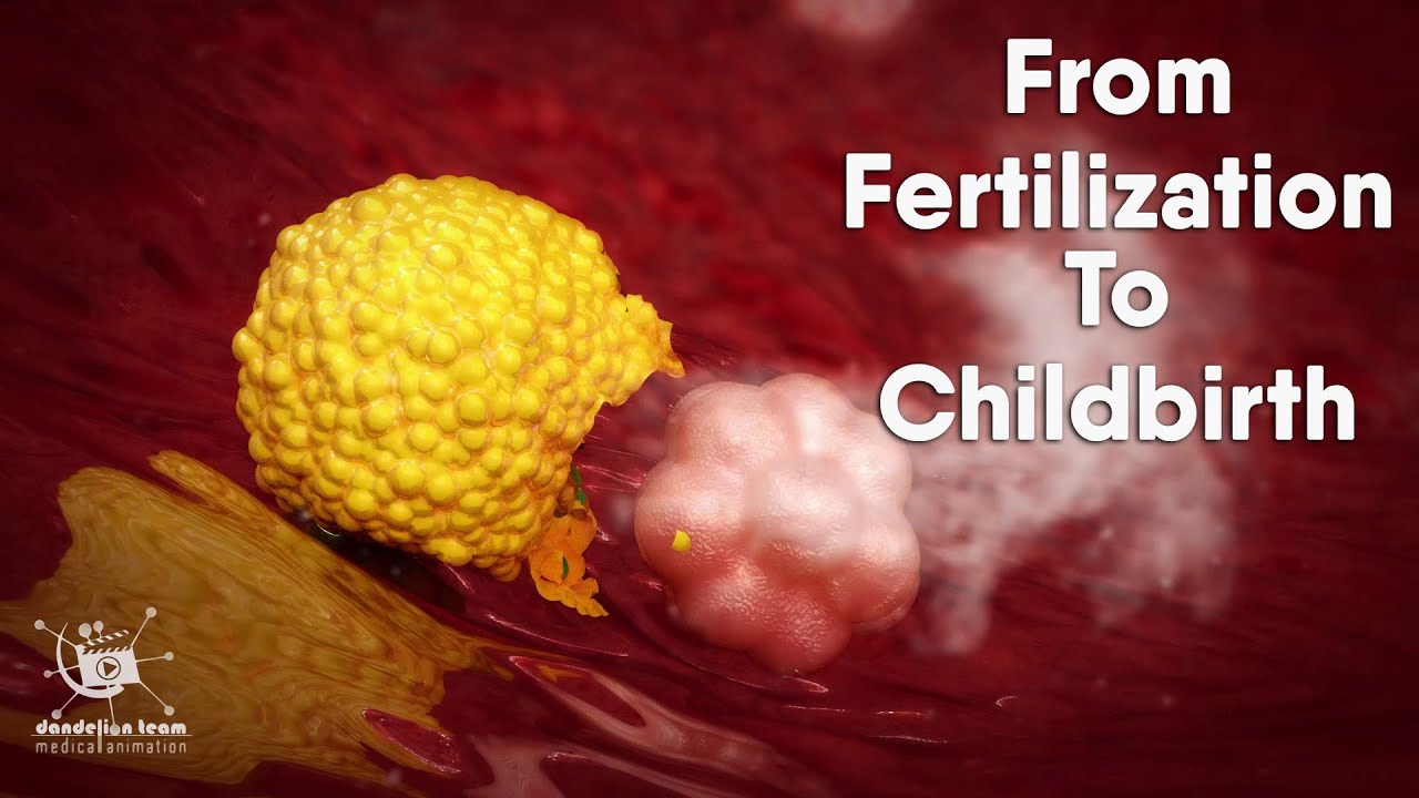
from fertilization to childbirth | 3d medical animation | by Dandelion Team

General Embryology Review in 20 minutes

EMBRIOLOGIA HUMANA | AULA 06 | PRIMEIRA SEMANA DO DESENVOLVIMENTO EMBRIONÁRIO

Anatomi Embriologi Manusia : Perkembangan Minggu Pertama

Fertilisasi dan Embriogenesis (Reproduksi Manusia)

Fertilisation and implantation
5.0 / 5 (0 votes)
