A-Level Biology - Cell Fractionation & Ultracentrifugation (2026/27 exams)
Summary
TLDRThis video explains the process of cell fractionation, a laboratory technique used to isolate and study the different organelles within cells. It outlines the four key steps: sample preparation, homogenization, filtration, and ultracentrifugation. The tutorial details how tissues are placed in an ice-cold isotonic buffer to protect organelles, then cells are broken open, debris removed, and organelles separated by density using a centrifuge. The video emphasizes the order of organelle separation from heaviest to lightest, making it especially useful for detailed study under an electron microscope. Additional learning resources are also highlighted.
Takeaways
- 😀 Cell fractionation is a laboratory technique used to isolate the different components of a cell, particularly organelles.
- 😀 The process begins by choosing an organism, like a plant, taking a tissue sample, and breaking open the cells.
- 😀 Organelles are then separated based on their size and density, which allows for more detailed study, especially under an electron microscope.
- 😀 There are four key steps in cell fractionation: sample preparation, homogenization, filtration, and ultra-centrifugation.
- 😀 Sample preparation involves placing the tissue in an ice-cold, isotonic, buffered solution to protect organelles and prevent damage.
- 😀 The solution is ice-cold to slow down enzyme activity, isotonic to maintain water balance, and buffered to keep the pH stable.
- 😀 Homogenization involves physically breaking open the cells, often using a blender, to release the organelles into the solution.
- 😀 Filtration removes larger debris and tissue fragments, with the organelles passing through gauze into the filtrate.
- 😀 Ultra-centrifugation separates organelles based on their density by spinning the filtrate at various speeds in a centrifuge.
- 😀 The process starts at a low speed to pellet the heaviest organelles (e.g., nuclei) and continues at higher speeds to separate lighter organelles, like chloroplasts, mitochondria, and ribosomes.
Q & A
What is cell fractionation?
-Cell fractionation is a laboratory technique used to isolate and study the different components of a cell, especially organelles, by breaking open cells and separating their components based on size and density.
Why is an ice-cold, isotonic, buffered solution used during sample preparation?
-The solution protects organelles by slowing enzyme activity (ice-cold), preventing water movement that could damage organelles (isotonic), and maintaining a constant pH to avoid protein denaturation (buffered).
What is the purpose of homogenization in cell fractionation?
-Homogenization physically breaks open cells to release organelles into solution, typically by using a blender or other mechanical method.
Why is filtration necessary after homogenization?
-Filtration removes large debris and tissue fragments that are not wanted in the sample, allowing only organelles to pass through into the filtrate for further processing.
How does ultracentrifugation separate organelles?
-Ultracentrifugation separates organelles based on size and density by spinning the sample at increasing speeds, causing heavier organelles to form pellets first, while lighter organelles remain in the supernatant.
What order are organelles separated in during ultracentrifugation of plant tissue?
-The organelles are separated from heaviest to lightest as follows: nuclei, chloroplasts, mitochondria, lysosomes, endoplasmic reticulum, and ribosomes.
Why is it important to maintain organelles intact during fractionation?
-Intact organelles are essential for accurate study of their structure and function, and for reliable biochemical or microscopic analysis.
How does isotonicity prevent organelle damage?
-An isotonic solution has the same water potential as the cells, preventing water from entering or leaving organelles, which could cause swelling or shrinkage and damage them.
What is the supernatant in the context of ultracentrifugation?
-The supernatant is the liquid portion that remains above the pellet after centrifugation, containing the lighter organelles that have not yet sedimented.
Why might electron microscopy be used after cell fractionation?
-Electron microscopy allows detailed visualization of isolated organelles, enabling researchers to study their structure and function more precisely than light microscopy.
What are some common methods to disrupt cells during homogenization?
-Common methods include mechanical grinding, blending, or using specialized homogenizers to physically break open cell membranes.
How does increasing centrifuge speed affect organelle separation?
-Increasing the centrifuge speed allows progressively lighter organelles to sediment and form pellets, ensuring separation from heavier organelles already pelleted.
Outlines

Cette section est réservée aux utilisateurs payants. Améliorez votre compte pour accéder à cette section.
Améliorer maintenantMindmap

Cette section est réservée aux utilisateurs payants. Améliorez votre compte pour accéder à cette section.
Améliorer maintenantKeywords

Cette section est réservée aux utilisateurs payants. Améliorez votre compte pour accéder à cette section.
Améliorer maintenantHighlights

Cette section est réservée aux utilisateurs payants. Améliorez votre compte pour accéder à cette section.
Améliorer maintenantTranscripts

Cette section est réservée aux utilisateurs payants. Améliorez votre compte pour accéder à cette section.
Améliorer maintenantVoir Plus de Vidéos Connexes

A Level Biology Revision "Cell Fractionation"

Differential Centrifugation (Animation)
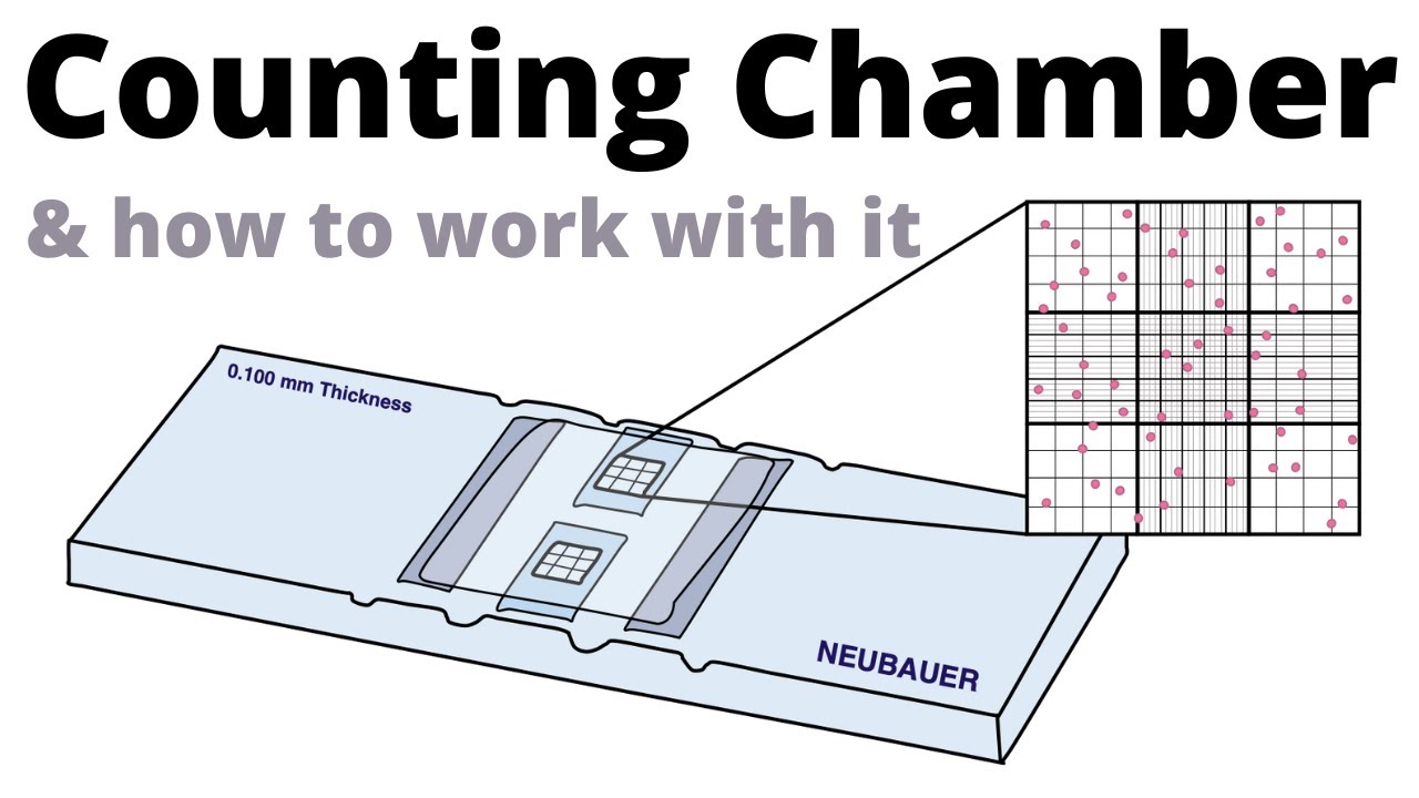
How to count cells with the Neubauer Counting Chamber

Estudo das Células 01 - Métodos de Estudo

BIOLOGI Kelas 11 - Struktur dan Organel Sel | GIA Academy
Pengenalan Sel. Mengenali Dunia Mikroskopis dalam tubuh Makhluk Hidup.@gururatna
5.0 / 5 (0 votes)
