CT Contrast Injection (Flow Rate, Delay and Duration)
Summary
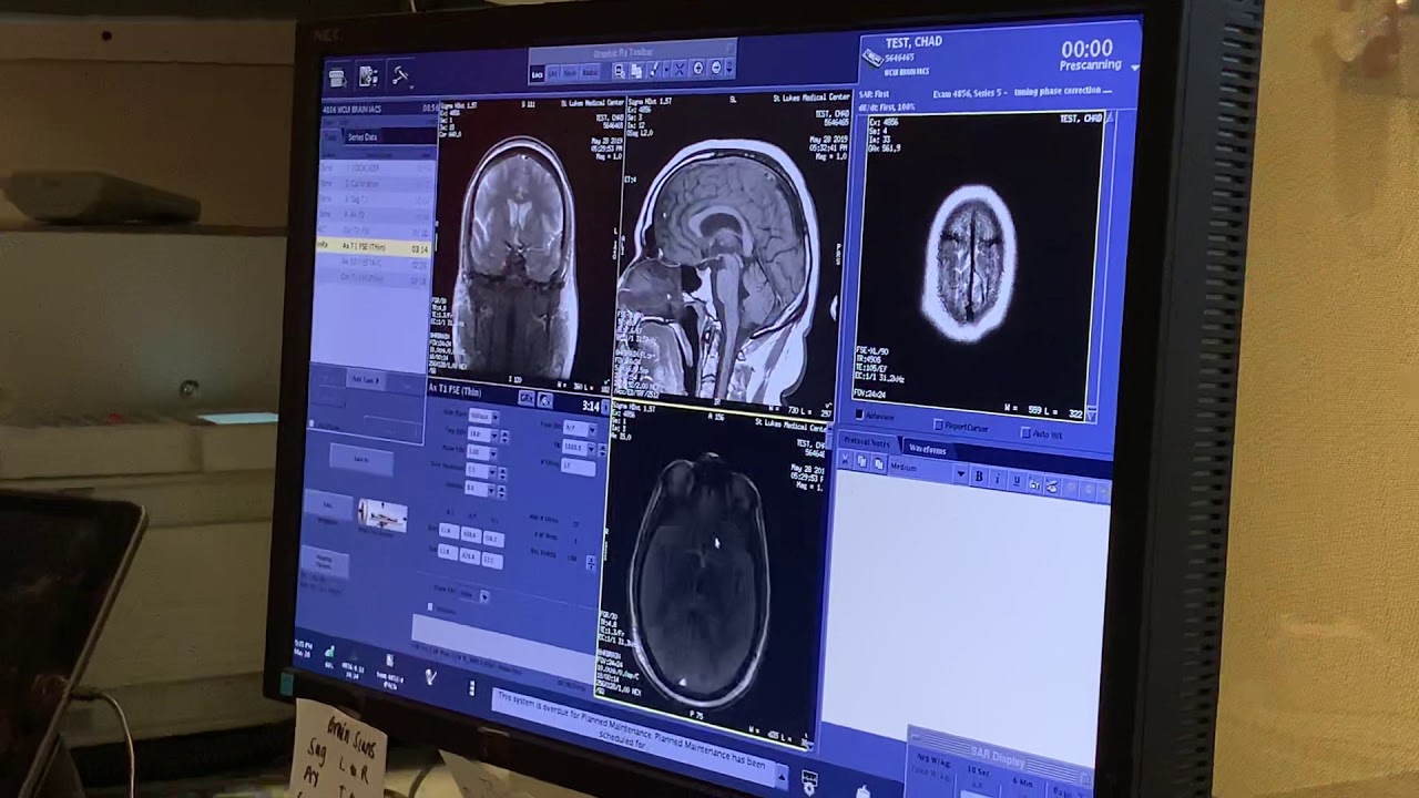
TLDRThis video explores the dynamics of contrast media in CT imaging, focusing on optimal timing and techniques to enhance visualization of organs and vessels. It explains how intravenous contrast travels through the circulatory system, the importance of arrival time, and methods like test bolus and bolus tracking to capture peak enhancement. Key factors affecting imaging quality—including patient size, cardiac output, contrast density, injection rate, and CT scanner settings—are discussed. The video also covers organ-specific contrast phases, from early arterial to excretory, providing practical guidance for achieving high-quality, diagnostically valuable CT scans.
Takeaways
- 😀Video script analysis Contrast media in CT scanning is injected intravenously to optimize safety and ease of access while reducing infection risk.
- 😀 Timing of contrast injection is crucial to achieve optimal enhancement in specific organs such as the heart, lungs, liver, and peripheral vessels.
- 😀 Contrast travels from veins → right heart → lungs → left heart → aorta → target organs; this pathway determines imaging timing.
- 😀 Different organs require imaging at specific contrast phases: early arterial, late arterial, portal venous, nephrogenic, and excretory phases.
- 😀 Time of arrival (T arrival) is the interval between contrast injection and its presence in the target vessel, which informs scan timing.
- 😀 Short injections produce sharp peaks and rapid decay in contrast enhancement, whereas longer injections create a plateau with gradual decay due to recirculation.
- 😀 Test bolus and real-time bolus tracking are techniques used to determine the optimal scanning window based on contrast arrival and threshold HU.
- 😀 Patient-specific factors like size, weight, and cardiac output significantly affect contrast enhancement, requiring dose adjustments for optimal imaging.
- 😀 Contrast media factors including density, injection rate,Key takeaways from script volume, and saline flush impact image quality, bolus tightness, and artifact reduction.
- 😀 CT scanner parameters such as tube voltage (kVp) and imaging phases influence contrast enhancement, with lower kVp increasing contrast but also noise.
- 😀 Proper diagnostic delays after contrast arrival ensure imaging occurs near peak enhancement for the best diagnostic quality.
- 😀 Understanding pharmacokinetics and organ-specific contrast dynamics allows radiologists to plan and execute CT protocols effectively.
Q & A
What is the primary purpose of using contrast in CT scanning?
-The primary purpose of using contrast in CT scanning is to enhance the visibility of blood vessels, organs, and tissues, allowing for clearer and more detailed images, especially for areas with poor natural contrast.
Why is venous contrast injection preferred over intra-arterial injection for CT scans?
-Venous contrast injection is preferred because it is easier to establish an IV line, involves a lower risk of infection, and is less invasive compared to intra-arterial injection.
What are some potential issues with contrast pooling in the veins?
-Contrast pooling in the veins can lead to artifacts, such as streaking or beam hardening, which result in image distortion. These artifacts can arise when there is a significant amount of contrast in the superior vena cava.
What does 'T arrival' refer to in CT contrast timing?
-'T arrival' refers to the time between the injection of contrast into the veins and the moment when the contrast reaches the target area (such as the heart or aorta) in the body, marking the beginning of the imaging phase.
How does the flow rate of contrast affect the timing of the scan?
-A faster flow rate will lead to a quicker rise in contrast levels and a sharper, more defined image, while a slower flow rate results in a more gradual buildup of contrast, which may require a longer scan duration.
What is the importance of the diagnostic delay in CT imaging?
-The diagnostic delay is crucial for ensuring that the CT scan is conducted at the optimal moment when the contrast has reached its peak. This prevents premature scanning, which might lead to suboptimal image quality.
How can a CT scanner automatically track contrast arrival and initiate the diagnostic scan?
-A CT scanner can use a test bolus or monitoring phase to track the arrival of contrast. Once the contrast reaches a predefined threshold (usually 50-150 Hounsfield units), the scanner automatically transitions into the diagnostic imaging phase.
What role does patient size play in contrast enhancement?
-Larger patients typically require more contrast to achieve the same level of enhancement as smaller patients. The patient's weight is often used as a surrogate for the total blood volume, affecting the amount of contrast needed for effective imaging.
Why is saline injected after the contrast in CT scanning?
-Saline is injected after the contrast to help push the contrast more efficiently through the veins, maintain a tighter bolus, reduce artifacts, and clear the contrast from the injection tubes.
How does the choice of kVp (kilovolt peak) impact contrast in CT scans?
-Lower kVp settings (such as 100 kVp) increase contrast by enhancing the photoelectric effect, which improves the absorption of X-rays by the contrast medium. However, this comes at the cost of higher noise and reduced penetration, which requires adjustments in other scan parameters.
Outlines

Cette section est réservée aux utilisateurs payants. Améliorez votre compte pour accéder à cette section.
Améliorer maintenantMindmap

Cette section est réservée aux utilisateurs payants. Améliorez votre compte pour accéder à cette section.
Améliorer maintenantKeywords

Cette section est réservée aux utilisateurs payants. Améliorez votre compte pour accéder à cette section.
Améliorer maintenantHighlights

Cette section est réservée aux utilisateurs payants. Améliorez votre compte pour accéder à cette section.
Améliorer maintenantTranscripts

Cette section est réservée aux utilisateurs payants. Améliorez votre compte pour accéder à cette section.
Améliorer maintenantVoir Plus de Vidéos Connexes


5.0 / 5 (0 votes)
