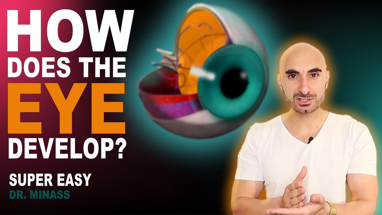
Development of EYE : Visual Learning: Easy learning
Summary
TLDRThis video explains the intricate development of the eye during embryogenesis. Beginning in the third week, the neural plate forms, eventually giving rise to the neural tube, which is the precursor to the brain and spinal cord. The optic vesicle and lens placode form, followed by the creation of the optic cup and the development of retinal layers, ciliary body, and iris. The lens forms from primary and secondary lens fibers, which continue to develop after birth. The video offers a detailed overview of how these structures come together to form the functional eye.
Takeaways
- 😀 The notochord secretes growth factors that stimulate the differentiation of the overlying ectoderm into the neural plate, initiating the development of the eye.
- 😀 Neural folds form from the lateral edges of the neural plate, which then fuse in the midline to create the neural tube, precursor to the brain and spinal cord.
- 😀 The neural tube formation is called neurulation and occurs by the end of the fourth week of development.
- 😀 By the fifth week, the cranial end of the neural tube develops three primary vesicles: prosencephalon (forebrain), mesencephalon (midbrain), and rhombencephalon (hindbrain).
- 😀 The prosencephalon further divides into the telencephalon and diencephalon, which will eventually form the brain's major structures.
- 😀 The optic vesicle forms from the diencephalon and comes into contact with the surface ectoderm, which thickens to form the lens placode.
- 😀 The lens placode sinks to form the lens vesicle, which eventually detaches from the surface ectoderm, forming the lens of the eye.
- 😀 The optic vesicle becomes the optic cup, which has a deficiency on the inferior side, forming the fetal fissure (or choroidal fissure).
- 😀 The mesenchyme surrounding the optic vesicle forms the fibrous outer layer (cornea and sclera) and vascular inner layer (choroid and ciliary body).
- 😀 The retina is derived from the inner and outer layers of the optic cup, with the outer layer forming the pigmented epithelium and the inner layer forming neural components such as rods, cones, and ganglion cells.
Q & A
What is the role of the notochord in the development of the eye?
-The notochord secretes growth factors that stimulate the differentiation of the overlying ectoderm into the neural ectoderm, initiating the formation of the neural plate, which is a crucial step in the development of the eye.
What happens during neural tube formation in eye development?
-The neural folds move towards each other and fuse at the midline, forming the neural tube, which is the precursor to the brain and spinal cord. This process is known as neurulation and is completed by the end of the fourth week.
What are the three primary vesicles formed from the cranial end of the neural tube?
-The three primary vesicles formed from the cranial end of the neural tube are the prosencephalon, mesencephalon, and rhombencephalon.
What are the secondary vesicles formed from the prosencephalon?
-The prosencephalon is divided into two secondary vesicles: the telencephalon and the diencephalon.
What is the role of the optic sulcus in eye development?
-The optic sulcus is a depression that forms along the lateral side of the diencephalon. Its distal part enlarges to form the optic vesicle, while the proximal part becomes constricted to form the optic stalk.
How does the lens placode form and contribute to eye development?
-The surface ectoderm overlying the optic vesicle thickens to form the lens placode. The placode sinks to form the lens vesicle, which eventually detaches from the surface ectoderm and inverts into the optic vesicle, contributing to the development of the lens.
What is the optic cup, and how is it formed?
-The optic cup is a double-layered structure formed from the invagination of the optic vesicle due to the presence of the developing lens. It gives rise to the retina and other eye structures.
What are the layers formed from the mesenchyme around the optic cup?
-The mesenchyme surrounding the optic cup forms two layers: the outer fibrous layer, which contributes to the sclera and cornea, and the inner vascular layer, which forms the choroid and ciliary body.
What is the origin of the retina's two layers?
-The retina's outer pigmented layer is derived from the outer wall of the optic cup, while the inner neural layer originates from the inner wall of the optic cup.
How is the lens formed during eye development?
-The lens is formed from the lens vesicle. The developing neural retina signals cells at the posterior end of the lens vesicle to elongate, forming primary lens fibers. These fibers fill the vesicle and form the lens nucleus, with additional secondary fibers being added later.
Outlines

Cette section est réservée aux utilisateurs payants. Améliorez votre compte pour accéder à cette section.
Améliorer maintenantMindmap

Cette section est réservée aux utilisateurs payants. Améliorez votre compte pour accéder à cette section.
Améliorer maintenantKeywords

Cette section est réservée aux utilisateurs payants. Améliorez votre compte pour accéder à cette section.
Améliorer maintenantHighlights

Cette section est réservée aux utilisateurs payants. Améliorez votre compte pour accéder à cette section.
Améliorer maintenantTranscripts

Cette section est réservée aux utilisateurs payants. Améliorez votre compte pour accéder à cette section.
Améliorer maintenantVoir Plus de Vidéos Connexes

Fertilisasi dan Embriogenesis (Reproduksi Manusia)

Muscles of the eye - extraocular muscles and movements

Embryology of the Eye (Easy to Understand)

Zebrafish embryo development animation

Early embryogenesis - Cleavage, blastulation, gastrulation, and neurulation | MCAT | Khan Academy

Anatomi Sistem Indera-Mata (Bagian 1)
5.0 / 5 (0 votes)
