Fetal Circulation by L. McCabe | OPENPediatrics

OPENPediatrics
16 Mar 202106:07

Summary

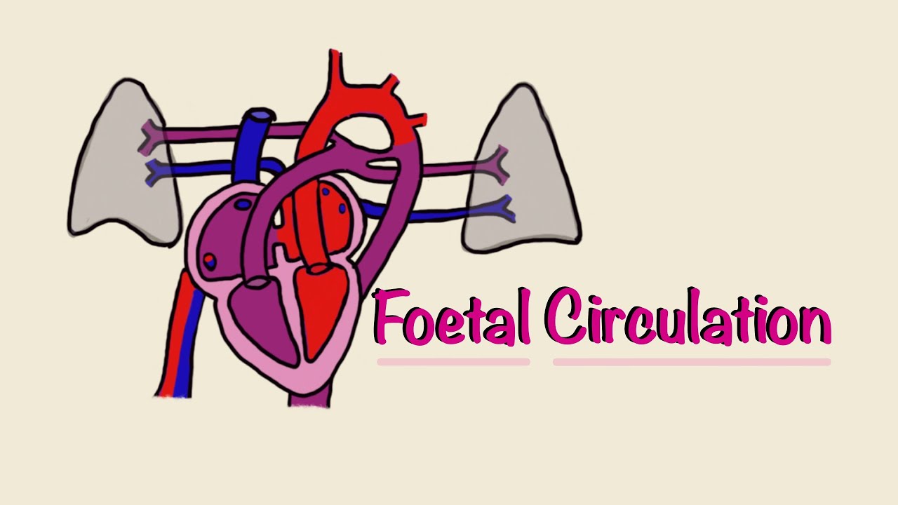
TLDRThis lecture by Lisa McCabe, a Clinical Nurse Specialist at Children's Hospital Boston, explains fetal circulation and the transition to postnatal circulation. Key structures involved in fetal circulation include the placenta, umbilical vein, ductus venosus, foramen ovale, ductus arteriosus, and umbilical arteries. Oxygenated blood from the placenta flows through these structures to nourish the fetus. After birth, the circulation changes as the lungs expand, the placenta is removed, and certain fetal structures close. The foramen ovale and ductus arteriosus normally close, transitioning the baby to postnatal circulation.

Takeaways

  • đŸ‘¶ Fetal circulation includes key structures: the placenta, umbilical vein, ductus venosus, foramen ovale, ductus arteriosus, and umbilical arteries.
  • 🌬 Blood rich in oxygen and nutrients flows from the placenta through the umbilical vein to the ductus venosus, then into the heart's right atrium.
  • 💖 The foramen ovale allows blood to bypass the lungs by flowing from the right atrium to the left atrium, then into the left ventricle, which pumps it to the body.
  • đŸ« Due to high resistance in the fluid-filled fetal lungs, most blood from the right ventricle flows through the ductus arteriosus into the aorta, bypassing the lungs.
  • đŸ©ž Only 8% of blood from the right ventricle reaches the lungs, which are not yet needed for gas exchange, as the placenta handles oxygenation.
  • đŸȘĄ After birth, the umbilical cord is clamped, and the placenta is removed from circulation, starting the transition to postnatal circulation.
  • đŸƒâ€â™‚ïž The increase in systemic vascular resistance and decrease in pulmonary pressure after birth allows blood to flow into the lungs for oxygenation.
  • đŸš« The foramen ovale closes as left atrial pressure rises, and in most cases, it permanently seals within three months.
  • 🔒 The ductus arteriosus and ductus venosus close shortly after birth, becoming the ligamentum arteriosum and ligamentum venosum, respectively.
  • 📊 While the foramen ovale closes in most people, studies show 15-25% of adults have a patent foramen ovale (PFO) that never fully closes.

Q & A

  • What are the main structures associated with fetal circulation?

    -The main structures associated with fetal circulation include the placenta, the umbilical vein, the ductus venosus, the foramen ovale, the ductus arteriosus, and the umbilical arteries.

  • What is the function of the umbilical vein in fetal circulation?

    -The umbilical vein carries blood rich in oxygen and nutrients from the placenta to the ductus venosus, which then directs the blood to the fetal heart.

  • How does blood flow between the right atrium and the left atrium in fetal circulation?

    -In fetal circulation, blood flows from the right atrium to the left atrium through the foramen ovale, allowing oxygenated blood to bypass the lungs and reach the left ventricle and aorta.

  • Why does only a small amount of blood flow to the fetal lungs?

    -Because the fetal lungs are fluid-filled and have high vascular resistance, only about 8% of the right ventricular output flows to the lungs, with most of the blood bypassing the lungs via the ductus arteriosus to the aorta.

  • What happens to the placenta after birth and how does this affect circulation?

    -After birth, the placenta is removed from the systemic circulation, causing systemic vascular resistance to rise and initiating the transition to postnatal circulation.

  • How does the baby’s first breath influence the pulmonary circulation?

    -With each breath, the baby’s alveoli expand and the surrounding blood vessels dilate in response to oxygen, which lowers pulmonary vascular resistance.

  • What is the role of the foramen ovale after birth?

    -After birth, the foramen ovale functionally closes as left atrial pressure increases, preventing blood flow between the atria. In most cases, it permanently closes within three months.

  • What happens if the foramen ovale doesn’t close after birth?

    -If the foramen ovale remains open, a condition called patent foramen ovale (PFO) occurs. While the PFO usually has a small shunt, autopsy studies show that 15-25% of adults may have a PFO that never closed.

  • When does the ductus arteriosus typically close after birth?

    -The ductus arteriosus begins to close shortly after birth and usually closes completely within 4-10 days.

  • What is the fate of the umbilical vessels after birth?

    -After birth, the umbilical vein and arteries are infiltrated with fibrin and eventually become ligaments within about a week.

Outlines

00:00

đŸ‘¶ Fetal Circulation and Key Structures

In this introduction, Lisa McCabe, a Clinical Nurse Specialist at Children's Hospital Boston, discusses fetal circulation and the major structures involved in the process. These structures include the placenta, umbilical vein, ductus venosus, foramen ovale, ductus arteriosus, and the umbilical arteries. She explains how oxygen and nutrient-rich blood from the placenta travels through the umbilical vein and ductus venosus into the inferior vena cava. The blood then flows into the right atrium, where it either moves to the right ventricle or crosses the foramen ovale to the left atrium, reaching the left ventricle and the aorta, supplying blood to the body. McCabe emphasizes the importance of the foramen ovale in ensuring oxygenated blood reaches the coronary arteries and brain.

05:05

đŸ«€ Blood Flow Distribution in Fetal Circulation

This paragraph provides a detailed explanation of how blood flows from the right ventricle into the pulmonary artery. Due to the high resistance in the lungs, most of the blood bypasses the lungs via the ductus arteriosus and flows into the aorta. The limited blood flow to the lungs nourishes developing tissues. Additionally, McCabe explains how blood pressure dynamics between the right and left atrium promote blood flow through the foramen ovale, with only 8% of the right ventricular output going to the lungs. The majority of the blood flows through the ductus arteriosus into the aorta, and the umbilical arteries carry blood back to the placenta for oxygenation.

đŸŒŹïž Transition from Fetal to Postnatal Circulation

This section focuses on the critical transition from fetal to postnatal circulation. After birth, the umbilical cord is clamped, and the placenta is removed from circulation, triggering systemic vascular resistance to rise. As the newborn breathes, pulmonary pressures begin to drop, though full normalization takes six to eight weeks. The foramen ovale and ductus arteriosus close in response to changes in pressure, effectively rerouting blood flow in postnatal circulation. McCabe highlights that although the foramen ovale usually closes, a small patent foramen ovale (PFO) may persist in some adults, allowing minimal blood shunting.

⏳ Closure of Ductus Arteriosus and Other Fetal Structures

This paragraph describes the closure process of the ductus arteriosus and other fetal structures after birth. Normally, the ductus arteriosus closes within 4-10 days, while the ductus venosus remains open initially, allowing central venous access through the umbilical vein. Over 3-7 days, fibrin infiltrates the ductus venosus, forming the ligamentum venosum. The umbilical vein and arteries also become ligaments within a week after birth. These structural changes are a crucial part of the newborn's adaptation to life outside the womb. McCabe concludes the lecture by thanking the audience.

Mindmap

Keywords

💡Fetal Circulation

Fetal circulation refers to the unique blood flow system in a developing fetus that allows oxygen and nutrients from the placenta to reach vital organs. In the video, it is described in detail, emphasizing the structures and pathways, like the umbilical vein, that facilitate the distribution of oxygenated blood throughout the fetus’s body.

💡Placenta

The placenta is an essential organ in fetal circulation that provides oxygen and nutrients to the developing fetus while removing waste products. In the script, the placenta supplies blood rich in nutrients and oxygen via the umbilical vein, making it a key part of the fetal circulatory system.

💡Ductus Venosus

The ductus venosus is a blood vessel in the fetal circulation system that directs oxygenated blood from the umbilical vein into the inferior vena cava, bypassing the liver. It is mentioned in the script as a critical pathway that enables efficient delivery of nutrient-rich blood to the heart.

💡Foramen Ovale

The foramen ovale is an opening between the right and left atria of the fetal heart that allows oxygenated blood to bypass the lungs, which are not yet in use. In the video, it plays a central role in ensuring that oxygenated blood from the placenta reaches important areas like the coronary arteries and cerebral circulation.

💡Ductus Arteriosus

The ductus arteriosus is a vessel that connects the pulmonary artery to the aorta in the fetus, allowing most of the blood to bypass the non-functioning lungs and flow directly into the systemic circulation. The video explains that this structure is vital in diverting blood away from the fluid-filled lungs to maintain proper circulation in the fetus.

💡Umbilical Vein

The umbilical vein carries oxygen-rich blood from the placenta to the fetus. It is one of the key vessels in fetal circulation, delivering essential nutrients and oxygen. The script explains its pathway to the ductus venosus, highlighting its role in sustaining fetal development.

💡Umbilical Arteries

The umbilical arteries are vessels that carry deoxygenated blood from the fetus back to the placenta for oxygenation and nutrient replenishment. These arteries are critical in maintaining the exchange process between the fetus and the placenta, as noted in the video.

💡Pulmonary Circulation

Pulmonary circulation refers to the movement of blood from the heart to the lungs and back, which is limited in the fetus due to fluid-filled lungs. The video emphasizes that, because of high resistance in the pulmonary vessels, most blood bypasses the lungs through the ductus arteriosus.

💡Postnatal Circulation

Postnatal circulation is the blood flow system that takes over after birth when the baby begins to breathe and the lungs take over oxygenation. The transition from fetal to postnatal circulation involves the closure of the foramen ovale and ductus arteriosus, as explained in the script.

💡Systemic Circulation

Systemic circulation refers to the part of the circulatory system that delivers oxygenated blood from the heart to the rest of the body and returns deoxygenated blood to the heart. The script discusses the low resistance in systemic circulation during fetal life, contrasting it with the high resistance in pulmonary circulation.

Highlights

Introduction to fetal circulation and its transition to postnatal circulation.

Key structures in fetal circulation: placenta, umbilical vein, ductus venosus, foramen ovale, ductus arteriosus, and umbilical arteries.

Oxygenated blood flows from the placenta through the umbilical vein to the ductus venosus, then into the inferior vena cava and right atrium.

The foramen ovale allows blood to bypass the lungs, moving from the right atrium to the left atrium, then into the left ventricle and aorta.

The foramen ovale plays a critical role in delivering oxygenated blood to the coronary arteries and brain in the fetus.

Due to the fluid-filled lungs in fetuses, blood is diverted from the pulmonary artery through the ductus arteriosus into the aorta.

The umbilical arteries carry deoxygenated blood back to the placenta for nutrient and oxygen exchange.

After birth, clamping the umbilical cord removes the placenta from circulation, initiating the shift to postnatal circulation.

Pulmonary pressures decrease as the baby breathes and the lungs expand, allowing more blood to flow through the lungs.

The foramen ovale functionally closes due to increased left atrial pressure after birth, with permanent closure within three months.

15-25% of adults retain a patent foramen ovale (PFO), though typically with minimal impact.

The ductus arteriosus closes 4-10 days after birth as the infant begins breathing air.

The ductus venosus closes within 3-7 days after birth, transforming into the ligamentum venosum.

The umbilical vein and arteries turn into ligaments within a week post-birth as they are infiltrated with fibrin.

Summary and conclusion of the transition from fetal to postnatal circulation.

Transcripts

play00:12

Fetal Circulation by Lisa McCabe.

play00:16

Hello, my name is Lisa McCabe.

play00:18

I'm a Clinical Nurse Specialist at Children's Hospital Boston

play00:22

in the Cardiovascular Program.

play00:24

We are now going to talk about fetal circulation

play00:27

and the transition to postnatal circulation that

play00:30

occurs after birth.

play00:32

The specific structures associated

play00:34

with fetal circulation include the placenta,

play00:38

the umbilical vein, the ductus venosus, the foramen ovale,

play00:44

the ductus arteriosus, and the umbilical arteries.

play00:49

Blood rich in nutrients and oxygen,

play00:51

supplied via the placenta, flows through the umbilical vein

play00:55

to the ductus venosus.

play00:57

Blood flows from the ductus venosus into the inferior vena

play01:01

cava, up to the right atrium.

play01:05

This blood mixes with blood returning

play01:07

to the heart from the upper body via the superior vena cava,

play01:12

and from the lower body via the inferior vena cava.

play01:16

Once in the right atrium, some of the blood

play01:20

flows to the right ventricle and some of the blood

play01:23

flows through the foramen ovale, through the left atrium,

play01:27

and into the left ventricle, where it is then pumped out

play01:32

into the aorta to the body.

play01:35

Point of Clarification. The flow of blood from the

play01:38

right atrium to the left atrium via the foramen ovale enables

play01:41

oxygenated blood from the placenta to reach the coronary arteries

play01:45

in cerebral circulation, the two most metabolic, active organs

play01:48

in the developing fetus.

play01:51

Blood that flows into the right ventricle

play01:54

is then pumped into the pulmonary artery.

play01:57

Because the lungs are fluid-filled instead

play02:00

of air-filled, the vessels in the lungs

play02:03

are narrow, creating higher resistance to blood flow

play02:07

into the lungs.

play02:09

Due to this high resistance to blood flow

play02:11

in the pulmonary circulation and the low resistance

play02:15

to blood flow in the systemic circulation,

play02:18

blood pumped into the pulmonary artery by the right ventricle

play02:23

is more likely to flow into the ductus arteriosus

play02:27

and then into the aorta.

play02:30

Due to the high blood flow from the superior vena

play02:32

cava and the inferior vena cava up to the right atrium,

play02:37

pressures in the right atrium are higher than

play02:40

in the left atrium.

play02:42

This promotes blood flow through the foramen ovale

play02:46

at the atrial level.

play02:48

Only 8% of right ventricular output flows into the lungs,

play02:53

providing nutrients for the developing lung tissues.

play02:58

Most of the blood passes through the ductus arteriosus

play03:02

into the aorta and out to the body.

play03:06

The umbilical arteries allow blood to flow from the body

play03:11

back to the placenta to be enriched

play03:14

with oxygen and nutrients.

play03:17

Once the baby is born and takes a breath,

play03:20

the umbilical cord is clamped and the placenta

play03:23

is removed from the systemic circulation.

play03:27

Immediately, the transition from fetal to postnatal circulation

play03:31

begins.

play03:32

With the elimination of the placenta from the circulation,

play03:36

systemic vascular resistance begins to rise.

play03:40

With each breath, more alveoli in the lungs

play03:44

expand, and the vessels surrounding

play03:46

them dilate in response to the presence of oxygen.

play03:51

Pulmonary pressures begin to decrease.

play03:54

Although pulmonary pressures are lower than systemic pressures,

play03:58

within minutes after birth it is six to eight weeks

play04:02

before pulmonary vascular resistance decreases to normal.

play04:08

In the postnatal circulation, blood no longer

play04:12

flows through the foramen ovale or the ductus arteriosus.

play04:16

The increase in left atrial pressure after birth

play04:20

forces the septum primum against the septum secundum,

play04:25

functionally closing the foramen ovale.

play04:28

Within three months, the foramen ovale permanently

play04:32

closes as fibrin deposits fuse the layers of septal wall

play04:37

together.

play04:40

Point of clarification.

play04:41

While the patent foramen ovale, or PFO, may close by this time,

play04:46

it also may not.

play04:47

Autopsy studies in adults indicate that 15 to 25%

play04:51

of adults have a patent foramen ovale which never closed.

play04:55

The shunt via the PFO after birth is usually small.

play05:00

The ductus arteriosus begins to close shortly after birth,

play05:05

once the infant begins to breathe.

play05:08

Normally the ductus closes completely in 4-10 days.

play05:13

The ductus venosus is open at the time of birth,

play05:16

making central venous access possible

play05:19

through the umbilical vein.

play05:22

As fibrin infiltrates the ductus venosus,

play05:25

it usually closes within 3-7 days.

play05:29

After it closes, the remnant is known as ligamentum venosum.

play05:34

Within a week after birth, the umbilical vein

play05:37

and umbilical arteries are infiltrated with fibrin

play05:42

and also become ligaments.

play05:45

Thank you for your time.

play05:46

This concludes our lecture on fetal circulation.

Rate This
★
★
★
★
★

5.0 / 5 (0 votes)

Étiquettes Connexes
Fetal CirculationPostnatal TransitionCardiovascularNewborn HealthPlacenta FunctionClinical EducationNurse SpecialistBlood FlowOxygenationInfant Care
Besoin d'un résumé en anglais ?