Abdominal organs (plastic anatomy)
Summary
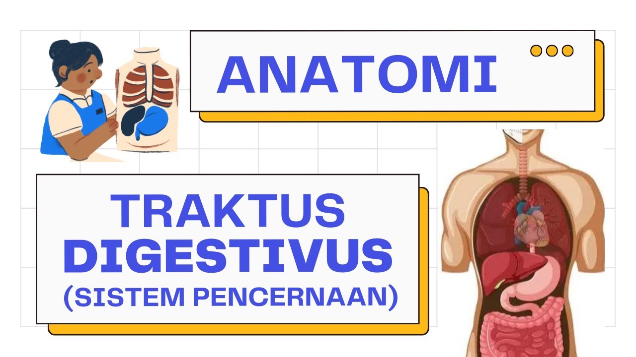
TLDRThis video provides an in-depth explanation of the human abdominal cavity and its organs. The speaker walks through each organ, including the liver, stomach, intestines, pancreas, and kidneys, explaining their locations, functions, and interrelations. The discussion touches on the complexities of anatomy, highlighting how organs are densely packed and how they interact with each other. The speaker also demystifies anatomical terms and concepts like peritoneum, visceral organs, and the role of the diaphragm, offering a detailed educational tour of the human body's internal structure.
Takeaways
- đ§ Anatomy Understanding: The speaker discusses their early curiosity about the anatomy of the human body and how things are tightly packed inside.
- đŹ Educational Insight: The need for anatomical education is emphasized, particularly the placement of organs like the stomach.
- đ Abdomen Defined: The abdomen is described as the region of the trunk between the thorax and the pelvis, with the diaphragm acting as a clear boundary between the thorax and abdomen.
- 𩮠Abdominal Boundaries: The abdomen's contents are enclosed in a peritoneum layer, and the distinction between the abdomen and pelvis is more nuanced due to the extension of the abdomen into the pelvis.
- đ Liver & Stomach: The liver is identified as the largest organ in the abdomen, extending across the midline, while the stomach nestles under it and connects to the esophagus.
- đïž Greater Omentum: The greater omentum is described as a connective tissue sheet hanging from the stomach, acting as a 'policeman' for the gut by preventing the spread of inflammation.
- đ« GI Tract Overview: The gastrointestinal tract is a long continuous tube from the esophagus to the anal canal, with the small and large intestines playing different roles in digestion and water absorption.
- đ Pancreas Functions: The pancreas has dual functions, producing hormones like insulin and secreting pancreatic juice into the duodenum to aid digestion.
- đ©ž Blood Supply: The abdominal aorta and its branches supply blood to various abdominal organs, including the pancreas, spleen, and liver.
- đ§Ș Kidney Role: The kidneys are retroperitoneal organs responsible for managing body fluids and electrolyte balance, with ureters transporting urine to the bladder.
Q & A
What is the primary function of the diaphragm in separating body cavities?
-The diaphragm is a muscular sheet that separates the thorax (chest) from the abdomen. It plays a key role in breathing by moving up and down, and it also physically separates the two cavities.
How do the contents of the abdomen interact with the pelvis?
-The abdominal contents are enclosed by a thin sheet called the peritoneum, which also extends down into the pelvis. This allows the abdominal contents to rest on top of the pelvic viscera. In certain conditions, like pregnancy or a full bladder, organs from the pelvis can push up into the abdominal space.
Where is the liver located in relation to the diaphragm and stomach?
-The liver is a large organ located on the right side of the abdomen, extending across the midline. It sits just underneath the diaphragm and overlaps with the stomach on the left side.
What is the function of the greater omentum in the abdominal cavity?
-The greater omentum is a connective tissue sheet that hangs down from the stomach and covers the abdominal organs. It helps prevent the spread of inflammation within the intestines and acts as a protective layer for the gut.
What role does the pancreas play in digestion and blood glucose regulation?
-The pancreas has two main functions: it produces hormones like insulin to regulate blood glucose levels, and it secretes pancreatic juice into the small intestine (duodenum) to aid in digestion.
How does the gallbladder assist in digestion?
-The gallbladder stores bile, which is produced by the liver. When needed, bile is released into the duodenum to help emulsify fats, aiding in digestion.
What is the role of the spleen in the body?
-The spleen plays a key role in the immune system, storing red blood cells, platelets, and immune cells like lymphocytes and macrophages. It helps recycle old red blood cells and can be activated to fight infections.
What is the significance of the mesentery in the gastrointestinal tract?
-The mesentery is a double layer of peritoneum that attaches the intestines to the posterior abdominal wall. It provides a pathway for blood vessels, nerves, and lymphatics to supply the intestines, allowing them to move around while staying connected.
How do the kidneys contribute to maintaining the bodyâs fluid balance?
-The kidneys filter the blood to regulate the bodyâs fluid balance by managing the amount of water and salts in the body. They produce urine to remove waste and excess fluid.
What are the adrenal glands, and what is their function?
-The adrenal glands sit on top of the kidneys and produce hormones like adrenaline and corticosteroids. These hormones are important for the bodyâs fight-or-flight response and for regulating metabolism and homeostasis.
Outlines
đ§ Introduction to Anatomy and Personal Experience
The speaker shares their early misunderstandings about how the body's organs fit together, explaining that their knowledge deepened during university when they began dissecting bodies. They reflect on how becoming an expert reveals how much there still is to learn. The focus then shifts to the topic of abdominal viscera, with a plan to discuss the arrangement of the organs and their relationships to one another.
đœïž Exploring the Abdominal Viscera
The speaker starts dissecting the anatomy of the abdomen, explaining how the abdominal organs are packed together under the diaphragm, which separates the abdomen from the thorax. The abdomenâs connection to the pelvis is discussed, emphasizing the role of the peritoneum, a thin sheet covering the abdominal contents. As the organs are removed, they examine the liver, stomach, and their interplay with the diaphragm.
đŠ The Greater Omentum and Gastrointestinal Tract
The speaker introduces the greater omentum, a connective tissue hanging from the stomach that protects and helps isolate inflammation in the abdominal cavity. They explain the gastrointestinal (GI) tract's structure, noting the large and small intestines, their movements, and their roles in digestion. The stomach connects to the small bowel, and food is pushed along for nutrient absorption, ending in the large bowel for water reabsorption.
đ©ž Functions of the Pancreas and Spleen
Detailed focus on the pancreas and spleen, highlighting the pancreasâs dual function in regulating blood glucose through hormones and aiding digestion through pancreatic juice. The pancreasâs relationship with the duodenum, the first part of the small intestine, is explained. The spleen, which supports immune function and stores red blood cells and platelets, also sends processed red blood cells to the liver.
đ« Liver, Blood Flow, and Digestive Roles
The liverâs key role in filtering blood from the gastrointestinal tract is explored, along with its anatomical connection to the inferior vena cava. The discussion includes the small intestine's segments (duodenum, jejunum, and ileum) and their distinct roles in digestion. The speaker also explains how the liver secretes bile into the small intestine to help digest fats, linking it to bile's effect on stool color.
đŹ Blood Supply and Abdominal Circulation
The speaker examines the major blood vessels in the abdomen, including the aorta and its branches, which supply blood to the abdominal organs. They also discuss the superior mesenteric artery and vein and how the liver processes blood before it re-enters circulation. The talk moves to the bladder, located in the pelvis, and the rectum, noting the digestive systemâs transition from colon to rectum.
đ§ Kidneys and Fluid Regulation
A look at the kidneys' role in filtering blood and maintaining fluid balance in the body. The speaker explains how the kidneys are retroperitoneal, located behind the peritoneum, and how they manage the body's salt and water content. They mention the adrenal glands sitting atop the kidneys, which produce hormones like adrenaline for stress responses and other steroids for maintaining homeostasis.
âïž Muscles and Blood Vessels of the Abdomen
The final focus is on the muscles of the posterior abdominal wall and their functions, such as hip flexion and lateral trunk movement. The speaker points out testicular arteries, which descend with the testes during development, and the aortaâs division into the iliac arteries, supplying blood to the lower limbs and pelvis. They conclude by summarizing how these layers interact to form the complex anatomy of the abdominal cavity.
đ Reflections and Closing Remarks
The speaker ends by reflecting on how much clearer their understanding of anatomy has become over time, though they once found it confusing as a child. They thank the audience for the suggestion and express satisfaction with the discussion, saying goodbye until the next session.
Mindmap
Keywords
đĄAbdomen
đĄDiaphragm
đĄLiver
đĄStomach
đĄPeritoneum
đĄGreater Omentum
đĄSmall Intestine
đĄPancreas
đĄSpleen
đĄKidneys
Highlights
Understanding the organization of organs in the body was challenging until hands-on experience in university.
The abdomen is the region of the trunk between the thorax and the pelvis, with the diaphragm acting as a clear separator.
The peritoneum acts like a bag covering the abdominal contents, with a soft distinction between the abdomen and pelvis.
The liver, the largest organ in the abdomen, extends across the midline and moves with the diaphragm.
The stomach is tightly nestled under the liver, with the esophagus passing through the diaphragm.
The greater omentum, known as the 'policeman of the gut,' helps prevent inflammation from spreading by covering abdominal organs.
The gastrointestinal (GI) tract consists of a continuous tube from the esophagus to the anal canal, with its main function being digestion and nutrient absorption.
The pancreas secretes hormones to regulate blood sugar and produces pancreatic juice for digestion in the small intestine.
The spleen plays a key role in the immune system, storing red blood cells, platelets, and macrophages.
The liver has over 500 functions, including processing blood from the GI tract and breaking down red blood cells from the spleen.
The duodenum is the first part of the small intestine and plays a crucial role in digestion, working in tandem with the pancreas and liver.
The mesentery allows mobility in the small intestine, while the large bowel's key role is water absorption.
The abdominal aorta supplies blood to the abdomen, pelvis, and lower limbs, while the inferior vena cava carries blood back to the heart.
Kidneys are retroperitoneal organs responsible for managing fluid and electrolyte balance in the body.
Adrenal glands, located on top of the kidneys, produce hormones like adrenaline, which are key in the fight-or-flight response.
Transcripts
[Music]
hi one of them was a kid I knew about
the organs of the body but I couldn't
work out how they all fit in I I think I
assumed they must be neatly organized
with the bit of space in between them
probably the the illustrations of the
time for children showed that sort of
thing I couldn't really work out he
wasn't even through all my school years
it wasn't until I got to University and
I started taking bodies apart that I
realized wow this stuff is really packed
in here we had a comment was he last
week somebody asked could I go through
all the abdominal viscera and I thought
and then I was teaching last week chat
to a student she wasn't entirely sure
where the stomach was so I thought
that's a good idea that we should go
through all of the abdominal viscera
[Music]
so yeah my father I've had an
interesting Anatomy for a very long time
and it hasn't really disappeared and I
think the stage I'm at now is the more I
the more we know the more I realize I
don't know and the more I try to hold in
my head the harder it is for me to hold
everything in my head
that makes any sense at all this but
same with other things you become an
expert you realize you don't know
they're very much a tool anyway right so
what is the abdomen and then what we'll
do is we're gonna take all these parts
away from the model so as we go we'll
point things out as we remove things
we'll try to see the pay attention to
what lies posterior to that organ or
near buys we've got an idea of where
things aren't relative to one another
also remember that whenever we're
talking about the body whenever we use
left or right we're talking about the
body is left and the body is right
okay not our view but the body's left
the body's right and we'll work our way
back to the posterior abdominal wall and
see what we see so then the abdomen the
abdomen is the region of the trunk
between the thorax and the pelvis is
your typical textbook description which
sounds nice and tidy now with a superior
and is nice and tidy what we're seeing
here is the diaphragm so the diaphragm
does cleanly separate the abdomen from
the four acts so the diaphragms got kind
of this is curvy shape to it was a
three-dimensional thing he was curving
over the liver here but the diaphragm is
a is a muscular sheet that separates the
thorax from the abdomen now in feelingly
it's a little bit harder because the
pelvis well here's the pelvis and the
pelvis has these bony parts so much of
the pelvic viscera is down here but this
is pelvis but the abdomen also extends
all the way down here so
what's happening is that the the
contents of the abdomen are covered in
the layer of peritoneum like a like a
thin sheet so it's almost like all of
these contents here like it's all in a
bag and when that bag sinks down into
the pelvis the abdominal contents have
sat on top of the the pelvic contents
the pelvic viscera and is the peritoneum
then that's really separating the pelvis
the pelvic viscera from the abdominal
viscera so then that distinction between
abdomen and pelvis is a little bit
softer down here and of course when
women get pregnant and the uterus
becomes in last or even when you buy
either filled with you and that becomes
enlarged those push from the pelvis up
into the abdomen so there's a bit of
interplay between the abdomen and the
pelvis okay what can we see so we see
some organs up here the biggest organ is
the liver is very big organ it's on the
right side of the abdomen but it's
extending across the midline to the left
side it was a very large organ and it's
just underneath the diaphragm so as the
diaphragm moves up and down as well the
liver will move up and down with us they
didn't interplay between the four legs
and the abdomen there so there's the the
liver and then nestling into the liver
on the left side this is the stomach so
the stomach as you can see it's pushed
up into the liver you know if we take
that knocked off if we lift the liver up
that's how we'd see the stomach nestling
into the liver there do you see -
they're very tightly stuck together
diaphragm diaphragm and of course the
esophagus passes down through the
diaphragm and I pop this over there's
the esophagus this who dives through the
diaphragm there's a hole in the
diaphragm now if I put the liver back
this here so if we if we're if we take
away the skin we take away the muscles
of the anterior abdominal wall and we
take away the peritoneum lining the
parietal peritoneum lining
Damo cavity we see this here and this is
the greater omentum this gets called the
the policeman of the gut now what it
does is it's a connective tissue sheet
that's hanging down from the greater
curvature of the stomach and it's
covering all of these organs down here
and it's again it's layers of peritoneum
it's got fat in it it's got blood
vessels its job seems to be is that if
there is some inflammation in the bowel
down here it will inherit my
inflammation and help prevent is
spreading too far everyday the greater
omentum off now we can see the GI tract
the GI tract will the gastrointestinal
tract is the bulk of four viscera that
we find within the abdominal cavity it's
a long continuous tube from the soffit
as' and stomach through the small
intestine the large intestine all the
way through to the rectum and the anal
canal the rectum is in the pelvis and
the anal canal is in the perineum
now what we see is we can see a little
bit of large bowel here this is the
ascending colon and we see a little bit
of large bowel here this is a transverse
colon and then we see a little bit of
large bowel around there the descending
colon so this is large colon or large
bowel this then is the small intestine
or the small bowel if we remove the you
see how the liver and the stomach a loss
superior and this stuff is squashed up
against it if I remove the liver here's
the stomach and the stomach is
continuous with all of this we we often
see in models and textbooks that the
transverse colon runs across here we
looks like a picture frame around the
small bowel in you if you're sat up your
transverse colon is probably dropping
drooping down a little bit like that if
you're looking at abdominal x-rays the
transverse colon probably is going to do
this it's probably going to be looping
down here because it's actually got a
mid-century so the knees entry is a
double fold of peritoneum with so that's
how the blood vessels and nerves and
lymphatics get to and from the bowel and
because the the transverse codons got a
meeting Terry means it can move around
the ascending colon and the descending
colon are fixed in place to the
posterior abdominal wall so they don't
move around the small bowel also has a
reason tree so it is fairly mobile and
of course what's happening is the food
is being passed into the stomach and
then it gets pushed into the small bowel
and peristalsis and the shortening and
lengthening of the small bowel pushes
the foods along the small intestine and
the nutrients get absorbed from it and
then it gets pushed into the large bowel
which largely agree absorbs water and
that sort of thing so it's helpful if it
if it moves around right okay so stomach
now if we take away this stomach we can
see some other viscera deep to that so
this here this is the pancreas so the
pancreas is doing two things it's making
hormones that it secretes into the blood
to steady blood glucose levels and it's
making an exocrine secretion of
pancreatic juice that it's going to
secrete into particularly the transverse
colon the duodenum here so the duodenum
is the first part of the small intestine
and it's forming a C shape it's curling
around the pancreas here so the pancreas
has got this duct and it's going to
secrete into the duodenum to help with
digestion of the contents that you just
eat and that the stomach is pushing into
the joint deal right pancreas duodenum
so out here
that's the spleen the spleen is at the
tail of the pancreas it's on the lateral
left side of the body
you got one pancreas you've got one
spleen so the spleen is a component of
the immune system it's a store of red
blood cells it's a store of platelets so
it can help with clotting and help with
replacing red blood cells if you lose
them if you have an injury losing your
loose of blood that sort of thing
there's also a site where you find lots
of lymphocytes and macrophages and it's
a site where parts of the immune system
are stored and where pass the immune
system can be activated in things like
that so it's got a couple of big roles
not least immunological in fact it sends
red blood cells once it's broken them
down to the liver which is nearby it's
got a blood vessel and it sends across
the liver of a liver has got 500 all
different functions it's got a huge
number of jobs it has a huge amount of
blood passing through all the blood from
the GI tract passes through the liver
and the liver is wrapped around the
inferior vena cava which we'll see more
of as we get deeper so blood from the
gut and the spleen passes through the
liver to the inferior vena cava back to
the rest of the circulation okay so
small bowel now the duodenum is the
first part of the small intestine it
passes food into the jejunum and the
judgment becomes the ileum so the small
intestine isn't the same along its
entire length food passes so the
judgment is out on this side and the
idea is over on this side and food
passes along the length from the
duodenum to the jejunum to the i liam
and if you look at the histology and the
structure of the small intestine as we
go along its length it changes slightly
and it has slightly different roles so
if we take the small intestine out we
can see that the small intestine is as I
said it's kind of suspended strung from
the posterior abdominal wall by
mesentery so this can all move around
now we've taken off the small intestine
we can see that Julie Dean and this is
the curve I was talking about here first
part second part third part that it goes
up again fourth part and you can see
here this is where the pancreas secretes
into the duodenum in fact the other
thing we've got here is you might have
spotted it it's the green bit there that
there is the gall bladder
so the google bladder is storing bile
and it's gonna pass that bile through
some ducts eventually down into this
point here so the common bile duct is
going to pass bile into the duodenum
here the same point of the pancreas
passed into the duodenum and the bile is
also going to help with digestion is
going to help emulsify fats and that
sort of thing and remember those five
hundred different jobs that the liver
does well some of the waste it produces
is going to get passed into the small
bowel to be removed from the body
through the common bile duct through the
same route
that's why behind is green and that's
what makes feces brown in fact it's it's
recycled red blood cells anyway so
duodenum here now what else can we see
so we can see the ascending colon and
the descending colon little bit better
we can see the posterior abdominal wall
and we can see some of the blood vessels
supplying blood to the large bowel here
now on the superior edge of the pancreas
so if I take this off we just jump ahead
we can see the major blood vessels this
is the a or to the abdominal aorta the
abdominal and so the aorta is supplying
blood to the four act to the abdomen and
eventually it's going to send off
branches to the pelvis and the lower
limbs and what-have-you that's the aorta
now the aorta has got three anterior
branches and those anterior branch is
going to supply blood to most of the
things in the abdomen so just superior
to the pancreas we have the celiac trunk
and you can see it's sending off
branches to the pancreas to the spleen
to the
gentleman off to the liver and that sort
of thing now only inferior side of the
pancreas we can see the superior
mesenteric artery and the superior
mesenteric vein the superior mesenteric
artery is a branch the aorta the
superior mesenteric vein is going to be
sending blood back to the portal vein
and the liver down here we can see the
bladder so the bladder is in the pelvis
normally that if we had peritoneum here
the bladder would be covered over by
peritoneum and that would mark the
inferior end of the abdominal cavity
that's where the abdomen end the
descending colon then becomes the
sigmoid colon here as it makes a bit of
an S shape and Wiggles around and when
it goes back there and straightens out
it becomes erect and that's what the
rectum means
rectum rectus means straight so the
rectum is the straight part of the large
bowel and the last part for the anal
canal all right if we take that off then
we can see the aorta and next to is the
inferior vena cava so this is going to
carry blood back from the abdomen the
liver is going to send blood inferior
vena cava and then that's going to go
through the diaphragm and straight back
into the heart into the right side of
the heart into the right atrium and then
off around the body
there's the superior vena cava up there
so inferior vena cava a Horta we can see
a whole bunch of blood vessels branching
from where the really important ones are
on here and of course we can see these 2
these are the kidneys so the kidneys are
posterior in the abdomen they're in the
posterior abdominal wall
they are posterior to the peritoneum
that connective tissue layer so they get
called retroperitoneal so the kidneys
are receiving a huge amount of blood
steak or some big blood vessels you see
the veins here because the the renal
veins are anteriors that the left renal
vein has to run quite a long way and
it's running it's running anterior to
the aorta but the the renal arteries are
posterior toes and they're gonna be
large is what's a large blood
supplying small organs mean that these
organs are doing something with the
blood and they're processing the blood
of course what they're doing is is
largely managing the amount of fluid
inside the body you are a bag of salty
water that's what your cells evolved
from and now you have a bag that you
carry that salty water around in you
have to maintain the right amount of
salty water and the microlight of salt
in it for yourselves to be happy to
function normally
so that's largely what the kidneys are
doing they're managing all of that so
then from each kidney we can see these
so these are the ureters and the ureters
then again s passed urine from the
kidney down to the bladder and then on
the posterior abdominal wall overlying
these muscle cells you see a number of
muscles making up the posterior
abdominal wall now in the superior pole
of each kidney we have a suprarenal
gland or an adrenal gland
now the adrenal glands make adrenaline
adrenaline and noradrenaline obviously
that's involved in the fight-or-flight
response in the adrenaline rush in that
activation of the sympathetic nervous
systems you can fight or flight fight or
fly fight off fly away run away and they
also make a whole bunch of steer or it's
deal with always glucocorticoids
mineralocorticoids that sort of thing
which manage you know manage homeostasis
manage metabolism and that sort of thing
so again related to the gut and energy
and food and that sort of thing so
you've still got the spleen out here so
can you see how the spleen is typically
it's a little bit of posterior but its
lateral can you see how it's nestling up
into the diaphragm whereas the kidneys
are a little bit more inferior and the
left kidney do you see it's a little bit
higher than the right kidney and that's
because of the liver so the liver is so
big it's actually pushing the right
kidney so it's a little bit lowering the
abdomen
so if you were to look at a transverse
section across here you might see just
one kidney on one side and the spleen
and all these other bits if you might
miss the other kidney go a bit lower
you'll get a section through both kids
using both on CT or mr you can see the
diaphragm then arching back there these
these thick muscles here these are the
psoas muscles the psoas major and it's
got a little psoas minor lying on the
top of it
this is iliacus and these two muscles
will join insert into the femur and give
hip flexion so now lift your knee toward
your chest these muscles here these are
quadratus lumborum and they're running
kind of what kind of in that direction
from the vertebral column to the pelvis
which will give you a little bit of
lateral flexion of your trunk all right
these arteries here these we were
talking about last week these are the
testicular arteries running from the
aorta in the abdomen all the way down to
the scrotum because the testes started
to form up here and then they descended
in the in the embryo and the fetus and
they trailed their blood vessels behind
and you can see the testicular veins
draining back the left one goes to the
left renal vein and the right would go
threatening the inferior vena cava
so the aorta ends when it divides into
left and right common iliac arteries
that's what we were seeing here the left
and right common iliac arteries then
divide again into an external iliac
artery which is going to run down to the
lower limb one then to the thigh in the
leg and then an internal iliac artery
which is going to go down into the
pelvis and supply blood to the vserver
of the pelvis and also to the external
genitalia and that sort of thing so I
take the stomach off there's the
pancreas and if I take the pancreas off
there's the kidney it's that's that
that's a sort of layering that we're
dealing with can you see the adrenal
glands they're just popping up now the
aorta you can't see it because he's just
running through a
gap in the diaphragm there but the the
inferior vena cava has just been cut
because we've removed the liver and
that's it I can't say kind of thing else
off if I was to take more off would be
at the muscles and I take the muscles
off and we be at the other side that's
what you find inside the abdominal
cavity if you take things out bit by bit
well I enjoyed that as a thank you for
that suggestion it did remind me of if I
was like when I was a kid kind of work
out how all this stuff was packed in and
where it all was exactly now I do kind
of take it for granted but there you go
okay see you guys next week
[Music]
Voir Plus de Vidéos Connexes
5.0 / 5 (0 votes)