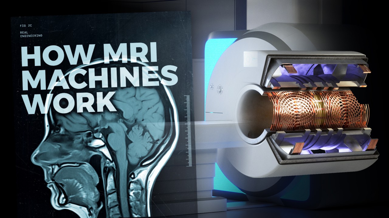
MRI Machine - Main, Gradient and RF Coils/ Magnets | MRI Physics Course | Radiology Physics Course#2
Summary
TLDRThis educational talk delves into the intricacies of MRI technology, focusing on the machine's components and their roles in creating MRI images. It explains how the main magnetic field is generated by the main coil, using superconductors like niobium titanium to maintain a strong, homogeneous field. Shims are introduced to refine field uniformity. Gradient coils are highlighted for their ability to spatially localize signals by altering magnetic field strength along different axes. The lecture also covers the radio frequency coil's function in exciting hydrogen protons, which is essential for signal detection and image formation. The talk promises further exploration into nuclear magnetic resonance in subsequent sessions.
Takeaways
- đ§Č The MRI machine is composed of multiple layers of magnets, each contributing to the creation of the magnetic field necessary for imaging.
- đ The main coil generates the primary magnetic field (B naught) along the longitudinal axis (Z-axis) of the patient, which is crucial for aligning hydrogen atoms.
- đĄ The strength of the main magnetic field is determined by the number of wire coils and the current flowing through them, with superconductors like niobium-titanium alloys used to maintain high current without resistance.
- âïž Liquid helium is circulated around the coils to keep them at temperatures below 4 Kelvin, ensuring the superconductivity necessary for strong magnetic fields.
- đ Shims, both passive (ferromagnetic metals) and active (with their own electric supply), are used to adjust the main magnetic field, making it as homogeneous as possible for accurate imaging.
- đ§Č Gradient coils apply a gradient to the magnetic field, allowing for spatial localization of signals within the MRI by manipulating the field strength along different axes.
- đ The gradient coils create differential magnetic field strengths, which in turn affect the precessional frequencies of hydrogen protons, enabling spatial encoding of the MRI signal.
- đĄ The radio frequency (RF) coil generates a magnetic field perpendicular to the main field, resonating with hydrogen protons at specific frequencies to selectively excite them.
- đ The RF pulse causes hydrogen protons to move from the longitudinal plane into the transverse plane, which is essential for signal measurement and image generation.
- đ The MRI process relies on the principles of nuclear magnetic resonance (NMR), where hydrogen atoms' alignment and precession within the magnetic field are manipulated to produce an image.
Q & A
What is the main purpose of the main coil in an MRI machine?
-The main coil in an MRI machine is responsible for generating the B naught or the main magnetic field along the longitudinal or Z axis, which is essential for aligning hydrogen atoms and creating the MRI signal.
How does the strength of the main magnetic field in an MRI machine depend on the number of coils and current?
-The strength of the main magnetic field is dependent on the number of coils of wire and the amount of current running through that wire. More coils and increased current result in a stronger magnetic field.
What is the role of superconductors in MRI machines, and why are they necessary?
-Superconductors are necessary in MRI machines to generate sufficient current for a strong magnetic field without resistance. Materials like niobium titanium alloys are used as they exhibit superconductivity at low temperatures, typically below 4 degrees Kelvin, allowing large currents to flow without resistance.
Why is liquid helium used in MRI machines, and what happens if the temperature rises above 4 degrees Kelvin?
-Liquid helium is used to maintain the temperature of the superconducting coils below 4 degrees Kelvin, which is necessary for superconductivity. If the temperature rises above this critical point, the superconductivity is lost, leading to increased resistance and heat generation, which can cause the liquid helium to expand and potentially lead to a quenching process where the helium is released as a gas.
What are shims in MRI imaging and how do they help in creating a homogeneous magnetic field?
-Shims in MRI imaging are used to manipulate the main magnetic field and make it as homogeneous as possible. They can be passive, made of ferromagnetic materials, or active, with their own electric supply, and are used to correct for imperfections in the magnetic field.
How do gradient coils contribute to spatial localization in MRI imaging?
-Gradient coils apply a gradient to the magnetic field, making it stronger or weaker along different axes (X, Y, Z). This differential magnetic field strength allows for spatial encoding of the signals, which is crucial for localizing the source of the MRI signal within the body.
What is the isocenter in the context of MRI imaging?
-The isocenter in MRI imaging refers to the point in the magnetic field where the magnetic field strength is equal to the background B naught magnetic field. It is the region where the magnetic field is most homogeneous andäžć gradient influence.
What is the function of the radio frequency (RF) coil in an MRI machine?
-The radio frequency coil generates an alternating magnetic field perpendicular to the main magnetic field. It selects specific hydrogen protons that are precessing at a set frequency and flips them into the transverse plane, allowing the MRI machine to measure the signal and generate images.
How does the radio frequency pulse affect hydrogen protons in an MRI scan?
-The radio frequency pulse matches the precessional frequency of hydrogen protons, causing them to gain energy, fan out, and come into phase. This results in a flip angle, which moves the net magnetization vector from the longitudinal plane to the transverse plane, enabling signal measurement.
What is slice selection in MRI imaging, and how does it relate to the radio frequency pulse?
-Slice selection in MRI imaging is the process of isolating a specific plane within the patient to acquire image data. It is achieved using the radio frequency pulse, which is tuned to match the precessional frequency of hydrogen protons in the desired slice, allowing for the selective excitation of these protons.
Outlines
đ Introduction to MRI Module and the Importance of Physics
The speaker welcomes the audience and reflects on the introductory MRI talk, which touched upon important topics but only superficially. The focus now is on understanding the physics behind MRI, starting with the machine itself. Specifically, the discussion centers on the different magnets within the MRI and their roles in creating the magnetic field needed for imaging. The main magnetic field (B naught) is generated by the outermost magnet, known as the main coil, using principles such as AmpĂšre's Law and superconductivity. The importance of materials like niobian titanium alloys and the role of liquid helium to maintain superconductivity are highlighted, emphasizing the critical temperature to avoid resistance and the quenching process.
đ§ Understanding MRI Shims for Field Homogeneity
This section introduces the concept of 'shims,' devices that help stabilize the magnetic field in MRI imaging. The goal of shims is to make the magnetic field as homogeneous as possible, given that imperfections in the machine disrupt the ideal uniform field. Two types of shims are explained: passive shims, made from ferromagnetic materials that adjust the field naturally, and active shims, which are powered by electricity to further fine-tune the magnetic field. Active shims may either be superconductive or resistive, both contributing to more accurate MRI signals by improving magnetic field uniformity.
đ Gradient Coils and Their Role in Spatial Localization
The gradient coils, located after the main and shim coils, are discussed in terms of their ability to manipulate the magnetic field strength along different planes (X, Y, and Z). The speaker explains how these coils can create a gradient in the magnetic field along the Z-axis by superimposing additional magnetic fields over the main field. This manipulation of field strength enables the MRI to vary the processional frequencies of hydrogen protons, which is crucial for spatial localization in MRI scans. By controlling gradients in the magnetic field, MRI machines can differentiate between signals from different parts of the body.
đ The Function of Gradient Coils in Signal Encoding
This part continues the discussion on gradient coils, focusing on how they affect the processional frequency of hydrogen protons based on their position within the magnetic field. By changing the magnetic field strength across different axes, the gradient coils help encode spatial information in the MRI signal. This encoding allows for the creation of detailed images by ensuring the MRI machine can differentiate between protons based on their location in the body. The concept of the 'iso-sensor,' where the magnetic field strength remains unchanged, is also introduced as a reference point for signal encoding.
đĄ Radio Frequency Coils and Their Role in Signal Measurement
This section introduces the radio frequency (RF) coils, which create an alternating magnetic field perpendicular to the main field. These coils are responsible for selecting hydrogen protons based on their processional frequency and flipping them into the transverse plane, a key step in measuring the MRI signal. The analogy of children swinging and being pushed by their fathers (representing protons and the RF pulse) is used to explain how only protons oscillating at the same frequency as the RF pulse will gain energy. This process aligns the protons, making them easier to measure and enabling MRI imaging through slice selection.
đ§Ș Summary and Transition to Nuclear Magnetic Resonance
The speaker recaps the different components of the MRI machine, including the main magnetic coil, shims, gradient coils, and radio frequency coils. These components work together to generate, manipulate, and measure the magnetic fields necessary for MRI imaging. Finally, attention shifts to the hydrogen atoms themselves, introducing the concept of nuclear magnetic resonance, which will be covered in the next talk. The speaker encourages viewers to check out curated question banks and ends the talk with a goodbye.
Mindmap
Keywords
đĄMRI machine
đĄMain coil
đĄSuperconductor
đĄShims
đĄGradient coils
đĄRadio frequency (RF) coil
đĄPrecession frequency
đĄFlip angle
đĄNuclear magnetic resonance (NMR)
đĄSpatial localization
Highlights
Introduction to the physics behind MRI imaging, focusing on the MRI machine and its components.
Explanation of how the main coil in the MRI machine creates the B naught magnetic field using Ampere's law.
The role of superconductors, like niobium titanium alloys, in generating strong magnetic fields without resistance.
The necessity of liquid helium to maintain the superconducting state of MRI machine coils at below 4 degrees Kelvin.
The concept of quenching as a safety feature to release helium gas if superconductivity is lost.
The function of shims in MRI to make the main magnetic field as homogeneous as possible.
Differentiation between passive shims, which are ferromagnetic metals, and active shims, which have their own electric supply.
The gradient coils' role in applying a gradient to the magnetic field for spatial localization of signals.
How gradient coils manipulate the magnetic field strength along different axes in the Cartesian plane.
The isocenter concept, where the magnetic field strength is the same as the background B naught magnetic field.
The radio frequency coil's function to generate a magnetic field perpendicular to the main magnetic field for signal measurement.
The process of matching the radio frequency pulse with the hydrogen protons' precession frequency for energy transfer.
The importance of the flip angle in moving the net magnetization vector from the longitudinal to the transverse plane.
Slice selection as a method to isolate specific hydrogen atoms within the patient using the radio frequency pulse.
Summary of the MRI machine components and their roles in generating the MRI image.
Anticipation of the next talk focusing on nuclear magnetic resonance and its application in MRI imaging.
Transcripts
hello everybody and welcome back I hope
you haven't been scared off by the first
talk the introductory talk in this MRI
module we covered many important topics
but only very superficially and we
haven't given any one of them really
enough time of day to understand the
underlying physics principles that go
into creating an MRI image now's our
turn to take a step back and look at
each one of those topics in a little bit
more depth and then hopefully bring that
knowledge together and ultimately get a
good understanding of the components in
MRI physics so today I want to focus on
the MRI machine itself the various
different magnets that go into creating
the magnetic field strengths that
ultimately create our MRI signal now you
can see that the MRI machine is made of
multiple different layers and each one
of these layers represents a different
magnet now we're going to start with the
main layer the outermost layer which is
known as the main coil now if you cast
your mind back to high school you'll
know that running a current through a
wire will generate a magnetic field
around that wire that's ampere's law the
right hand rule and if we run these
wires in a coil the magnetic fields will
superimpose along one another and they
will ultimately create a large single
magnetic field that runs through the
center of that coil and that's exactly
what's happening in the main coil in our
MRI machine we create what is known as
the B naught or the main magnetic field
that runs along the longitudinal or Z
axis in the MRI machine
now the strength of that main magnetic
field is dependent on two factors it's
dependent on the number of coils of wire
and the amount of current that is
running through that wire
now as we increase current more and more
we will get increased resistance within
that wire and we need to use what is
known as a superconductor in order to
generate sufficient current that will
allow us to have a strong enough
magnetic field strength now in order to
do this we need to utilize the principle
of superconductivity there are certain
materials that add a low enough
temperature will act as a superconductor
one of such material is niobian titanium
Alloys that's generally what's used in
MRI machines now in order to keep the
temperature low enough we circulate
liquid helium around these coils the
liquid helium is generally below 4
degrees Kelvin now when electrons are
running through the wire current flowing
through a wire they interact with the
lattice of that wire now the hotter the
temperature in that wire the more the
lattice is vibrating and the more it
interferes with those flowing electrons
as we cool that temperature the lattice
vibrates less and less and those
electrons can pass with less resistance
now if we're looking at a
non-superconductor the lower we make the
temperature the lower the resistance but
there will always be some form of
resistance even at zero degrees Kelvin
in a superconductor there's an Abrupt
drop-off that's what's known as our
critical temperature now the critical
temperature is generally around four
degrees Kelvin and if we keep the wire
temperature below 4 degrees Kelvin we
will actually in fact get no resistance
in that wire it allows us to pass a
large current through those wires
now in the MRI machine we need to
constantly replace that liquid helium in
order to keep that temperature below 4
degrees Kelvin if the temperature ever
gets above 4 degrees Kelvin we will
suddenly get a large amount of
resistance within that wire and we will
generate heat within the wire as we
generate heat we get more and more
resistance and this liquid helium will
now become a gas and that gas will
expand we know that gas is expand to
full space and we get a process known as
quenching where that liquid helium is
then released off as gaseous helium into
the room and this is a safety feature in
an MRI machine we need to be able to
release that helium off if we no longer
get superconductivity Within These wires
that process is known as quenching
so the function of the main magnetic
coil is to apply this B naught magnetic
field along the longitudinal axis of our
patient and we've seen that if we apply
a large magnetic field along the z-axis
of our patient it will cause hydrogen
atoms free hydrogen atoms within that
patient to align with that magnetic
field and each one of those hydrogen
protons will process at a set frequency
now the precession frequency is
determined by the atom itself hydrogen
has a set what's known as a gyromagnetic
ratio which we're going to look at in
our next talk when we look at nuclear
magnetic resonance and it's determined
by the strength of the magnetic field
the stronger the magnetic field the
faster the hydrogens will process around
their axis along the parallel direction
of that main magnetic field so we can
see that manipulating the current within
the main magnetic coil will manipulate
the magnetic field strength and that
change in magnetic field strength will
change the processional frequency of the
hydrogen protons within the patient now
when we are looking at the diagram of
the magnetic coil you may have noticed
these two structures sitting within that
main coil now these are what is known as
shims now what exactly is a shim well a
shim in woodworking is a triangular
piece of wood that you can wedge between
two objects in order to make those
objects more steady if you're at a table
that's wobbly you will place a shim
under the shortest leg in order to
prevent that wobble within the table now
shims in MRI imaging are doing the same
thing they are manipulating that main
magnetic field in an Ideal World we want
a perfectly homogeneous and magnetic
field running along the longitudinal
axis of the patient but because of the
various different components of the MRI
machine itself and the way in which
magnetic fields are generated we don't
actually get a perfect homogeneous
magnetic field along the z-axis now the
shims will alter that magnetic field in
order to try and make the main magnetic
field as homogeneous as possible now as
you can see there are two separate types
of shims the first type of shim is
what's known as a passive shim that's a
magnetic sheet or a ferromagnetic metal
that is placed within the bore of the
MRI machine that will passively
manipulate the magnetic field we know
that ferromagnetic substances will
manipulate magnetic fields that run past
them the second type of shim is known as
an active shim now active shims have
their own electricity Supply their own
current running through the coils Within
These shims and we can change that
current in order to manipulate the main
magnetic field again shims are trying to
make that magnetic field as homogeneous
as possible the more homogeneous that
main magnetic field the more accurately
we can localize those signals generated
in MRI imaging now active shims can
either be superconductive be within our
helium or they can be resistive shims
that lie within the bore the magnet
themselves they have their own
electricity Supply and we can manipulate
the amount that they influence the main
magnetic field
now let's move on and look at the next
layer of the MRI machine itself this
purple layer here and this is what's
known as the gradient coils now the
gradient coils do exactly that they
apply a gradient along the magnetic
field now as you see here the gradient
coils lie perpendicular to one another
here in both the Y plane and the X-Plane
they also lie along the Z plane now this
becomes really important when we get to
spatial localization of signals within
MRI
now if we take away the two cents of
gradient coils and we're left with the
flanking gradient coils along the z-axis
here we know that we are applying a b
naught or a main magnetic field along
the z-axis now we know that these
hydrogen atoms are going to process at a
frequency that is determined by the B
naught magnetic field and generally in
clinical Imaging we're talking about a
magnetic field strength between 1 and 3
Tesla now the gradient coils can create
their own magnetic field we can take
this gradient coil here and run current
through that coil if we run current
through the coil like this what we're
going to do is create a magnetic field
in the left to right direction here a
magnetic field that superimposes along
the same direction of our B naught
magnetic field
we can do the opposite in this gradient
call and run a current in the opposite
direction we are then going to create a
magnetic field that runs in the opposite
direction to the main B naught magnetic
field so we've generated two separate
magnetic fields from these gradient
coils that are ultimately going to
influence the main magnetic field that
is running through the z-axis here
if the magnetic field generated by this
gradient coil is superimposed over the
main magnetic field we will get a
reduction in the magnetic field at this
end of the MRI scanner if we superimpose
this gradient coil over the main
magnetic field we are adding to the
magnetic field strength at this end of
the MRI scanner and you can see how
superimposing these gradient coil
magnetic fields will manipulate our B
naught magnetic field and we get
manipulation of this main magnetic field
that was constant along the z-axis to
become a gradient between those two
gradient coils we are now applying a
differential magnetic field strength to
these various protons within the field
we can see that the field strength on
this end of the z-axis will be less than
the field strength at this end now
importantly this is not a vector here we
are not changing the direction of that
mean magnetic field we're changing the
strength of the magnetic field as we
head along the z-axis what we've created
now is a gradient where the magnetic
field strength is stronger at the Z axis
here than it is at the Z axis here now
because the magnetic field strengths
differ along the z-axis now the
processional frequencies of those
hydrogen protons will also differ as the
gradient gets stronger or as a magnetic
field strength is stronger the
processional frequency gets faster now
that is the main function of gradient
coils they change or manipulate the
magnetic field strength along the
separate axes in the Cartesian plane not
only can we change processional
frequency along the z-axis we can do the
same along the x-axis and y-axis and
this is a foundation for spatial
encoding of the signal that we are
generating in MRI imaging
now the part of the magnetic field that
is unchanged is what is known as the iso
sensor that's the part of the magnetic
field that has the same magnetic field
strength as that background B naught
magnetic field that we've created
now when we look at the gradient coil
itself we can see that they lie
perpendicular to one another and we can
use these coils here to generate
gradients along both the x-axis and the
y-axis now let's move on to the last
magnet within the MRI machine the radio
frequency coil now the radio frequency
coil generates a magnetic field that is
perpendicular to the main magnetic field
we've seen that hydrogen protons will
process at a specific frequency that is
dependent on the strength of the
external magnetic field that is being
applied and we've seen how we can apply
a gradient along an axis within the
patient now that gradient is causing
these hydrogen protons to process at
different frequencies now the radio
frequency coil generates an alternating
magnetic field in the perpendicular or
transverse axis here the X Y plane now
if you think of each one of these
hydrogen protons or net magnetize
vectors as being children swinging on a
swing at a set frequency they're all
swinging at different frequencies the
radio frequency coil is generating
magnetic pulses in the transverse plane
here they're like the dads at the swing
pushing their children
now the dads are closing their eyes and
pushing at a set frequency that's the
radio frequency pulse only the children
that are swinging at the frequency that
the dads are pushing will get more and
more energy and swing further and
further out those children that are
swinging naturally at a different
frequency to what the dad is pushing
they won't end up matching up with the
dad and they won't gain any energy from
the dad pushing the same thing happens
with the radio frequency pulse only the
hydrogen atoms that are processing at
the exact same frequency as the radio
frequency poles will gain more and more
energy we've seen that hydrogen atoms
are processing at a set frequency and if
the radio frequency pulse is the same as
that processional frequency two things
will happen first the protons will start
to Fan out more and more and second
those protons will now become in Phase
with one another initially they were out
of phase the radio frequency pulse
causes them to become in Phase they
become in phase and they fan more and
more out at a bigger and bigger angle
known as the flip Angle now the longer
we generate rate that radio frequency
pulse the bigger and bigger that flip
angle becomes and that movement out of
the longitudinal plane into the
transverse plane is what allows us to
measure that signal that's the main
function of the radio frequency pulse is
it allows us to change our net
magnetization Vector from the
longitudinal plane to the transverse
plane now in this example if we match
our radio frequency pulse with the
central frequency here we will see that
only that net magnetization Vector will
flip over in 90 degrees in this case
here and it's the radio frequency pulse
that allows us to isolate specific
hydrogen atoms within the patient and
this is what's known as slice selection
which we're going to look at in Signal
localization later so to summarize we've
looked at the main coil that generates
the main magnetic field along the
longitudinal axis of our patient and we
can use both active and passive shims to
manipulate that magnetic field to make
it as homogeneous as possible we we can
then apply a gradient field strength
along that magnetic field altering the
strength of the magnetic field either in
the Z x or y axes and that's the
responsibility of the gradient coils
we've then looked at the radio frequency
coils which select specific hydrogen
protons that are processing at a set
frequency and then flips those hydrogen
protons into the transverse plane
allowing us to measure signal in the
transverse plane and ultimately
generates our MRI image now we're going
to shift our attention to the hydrogen
atoms itself and look at nuclear
magnetic resonance why nuclear magnetic
resonance occurs and how we can use
nuclear magnetic resonance in order to
generate signal in the image again if
you're studying for a radiology Physics
exam check out the curated question
banks that I've Linked In The Top Line
in the description below otherwise I'll
see you all in the next talk goodbye
everybody
Voir Plus de Vidéos Connexes
Spin, Precession, Resonance and Flip Angle | MRI Physics Course | Radiology Physics Course #3
T2 Relaxation, Spin-spin Relaxation, Free Induction Decay, Transverse Decay | MRI Physics Course #4
How does an MRI machine work?
MRI Physics | Magnetic Resonance and Spin Echo Sequences - Johns Hopkins Radiology
How does an MRI machine work?
Proses di Dalam Tubuhmu kalau Kamu Diperiksa dengan Mesin MRI
5.0 / 5 (0 votes)