How does an MRI machine work?
Summary
TLDRThis script delves into the workings of MRI technology, which uses magnetic fields and radio frequency pulses to create detailed images of internal body tissues. It explains how water composition in the body, particularly the hydrogen atoms, is crucial for MRI imaging. The script also touches on the differences between healthy and diseased tissues, the role of gradient coils in targeting specific areas, and the importance of RF coils in generating the diagnostic images. The video aims to educate viewers on the principles behind MRI, making complex medical technology more accessible.
Takeaways
- 🧲 MRI scans use magnetic fields and radio pulses to create detailed images of internal body structures, aiding in diagnosis of conditions like brain hemorrhage or blood clots.
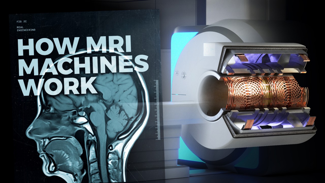
- 🌀 MRI machines consist of strong electromagnets, gradient coils, and RF (radio frequency) coils to generate the necessary magnetic fields and radio frequency pulses.
- 💧 The human body's water composition is crucial for MRI imaging, as the magnetic field realigns water molecules, which then emit signals when perturbed by radio pulses.
- 🔍 The difference in water composition between healthy and unhealthy tissues allows MRI to distinguish between them, providing clear images for medical analysis.
- 🌀 Hydrogen atoms in the body, which have a small magnetic field, are aligned by the MRI's magnetic field and then flipped by radio frequency pulses, emitting signals as they return to their original state.
- 🔧 The resonance frequency, determined by the Larmor equation, is key to MRI imaging, as it dictates the frequency at which hydrogen atoms will flip in response to the RF pulses.
- 📊 Phase and frequency encoding, along with Fourier transformation, are used to generate detailed 2D images from the signals emitted by hydrogen atoms.
- 🧲 Gradient coils in the MRI machine create varying magnetic fields to select and image specific parts of the body, with different sets of coils for the X, Y, and Z directions.
- 🛠️ The electromagnet in an MRI machine is a critical component, often superconducting and much stronger than common household magnets.
- 🔄 RF coils come in various designs, categorized as surface or volume coils, and are tuned to resonate at the same frequency as the part of the body being imaged.
- 📚 The script also mentions the importance of supporting the creators on Patreon, indicating an educational and community-driven aspect of the content.
Q & A
What is the primary use of MRI scans in the medical field?
-MRI scans are primarily used for diagnosis in cases such as detecting brain hemorrhages or blood clots deep inside the brain where direct observation is not possible.
How do MRI machines create detailed images of the human body's organs and tissues?
-MRI machines use varying magnetic fields and radio frequency pulses to create detailed images of the organs and tissues in the human body.
What role does water composition play in MRI imaging?
-Water composition is crucial in MRI imaging because the human body is made up of about 60% water, and the water molecules in the body are temporarily realigned by the magnetic field during an MRI scan.
How does the MRI scanner operator manipulate the hydrogen atoms within the body during an MRI scan?
-The scanner operator uses RF coils to send radio pulses that cause the aligned hydrogen atoms to flip, and then these atoms re-emit RF signals as they realign with the magnetic field, which are used to create MRI images.
What is the significance of the resonance frequency in MRI imaging?
-The resonance frequency, determined by the Larmor equation, is significant in MRI as it is the frequency at which the hydrogen atoms resonate and flip in response to the RF pulses.
How do MRI images differentiate between healthy and unhealthy tissues?
-MRI images differentiate between healthy and unhealthy tissues by analyzing the unique water composition of each tissue; changes in water composition due to a blood clot, for example, allow for clear distinction between the two.
What is the role of gradient coils in MRI imaging?
-Gradient coils are used to select specific parts of the brain for imaging by producing varying magnetic fields in the X, Y, and Z directions, which helps to localize the area of interest.
How do the RF signals emitted by hydrogen atoms contribute to the formation of MRI images?
-The RF signals emitted by hydrogen atoms as they return to their normal orientation are received by the computer, which converts these signals into images using phase and frequency encoding with the help of Fourier transformation.
What are the two main types of RF coils used in MRI scanners?
-The two main types of RF coils used in MRI scanners are surface coils, which rest on the surface of the object being imaged, and volume coils, which are designed to cover a larger volume.
Why are superconducting magnets commonly used in MRI machines?
-Superconducting magnets are used in MRI machines because they provide a strong and constant magnetic field, which is essential for high-quality imaging. They are typically much stronger than household magnets.
How does the FEA simulation help in understanding the MRI process?
-FEA (Finite Element Analysis) simulation helps visualize the magnetic fields produced by electromagnets and gradient coils, as well as the magnetic flux density in RF coils, providing a deeper understanding of how MRI machines function.
Outlines
🧠 MRI Technology and Its Medical Applications
This paragraph introduces the use of MRI scans in diagnosing medical conditions such as brain hemorrhages and blood clots. It explains the MRI process, which involves using magnetic fields and radio pulses to create detailed 2D images of organs and tissues. The human body's water composition plays a crucial role in MRI imaging, as the magnetic field realigns water molecules, and radio frequency pulses cause these molecules to emit signals that are captured to form images. The paragraph also touches on the differences in water composition between healthy and unhealthy tissues, which MRI can discern, and the role of hydrogen atoms in this imaging technique.
🛠️ Components of an MRI Machine and Their Functions
The second paragraph delves into the components of an MRI machine, including the electromagnet, which generates a strong magnetic field, and the RF coils, which are categorized into surface and volume coils. The electromagnet is a key element, with superconducting magnets ranging from 0.5 to 3 Tesla being common. The paragraph also discusses the importance of gradient coils, which provide linear gradients of the magnetic field to allow for the imaging of specific body regions. The use of FEA simulations to analyze the magnetic field produced by the x-axis gradient coil is mentioned, showing the variation in the magnetic field and how it is utilized in MRI imaging.
Mindmap
Keywords
💡MRI scans
💡Magnetic fields
💡Radio frequency (RF) pulses
💡Gradient coils
💡Water molecules
💡Hydrogen atoms
💡Resonance frequency
💡Phase and frequency encoding
💡Fourier transformation
💡Superconducting magnet
💡RF coils
Highlights
MRI scans are crucial in diagnosing conditions like brain hemorrhages and blood clots that are not visible to the naked eye.
MRI imaging uses magnetic fields and radio pulses to create detailed 2D images of internal organs and tissues.
The human body's water composition plays a vital role in MRI technology due to the magnetic properties of hydrogen atoms.
When inside an MRI machine, water molecules in cells are realigned by the magnetic field, aiding in imaging.
Radio frequency (RF) signals are used to flip the aligned hydrogen atoms, which then emit signals as they realign.
Each tissue has a unique water composition, allowing MRI to differentiate between healthy and damaged tissues.
The resonance frequency of hydrogen atoms, determined by the Larmor equation, is key to MRI imaging.
The MRI process involves striking an RF pulse to flip hydrogen atoms, which then emit signals detected by the machine.
Unhealthy tissue with more hydrogen atoms generates different RF signals, aiding in diagnosis.
Phase and frequency encoding with Fourier transformation is used to generate detailed MRI images.
Gradient coils in MRI machines select specific parts of the body for imaging by altering magnetic field strength.
The main electromagnet in MRI machines provides a constant magnetic field, with gradient coils adjusting it for imaging.
Superconducting magnets, ranging from 0.5 to 3 Tesla, are used in MRI machines for their strong magnetic fields.
RF coils in MRI scanners come in various designs, categorized into surface and volume coils.
Surface coils in MRI lie on the surface of the object being imaged, enhancing the magnetic flux density.
FEA simulations demonstrate the distribution of magnetic flux density in RF coils during MRI.
The resonant frequency of the RF coil is tuned to match the part of the body being imaged for optimal results.
Transcripts
you might have seen this on TV or some
of you might have even experienced this
strange procedure firsthand
MRI scans are used in the medical field
for diagnosis
in cases such as a brain hemorrhage a
blood clot occurs deep inside the brain
where doctors can't see
an MRI scan produces a very detailed 2D
image of the brain allowing doctors to
analyze the damaged tissue and then
perform their targeted surgery
let's take a deep dive into how MRI
imaging works and the principle behind
it
MRI is a technique that uses varying
magnetic fields and radio pulses to
create detailed images of the organs and
tissues in the human body
MRI machines have tube-shaped strong
electromagnets gradient coils and RF
coils to generate strong varying
magnetic fields and radio frequency
pulses
using the help of em Works in this FEA
simulation we can see the magnetic
fields produced by electromagnets
uniform magnetic fields are produced
inside the bore
now let's focus on the effect of
magnetic fields and radio frequency
signals on the body
the human body is made up of 60 water
and water composition surprisingly plays
an important role in this technique
when you lie inside an MRI machine
the magnetic field generated by the
electromagnets and gradient coil
temporarily realigns most of the water
molecules in your cells in the direction
of the magnetic field
the scanner operator commands RF coils
to send radio pulses causing these
aligned atoms to flip
within a few seconds the atoms realign
with the magnetic field emitting an RF
signal
this RF signal is used to create
cross-sectional 2D MRI images
we'll expand on this brief explanation
of MRI imaging technology later on
first let's explore the difference
between healthy and unhealthy tissues in
the human body each tissue has its own
unique water composition
when a blood clot happens in a certain
tissue it changes the water composition
of that area
this allows us to clearly see the
difference between healthy and damaged
tissues
let's narrow our focus on the water
molecules inside our tissues
the water molecule has two hydrogen
atoms
this hydrogen atom has a small magnetic
field and it acts as a tiny bar magnet
however this magnetic field keeps on
spinning since the atom is spinning
naturally the axis of the spinning
magnetic field is oriented randomly in
the human body
when this hydrogen atom comes into
contact with an external magnetic field
the orientation of the magnetic field of
the hydrogen atom changes and aligns
with the direction of the field
Additionally the rate of magnetic field
spin also changes
in this case the rate of the spin is
known as the resonance frequency and it
varies according to the strength of the
external magnetic field
this resonance frequency is quite
important in MRI and is determined using
the laramore equation
let's move on to the next step
so far we've aligned all the hydrogen
atoms in the direction of the magnetic
field
let's strike a radial pulse on a
hydrogen atom as a result the hydrogen
atom changes its orientation and flips
90 degrees with respect to the main
magnetic field
it's important to remember that the
frequency of an RF signal should be the
same frequency as the hydrogen atom's
resonance frequency
only then will the hydrogen atom flip
when we remove the RF pulses the atom
loses its energy and returns to its
normal orientation
the energy it emits is in the form of RF
pulses
however the time it takes each atom to
return to normal is different meaning
that the duration of emission of RF
signals is also different
the computer receives these signals and
converts them into images
since the unhealthy tissue has more
hydrogen atoms it generates different RF
signals
but how exactly can we generate a
detailed image the secret lies in phase
and frequency encoding with the help of
Furrier transformation
now that you know how an MRI machine
generates 2D images of a particular part
of the brain you may be wondering how
the machine is able to select the
specific part of the brain for Imaging
this work is done with the help of
gradient coils
the main electromagnet is constant and
the pair of gradient coils produce
opposite magnetic fields to each other
one coil increases the magnetic field
and the other side coil decreases the
magnetic field due to this gradient of
the magnetic field formed in between the
two coils
there are three sets of coils for the X
Y and Z directions let's see the
magnetic field produced by the x-axis
gradient coil with the help of FEA
results produced by em works
these results show us that variation in
the magnetic field gradually increases
from one coil to the other
gradient coils need to provide linear
gradations of the magnetic field as we
see in this FEA result
as a result the resonance and frequency
of hydrogen atoms will change from head
to toe gradually in order to image the
body region of Interest we must simply
use the specific radio frequency in RF
coils to excite just that part of
hydrogen atoms that provide a signal
as you might imagine the main component
of an MRI machine is the electromagnet
which provides a strong magnetic field
most magnets are of the superconducting
type ranging from 0.5 to 3 Tesla
the superconducting magnet is
continuously on from the time of
installation to the time of
decommissioning
the magnet is 100 times stronger than
small household magnets such as those
found on refrigerator doors or in
children's toys
another main component of an MRI scanner
is the RF coils
many coil designs exist but they all
fall into two main categories surface
coils and volume coils
as the name suggests a surface coil
rests on the surface of the object being
imaged Let's do an FEA simulation once
again of a Birdcage brain RF coil with
the help of em works
this result shows that there is more
magnetic flux density on the side
conductors and less on the end rings
in its simplest form it's a coil of wire
with a capacitor in parallel the
inductance of the coil and the
capacitance from a resident circuit are
tuned to have the same resonant
frequency as the part being imaged
thank you for watching we hope you
enjoyed this video please don't forget
to support lezix on patreon
Browse More Related Video
Proses di Dalam Tubuhmu kalau Kamu Diperiksa dengan Mesin MRI
MRI Machine - Main, Gradient and RF Coils/ Magnets | MRI Physics Course | Radiology Physics Course#2
Wie funktioniert MRT? Die Basics
T2 Relaxation, Spin-spin Relaxation, Free Induction Decay, Transverse Decay | MRI Physics Course #4
Wie funktioniert MRT? T1 und T2 Wichtung
Spin, Precession, Resonance and Flip Angle | MRI Physics Course | Radiology Physics Course #3
5.0 / 5 (0 votes)