Implantation | Behavior | MCAT | Khan Academy
Summary
TLDRThis script narrates the journey of a fertilized egg from the zygote stage to implantation and the formation of the placenta. It details the blastocyst's arrival in the uterus, the disintegration of the zona pellucida, and the interaction with the endometrial lining. The script explains the roles of trophoblasts and syncytiotrophoblasts in embedding the embryo and forming villi, which facilitate nutrient and waste exchange with the mother's blood. The process culminates in the development of the placenta, critical for fetal growth.
Takeaways
- đ± A week post-fertilization, the fertilized egg, now a blastocyst, has traveled to the uterus from the fallopian tube.
- đ The blastocyst begins to interact with the endometrial lining of the uterus, preparing for implantation.
- đ„ The zona pellucida, a protective glycoprotein layer, starts to disintegrate, allowing the blastocyst to 'hatch'.
- đż The endometrium is actively preparing for the blastocyst's arrival by proliferating and forming valleys or crypts.
- đ€ The blastocyst comes to rest in a crypt, making initial contact with the endometrial lining in a process called apposition.
- đȘïž Trophoblasts, the outer cell layer of the blastocyst, multiply and invade the endometrial tissue, leading to adhesion.
- đĄïž The uterine endometrium continues to change, with blood vessels enlarging and forming pools of blood.
- đ€° Syncytiotrophoblasts form from the fusion of trophoblasts, creating a unique structure for nutrient and waste exchange.
- đ Cytotrophoblasts, the non-fused trophoblasts, line the edges of villi that project from the syncytiotrophoblasts into the endometrium.
- đ The developing fetal blood vessels within the villi come into close proximity with the uterine blood vessels, facilitating nutrient and waste exchange.
Q & A
What significant change occurs approximately a week after fertilization?
-About a week after fertilization, the fertilized egg, which was once an egg, has gone through the zygote stage, cleavage, and has finally become a blastocyst.
What is the role of the zona pellucida in the early stages of development?
-The zona pellucida is a thick layer of glycoproteins that surrounds the blastocyst, protecting it until it is ready to implant into the uterine wall.
What happens to the zona pellucida as the blastocyst approaches the uterine wall?
-As the blastocyst nears the uterine wall, the zona pellucida starts to disintegrate, allowing the outer cells of the blastocyst to become exposed.
What is the term for the process where the blastocyst's outer cells come in direct contact with the endometrial lining?
-The process where the blastocyst's outer cells come in direct contact with the endometrial lining is called apposition.
How does the endometrium change in anticipation of the blastocyst's arrival?
-The endometrium proliferates and develops valleys, known as crypts, where the blastocyst can rest and begin the process of implantation.
What are trophoblasts and what is their function during implantation?
-Trophoblasts are the outer cell layer of the blastocyst that multiply and invade the endometrial tissue, helping the blastocyst to adhere to the uterine wall.
What is the term used to describe the large, multi-nucleated cells that form from trophoblasts?
-The large, multi-nucleated cells that form from trophoblasts are called syncytiotrophoblasts.
What is the difference between syncytiotrophoblasts and cytotrophoblasts?
-Syncytiotrophoblasts are large, fused, multi-nucleated cells that grow into the endometrium, while cytotrophoblasts are the non-fused, single cells that maintain their individuality and line the edges of the villi.
What are villi and how do they function in nutrient transfer?
-Villi are finger-like projections of the syncytiotrophoblasts that extend into the endometrium. They facilitate nutrient transfer from the uterine blood vessels to the fetal blood vessels and waste transfer in the opposite direction.
How does the structure that forms from the trophoblasts and endometrial changes evolve into the placenta?
-Over time, the structure formed by the trophoblasts and endometrial changes grows, with more cytotrophoblasts lining the villi and fetal blood vessels developing within them. This structure, which facilitates nutrient and waste exchange, eventually becomes the placenta.
Outlines
đ± Early Stages of Implantation
This paragraph describes the journey of a fertilized egg, now a blastocyst, from its fertilization to the process of implantation in the uterus. The blastocyst, previously protected by the zona pellucida, begins to hatch and make contact with the endometrial lining of the uterus. The endometrium is actively preparing for the embryo's arrival by proliferating and forming valleys or crypts where the blastocyst can rest. The outer cells of the blastocyst, known as trophoblasts, start to multiply and invade the endometrial tissue, leading to adhesion. Meanwhile, the endometrial cells continue to divide and grow, and the blood vessels within the endometrium start to change, forming pools of blood that will eventually facilitate nutrient transfer.
Mindmap
Keywords
đĄFertilization
đĄZygote
đĄCleavage
đĄBlastocyst
đĄEndometrial Cavity
đĄZona Pellucida
đĄImplantation
đĄTrophoblasts
đĄSyncytiotrophoblasts
đĄCytotrophoblasts
đĄVilli
đĄPlacenta
Highlights
A week post-fertilization, the egg has transformed into a blastocyst.
The blastocyst has reached the endometrial cavity within the uterus.
The zona pellucida, a glycoprotein shell, begins to disintegrate, allowing the blastocyst to hatch.
The endometrium is actively proliferating in anticipation of the blastocyst's arrival.
The blastocyst rests in a valley of the endometrium known as a crypt.
Apposition occurs as the blastocyst's outer layer comes into direct contact with the endometrium.
Trophoblasts, the outer cell layer, begin to multiply and invade the endometrial tissue for adhesion.
Uterine endometrial cells continue to divide and grow, embedding the blastocyst.
Blood vessels within the endometrium form pools of blood, facilitating nutrient transfer.
Trophoblasts differentiate into syncytiotrophoblasts, which are multinucleated cells.
Cytotrophoblasts maintain their unicellularity and line the edges of developing villi.
Villi, finger-like projections, extend from the syncytiotrophoblasts into the endometrium.
Fetal blood vessels within the villi come into close contact with uterine blood vessels for nutrient exchange.
The placental structure grows with the developing embryo, allowing for increased nutrient and waste transfer.
The placenta eventually lines almost the entire inside of the uterine cavity, supporting the embryo.
Transcripts
- [Voiceover] So it's been about
a week since you've been fertilized.
You used to be an egg until a sperm found you,
you went through a zygote stage
and you cleaved, dividing into multiple cells,
and you've finally become a blastocyst.
You've been bouncing around in the fallopian tube
for a little while, but you finally made it
into the endometrial cavity within the uterus,
and you start bumping against this
nice endometrial lining, and that looks like
a great place to get nutrients.
In fact, you'd like to start the
process of implantation.
But at this point, you're still stuck
inside this shell, this thick layer
of glycoproteins that are surrounding
you called the zona pellucida.
Well a good thing, about this time
your zona pellucida is starting to finally
disintegrate away, so I'm going
to erase bits of it here, you can see
it's starting to fall apart, and your
outer cells are becoming exposed,
you're hatching out of your zona pellucida.
But the endometrium isn't sitting still either,
in fact, it's anticipating your arrival,
and the endometrial lining is proliferating.
In fact, it's developing these valleys here,
and you come to rest in one of these
valleys, and that's called a crypt.
And here, your zona pellucida is still
disintegrating away, and your outer layers
of cells can come directly in contact,
and this contact between the two
cell layers is called apposition.
Now at this point, you're not really
firmly embedded at the endometrium,
you're just kind of resting on top of it.
You could be easily dislodged.
What you'd like to do is get really
stuck in there, so that you can start
the process of nutrient transfer.
And this outer cell layer that we've
mentioned before, these are called trophoblasts.
They start to multiply, in fact,
they don't only multiply, but they start
invading in, and you can see them here,
invading into the endometrial tissue.
Now this gets you good and stuck,
and this is called adhesion.
But the uterine endometrial cells
aren't sitting still either.
They continue to divide, they get larger
and larger, and pretty soon, you're entirely
embedded within the endometrium, but that's not
the only thing that your endometrium is doing.
Your endometrium has these blood
vessels within it, and really what
they are is actually just collections
of blood that's slowly moving around.
They are fed by blood vessels from
the uterine arteries, but as they get bigger,
they become somewhat irregular,
and then they they start to coalesce,
and they form these large pools of blood.
And at the same time, your trophoblasts
keep dividing, but something interesting
is happening to your trophoblasts,
you can see I'm drawing them here in pink.
They're starting to get bigger.
They're actually starting to fuse,
and you get these large cell conglomerations,
and these are actually multi-nucleated cells
that are growing out into the endometrium.
And because they look so different,
we've got to give them a different name,
so we call them syncytiotrophoblasts.
Now I know that's a mouthful, but remember
we started with trophoblasts, so we've
got that as part of the name, and syncytio-
is just a root that means 'combined'
or 'fused', and a syncytium is just
a fusion of a bunch of cells, and so here
you can have a syncytium of trophoblasts,
and so we call it a syncytiotrophoblast.
But that leaves us with the problem
of what we call our original cells,
and we've got to give them a new name too,
so these ones that haven't formed
a syncytium we call cytotrophoblasts.
And same thing, they come from trophoblasts,
and we add the prefix cyto- just to remind us
that they've maintained their unicellularity.
Now, our syncytiotrophoblasts continue to grow,
and they continue to grow,
and they form these finger-like projections
that go out into the endometrium, and these
finger-like projections are called villi.
But the uterine blood vessels, which
are now really just pools of blood,
continue to grow and fuse as well.
So over time, these structures continue to grow.
You get more cytotrophoblasts that line
the edges of these villi as they
creep out into the endometrium.
And within these villis, you start
to develop little fetal blood vessels.
And as you can see, the fetal blood
vessels are in really close contact
to the uterine blood vessels.
Now they're not actually mixing together,
because there's this membrane of trophoblasts
in between them, but they're close enough
that nutrients from the uterine blood
can diffuse into the fetal blood,
and waste products from the fetal blood
can diffuse out into the uterine blood.
And over time, this structure continues
to grow with the developing embryo,
and as it gets larger, more and more nutrients
can be transferred, and waste can be transferred,
and the structure gets bigger and bigger,
and eventually lines almost the
entire inside of the uterine cavity,
and this structure is known as the placenta.
Voir Plus de Vidéos Connexes
Human #female reproductive system |sexual reproduction| 10th Biology :CBSE | ncert class 10 |Science
development of the placenta-labor and delivery - birth-embryology-placental maternal side formation
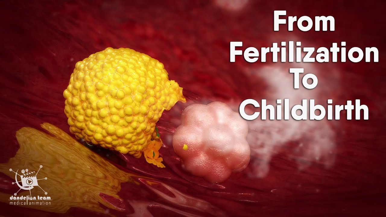
from fertilization to childbirth | 3d medical animation | by Dandelion Team
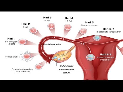
Pregnancy - How a Wonder is Born! (Animation)
Embryology: Development of the Placenta and Fetal Circulation, Animation
Early embryogenesis - Cleavage, blastulation, gastrulation, and neurulation | MCAT | Khan Academy
5.0 / 5 (0 votes)