Ischemic Stroke - causes, symptoms, diagnosis, treatment, pathology
Summary
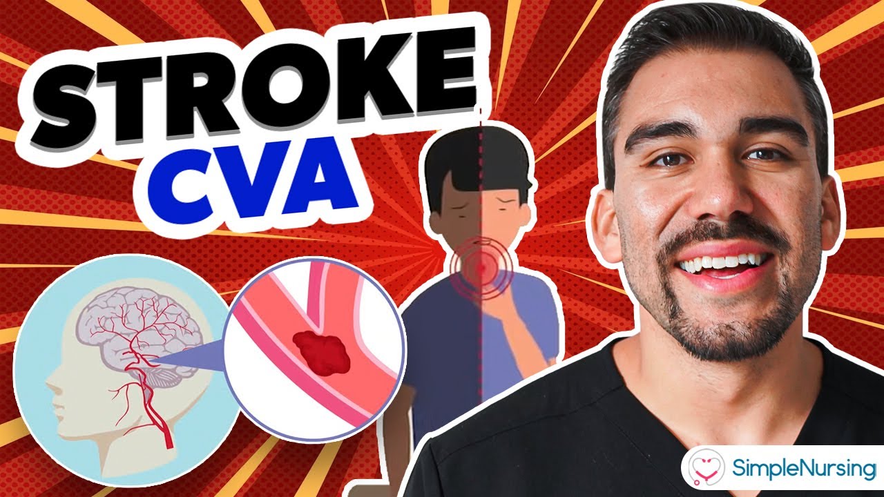
TLDRThis video script discusses two main types of strokes: ischemic, caused by a blocked artery, and hemorrhagic, caused by a ruptured artery. Ischemic strokes are more common, and their severity depends on how much blood flow is reduced. The script explains brain anatomy, blood flow, and common stroke symptoms, using the acronym FAST: Facial drooping, Arm weakness, Speech difficulties, and Time. Treatment options focus on restoring blood flow and preventing further damage. It also touches on prevention strategies, like quitting smoking and managing risk factors such as high blood pressure and cholesterol.
Takeaways
- 🧠 There are two types of stroke: ischemic (blocked artery) and hemorrhagic (artery breaks), with ischemic strokes being more common.
- 🕒 If stroke symptoms resolve within 24 hours, it's known as a transient ischemic attack, which usually has minimal long-term effects.
- 🧬 Basic brain anatomy includes the cerebrum, cerebellum, and brainstem, each with distinct functions and roles in motor control, sensory processing, and vital functions.
- 🔄 The brain's blood supply comes from the internal carotid and vertebral arteries, forming the Circle of Willis, which allows for alternative blood flow in case of blockage.
- 🚫 Ischemic strokes often occur due to endothelial cell dysfunction leading to atherosclerosis, where plaques can block blood flow.
- 🩸 Embolism is another cause of ischemic stroke, where a blood clot breaks off and lodges in a smaller artery, often originating from atherosclerosis or heart conditions.
- 🌊 Lacunar strokes are a type of ischemic stroke affecting deep branches of the middle cerebral artery, often due to hyaline arteriolosclerosis related to hypertension or diabetes.
- 🌊 Watershed infarcts occur when global blood flow reduction damages the brain's 'furthest downstream' tissues, typically at the border of two blood supplies.
- 🏥 Diagnosis of ischemic stroke involves medical imaging like CT, MRI, and angiography to locate and size the stroke, and differentiate between new and old injuries.
- 💊 Treatment aims to reestablish blood flow quickly, using thrombolytic enzymes like TPA, aspirin, or surgical procedures to remove clots and prevent further damage.
Q & A
What are the two main types of stroke mentioned in the script?
-The two main types of stroke are ischemic stroke, caused by a blocked artery that reduces blood flow to the brain, and hemorrhagic stroke, caused by a broken artery in the brain that leads to a pool of blood, damaging the brain.
Which type of stroke is more common, and what factors influence the amount of damage it causes?
-Ischemic strokes are more common. The amount of damage they cause depends on the parts of the brain affected and how long the brain suffers from reduced blood flow.
What is a transient ischemic attack (TIA), and what are its typical long-term effects?
-A transient ischemic attack (TIA) occurs when stroke symptoms self-resolve within 24 hours. It usually results in minimal long-term problems.
What are the main functions of the brain's lobes and other key regions mentioned in the script?
-The frontal lobe controls movement and decision-making (executive function), the parietal lobe processes sensory information and guides movement in 3D space, the temporal lobe is involved in hearing, smell, memory, and visual recognition, and the occipital lobe handles vision. The cerebellum helps with muscle coordination and balance, while the brainstem controls heart rate, blood pressure, breathing, and consciousness.
How does the Circle of Willis contribute to the brain’s blood supply?
-The Circle of Willis allows blood to circulate from one side of the brain to the other in case of a blockage, providing alternative ways for blood to bypass an obstructed vessel, helping maintain blood flow.
What are the two main mechanisms through which ischemic strokes occur?
-Ischemic strokes occur either due to endothelial cell dysfunction, where atherosclerosis causes a plaque to block an artery, or through an embolism, where a blood clot forms and travels to block an artery downstream.
What is the difference between a thrombus and an embolism in ischemic stroke formation?
-A thrombus is a clot that forms at the site of atherosclerosis and blocks the artery, while an embolism refers to a clot that breaks off from another location and travels through the bloodstream to lodge in a smaller artery or capillary.
What is a lacunar stroke, and how does it differ from other types of ischemic strokes?
-A lacunar stroke affects the deep branches of the middle cerebral artery, often resulting in small, fluid-filled cysts in the brain tissue. It is typically caused by hyaline arteriolosclerosis due to hypertension or diabetes, which thickens the arteriole walls.
What is the 'ischemic penumbra,' and why is it important during a stroke?
-The ischemic penumbra is the area of brain tissue surrounding the ischemic core that is still viable for a period of time due to collateral circulation. If blood flow is restored quickly enough, the penumbra may survive, reducing brain damage.
What treatments are available to restore blood flow in ischemic stroke cases, and how do they work?
-Treatments include thrombolytic enzymes like tissue plasminogen activator (TPA), which activate the body's natural clot-busting mechanisms, and surgical procedures like MERCI or suction removal to physically remove clots from arteries.
Outlines
🧠 Types and Effects of Stroke
The first paragraph introduces the two main types of stroke: ischemic and hemorrhagic. Ischemic strokes, caused by blocked arteries, are more common, while hemorrhagic strokes occur due to artery rupture in the brain. The severity of the stroke depends on the brain areas affected and the duration of reduced blood flow. A transient ischemic attack (TIA) occurs when symptoms resolve within 24 hours, usually with minimal long-term effects. The paragraph also provides a basic overview of brain anatomy, describing key regions like the cerebrum, cerebellum, brainstem, and the roles of various lobes in motor control, sensory processing, memory, and vital functions.
🩸 Mechanisms of Ischemic Stroke
The second paragraph delves into the mechanisms behind ischemic strokes. It explains how endothelial cell dysfunction, often triggered by irritants like tobacco toxins, leads to atherosclerosis—a buildup of plaque in arteries that restricts blood flow. The paragraph describes how the rupture of these plaques can cause a sudden and complete blockage, leading to stroke. It also discusses embolic strokes, where a clot from another part of the body, such as the heart, travels to the brain, causing blockage. Additionally, lacunar strokes, which involve small arteries in the brain, are covered, along with the concept of watershed infarcts caused by reduced blood flow in the body.
⏰ Stroke Symptoms, Diagnosis, and Treatment
The third paragraph focuses on stroke symptoms, diagnosis, and treatment. It explains how symptoms vary depending on the affected brain region, with specific examples like speech difficulties from strokes in Broca’s or Wernicke’s areas, and vision problems from posterior cerebral artery strokes. The acronym FAST (Facial drooping, Arm weakness, Speech difficulties, Time) is introduced as a mnemonic for recognizing stroke symptoms and the urgency of seeking help. The paragraph also covers diagnostic tools like CT, MRI, and angiography, and describes treatments aimed at restoring blood flow, such as thrombolytic enzymes, aspirin, and surgical procedures like MERCI. Finally, it highlights the importance of minimizing risk factors, such as smoking and high blood pressure, to prevent future strokes.
Mindmap
Keywords
💡Ischemic stroke
💡Hemorrhagic stroke
💡Cerebrum
💡Lobe
💡Cerebellum
💡Brainstem
💡Circle of Willis
💡Atherosclerosis
💡Embolism
💡Lacunar stroke
💡FAST
Highlights
There are two main types of stroke: ischemic stroke and hemorrhagic stroke.
Ischemic strokes are more common and occur when an artery to the brain is blocked.
Transient ischemic attacks (TIA) resolve within 24 hours and usually cause minimal long-term damage.
The cerebrum, divided into two hemispheres, controls different functions depending on the region affected.
The frontal lobe controls movement and decision-making, while the parietal lobe processes sensory information.
The temporal lobe is involved in hearing, memory, and recognition of faces and languages.
The occipital lobe primarily handles vision, while the cerebellum coordinates muscle movement and balance.
The Circle of Willis allows blood to flow between arteries, offering alternative circulation in case of blockage.
Atherosclerosis can lead to strokes by causing plaque buildup in arteries, which can block blood flow to the brain.
Small plaques with softer fibrous caps are more prone to rupture, leading to rapid clot formation.
Embolic strokes occur when a clot breaks off, travels, and lodges in a smaller artery.
A lacunar stroke involves the deep branches of the middle cerebral artery, often caused by hypertension or diabetes.
Ischemic stroke treatment focuses on quickly restoring blood flow to prevent further brain damage.
Thrombolytic agents like tissue plasminogen activator (TPA) can be used to break down clots, but they have time limits.
The FAST acronym is used to identify stroke symptoms: Facial drooping, Arm weakness, Speech difficulties, and Time.
Transcripts
There are two main types of stroke: an ischemic stroke which is when there’s a blocked artery
that reduces blood flow to the brain and a hemorrhagic stroke which is when an artery
in the brain breaks, creating a pool of blood that damages the brain.
Of the two, ischemic strokes are much more common, and the amount of damage they cause
is related to the parts of the brain that are affected and how long the brain suffers
from reduced blood flow.
Now if symptoms self-resolve within 24 hours, it’s called a transient ischemic attack
and there are usually minimal long-term problems.
OK - let’s start with some basic brain anatomy.
The brain has a few regions - the most obvious is the cerebrum, which is divided into two
cerebral hemispheres, each of which has a cortex - an outer region - divided into four
lobes including the frontal lobe, parietal lobe, temporal lobe, and the occipital lobe.
There are also a number of additional structures - including the cerebellum, which is down
below, as well as the brainstem which connects to the spinal cord.
The right cerebrum controls muscles on the left side of your body and vice versa.
The frontal lobe controls movement, and executive function, which is our ability to make decisions.
The parietal lobe processes sensory information, which lets us locate exactly where we are
physically and guides movements in a three dimensional space.
The temporal lobe plays a role in hearing, smell, and memory, as well as visual recognition
of faces and languages.
Finally there’s the occipital lobe which is primarily responsible for vision.
The cerebellum helps with muscle coordination and balance.
And finally there’s the brainstem plays a vital role in functions like heart rate,
blood pressure, breathing, gastrointestinal function, and consciousness.
The brain receives blood from the left and right internal carotid arteries, as well as
the left and right vertebral arteries, which come together to form the basilar artery.
The internal carotid arteries turn into the left and right middle cerebral arteries which
serve the lateral portions of the frontal, parietal, and temporal lobes of the brain.
Each of the internal carotid arteries also give off branches called the anterior cerebral
arteries which serve the medial portion of the frontal and parietal lobes and connect
with one another with a short little connecting blood vessel called the anterior communicating
artery.
Meanwhile, the vertebral arteries and basilar artery gives off branches to supply the cerebellum
and the brainstem.
In addition, the basilar artery divides to become the right and left posterior cerebral
artery which mainly serve the occipital lobe and some of the temporal lobe as well as the
thalamus.
Finally, the internal carotid arteries each give off a branch called the posterior communicating
artery which attaches to the posterior arteries on each side.
So together, the main arteries and the communicating arteries complete what is called the Circle
of Willis - a ring where blood can circulate from one side to the other in case of a blockage.
The Circle of Willis offers alternative ways for blood to get around an obstructed vessel.
In general, the brain can get by on diminished blood flow - especially when it happens gradually
because that allows enough time for collateral circulation to develop, which is where a nearby
vessel starts sending out branches of blood vessels to serve an area that’s in need.
But once the supply of blood flow is reduced to below the needs of the tissue - it causes
tissue damage, which we call an ischemic stroke.
There are two main ways that an ischemic stroke happens.
One mechanism is endothelial cell dysfunction, which is when something irritates or inflames
the slippery inner lining of the artery—the tunica intima.
One classic irritant is the toxins found in tobacco which float around in the blood damaging
the endothelium.
That damage becomes a site for atherosclerosis, which is where a plaque forms.
This is when a buildup of fat, cholesterol, proteins, calcium, and immune cells forms
and starts to obstruct arterial blood flow.
This plaque has two parts to it, the soft cheesy-textured interior and the hard outer
shell which is called the fibrous cap.
Branch points in arteries and particularly the internal carotid and middle cerebral arteries
are the most common spots for atherosclerosis.
Usually, though, it takes years for plaque to build up, and this slow blockage only partially
blocks the arteries, and so even though less blood makes it to brain tissue, there’s
still some blood.
So Strokes happen when there’s a sudden and complete or near-complete blockage of
an artery—so let’s see how that can happen.
Since plaques sit in the lumen of the blood vessel, they’re constantly being stressed
by mechanical forces from blood flow, and interestingly it’s often the smaller plaques
that are more dangerous.
Their fibrous caps are softer than the larger ones and are prone to getting ripped off.
Once that happens, the inner cheesy filling is exposed to the blood and is thrombogenic,
which means that it tends to form clots very quickly.
Platelets adhere to the exposed cheesy material, and they release chemicals that enhance the
clotting process.
Within a minute that artery can be fully blocked.
Another mechanism for ischemic stroke formation is an embolism.
An embolic stroke typically happens when a blood clot breaks off from one location, travels
through the blood, and gets lodged in an artery downstream, typically an artery, arteriole,
or capillary with a smaller diameter.
These blood clots typically emerge from atherosclerosis, but they can also form in the heart.
For example stagnant blood can form a clot, and blood can stagnate due to an atrial fibrillation
or after a heart attack.
If a clot forms in the left atrium, it moves into the left ventricle and from there it
has a direct route to the brain.
On the other hand, if a clot forms in the low-pressure veins or right atrium, then it
goes into the right ventricle and gets lodged in the pulmonary capillaries - with no way
of getting to the brain.
An important exception is if a person has a heart defect like an atrial septal defect
that allows blood and potentially a blood clot to wander from the right side of the
heart over to the left side of the heart.
In that situation, a venous or right atrial blood clot will have bypassed the pulmonary
circulation and established a route to the brain.
One specific type of ischemic stroke is called a lacunar stroke, and they typically involve
the deep branches of the middle cerebral artery that feed the basal ganglia.
Lacunar refers to “lake”, and is called that since after a lacunar stroke the damaged
brain tissue develops fluid filled pockets called cysts that look like little lakes under
a microscope.
Lacunar strokes classically develop as a result of hyaline arteriolosclerosis which is when
the arteriole wall gets filled with protein.
This can happen as a result of hypertension or diabetes, and can make the artery wall
quite thick, reducing the size of the lumen.
In addition to problems specific to an artery, something like shock can lead to a reduction
in blood flow throughout the entire body.
In these cases, the tissues that are the furthest downstream are affected the most.
This is because healthy tissue continues to extract what it needs from the blood flowing
by, leaving little or no oxygen and nutrients for the tissue furthest away.
The “furthest downstream” tissues in the brain are at the border of two different blood
supplies.
When the blood flow throughout the body is diminished for any reason, they get damaged,
and this pattern of injury is called a watershed infarct.
Regardless of the mechanism of an ischemic stroke, it’s helpful to remember that there’s
an ischemic core, which is the brain tissue that will likely die from ischemia, and then
there’s tissue around the core, called the ischemic penumbra, which is preserved for
a period of time by collateral circulation and has a chance to survive if blood flow
is restored quickly enough.
Regardless of the type of ischemic stroke, without a steady supply of glucose and oxygen,
cells run out of energy within minutes and you get a high buildup of sodium and calcium
levels.
High sodium levels draws water into the cell making it swell, this is called cytotoxic
edema.
And high calcium leads to the buildup of reactive oxygen radicals that react with lipids in
the membranes of mitochondria and lysosomes.
Damage to these organelles allows apoptosis-inducing factors and degradative enzymes to seep out
of the cell.
Over a period of 4-6 hours, immune cells begin to haul away damaged cells and the resulting
inflammation damages the blood brain barrier allowing fluid and proteins to get into the
brain tissue causing swelling or vasogenic edema.
Because the skull creates a fixed volume the swelling leads to a mass effect where the
swollen brain tissue pushes into the unaffected side of the brain-called cingulate or uncal
herniation, or slips down and out of the base of the skull - called cerebellar tonsil herniation,
which is particularly dangerous because it can push onto the brainstem and affect breathing
and consciousness.
Stroke symptoms depend on the exact part of the brain that is affected.
For example, an anterior or middle cerebral artery stroke can cause numbness and sudden
muscle weakness.
If a stroke affects the Broca’s area, which is usually in the left frontal lobe, or Wernicke’s
area, which is usually in the left temporal lobe, then it can cause slurred speech or
difficulty understanding speech, respectively.
If there’s a posterior cerebral artery stroke, then it can affect vision.
An acronym to remember some common stroke symptoms is FAST - Facial drooping, Arm weakness,
Speech difficulties, and Time.
Time is obviously not a symptom but just a reminder to get help as quickly as possible
to minimize cell injury and maximize the chance of a full recovery.
To diagnose and confirm the location and size of an ischemic stroke, medical imaging with
a CT or MRI can be used.
Also, angiography, which uses contrast injected into the blood, can help to visualize the
exact location where blood flow is blocked within an artery.
In addition, using FLAIR sequence MRIs, it’s possible to distinguish a new stroke injury
from an old one.
In an ischemic stroke the ultimate treatment is to reestablish blood flow as quickly as
possible to prevent further cell death, particularly in the penumbra - every minute counts.
So thrombolytic enzymes, like tissue plasminogen activator or TPA, are used to activate the
body’s natural clot busting mechanisms, but TPA does have a time limit of when it
can be used.
Aspirin is also used to prevent platelets from forming additional clots.
If TPA is unsuccessful, surgical procedures can be used that push a wire through the artery
and physically remove the clot.
In mechanical embolus removal in cerebral ischemia, called MERCI for short, the wire
grabs on to the clot and draws it out of the artery.
In suction removal, the wire is used to physically break down the clot and clot fragments are
removed with suction.
After a stroke has occurred, there is an elevated risk of having additional strokes so it’s
important to minimize risk factors - the main one being quitting smoking, but others include
having a healthy blood pressure, normal LDL cholesterol levels, and controlling other
diseases like diabetes.
Occasionally, a surgery may be necessary to help clean arteries obstructed by severe atherosclerosis.
For example, in a carotid endarterectomy, the internal carotid artery is opened up and
atherosclerotic plaque is removed.
Alternatively, a stent may be placed to keep the artery opened up.
Okay, a quick recap: An ischemic stroke occurs when there’s an acute decrease in the arterial
blood supply.
It can be due to atherosclerosis, a thrombus, an embolus,, or a global reduction in blood
flow.
The goal is to identify symptoms and reestablish blood flow to prevent long-term damage - to
remember this a common acronym is FAST - Facial drooping, Arm weakness, Speech difficulties,
and Time.
Voir Plus de Vidéos Connexes
Brain Stroke, Types of, Causes, Pathology, Symptoms, Treatment and Prevention, Animation.
What happens during a stroke? - Vaibhav Goswami
Coronary Artery Disease Animation
Stroke Pathogenesis (with a focus on Ischemic Stroke)
Recognizing Warning Signs and Symptoms of a Stroke | In Case of Emergency | Mass General Brigham
This exercise boosts blood flow to the brain by 576 times and instantly makes you feel younger!
5.0 / 5 (0 votes)