Staining of Nucleic Acid by Acetocarmine
Summary
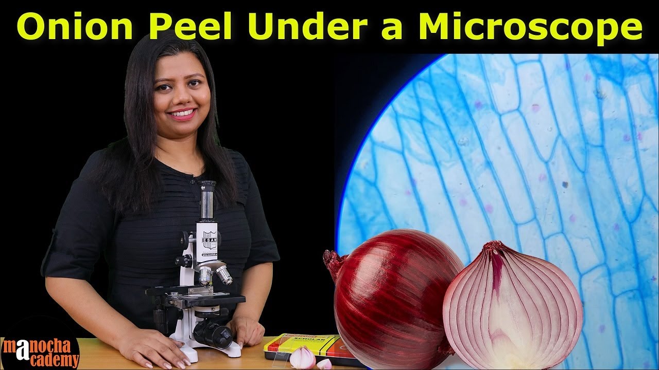
TLDRThis video demonstrates the process of staining nucleic acids in onion cells using aceto-carmine. The procedure involves preparing a fresh onion peel, applying water and aceto-carmine stain, and gently heating the slide to avoid drying. After adding a cover slip and removing excess stain, the slide is examined under a compound microscope. Observations reveal that the onion cells are rectangular or hexagonal, each with a distinct nucleus at the periphery. The cells also have a cell wall, lightly stained cytoplasm, and a large vacuole, providing a clear view of cell structure.
Takeaways
- 😀 The procedure involves using onion peel for staining nucleic acids.
- 😀 Aceto Carmine stain and water are required for the process.
- 😀 Tools used include forceps, a glass slide, a cover slip, dropper, needle, filter paper, spirit lamp, and a compound microscope.
- 😀 The onion peel is carefully placed on a glass slide with a few drops of water to prevent desiccation.
- 😀 Two drops of Aceto Carmine stain are added to the peel for staining.
- 😀 The glass slide is gently heated over a spirit lamp to facilitate the staining process.
- 😀 A cover slip is placed over the peel, avoiding air bubbles and wrinkles.
- 😀 Excess stain is removed with filter paper after the cover slip is applied.
- 😀 The sample is examined under a compound microscope for observation.
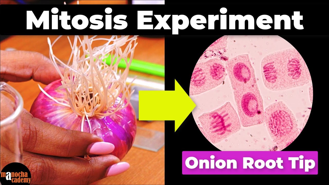
- 😀 Under the microscope, the nucleus is seen as uninucleate, with cells arranged in rectangular or hexagonal shapes.
- 😀 The cells contain distinct nuclei at their periphery, along with cytoplasm and large vacuoles.
Q & A
What is the purpose of using aceto-carmine stain in this procedure?
-The aceto-carmine stain is used to stain nucleic acids, allowing the nucleus and other cell structures to become visible under a microscope.
What materials are needed for this procedure?
-The materials required include onion, aceto-carmine stain, water, forceps, glass slide, cover slips, dropper, needle, filter paper, spirit lamp, and a compound microscope.
How should the onion peel be prepared for staining?
-A small piece of the epidermal peel should be cut from a fresh onion using a needle and forceps, and placed onto a glass slide.
Why is water added to the onion peel on the glass slide?
-Water is added to prevent the onion peel from drying out during the procedure.
What is the purpose of gently heating the slide over a spirit lamp?
-Heating the slide gently helps to fix the aceto-carmine stain onto the onion peel and facilitates better staining.
Why is it important to avoid air bubbles and wrinkles when placing the cover slip?
-Avoiding air bubbles and wrinkles ensures that the onion peel remains flat and the stain is distributed evenly, allowing for clear microscopic observation.
What should be done with the excess stain after applying it to the onion peel?
-The excess stain should be wiped away using filter paper to prevent interference with the microscopic observation.
What is observed under the microscope in terms of cell structure?
-Under the microscope, the cells appear rectangular or hexagonal in shape with a large nucleus positioned on the periphery of each cell. The cells are uninucleate and contain a large central vacuole, cytoplasm, and a cell wall.
What is the shape of the cells observed in the onion peel?
-The cells in the onion peel are rectangular or hexagonal in shape.
What can be inferred about the onion cells from the observation under the microscope?
-The onion cells are regular in shape and exhibit a clear, distinct nucleus on the periphery of each cell. The presence of a large vacuole and cytoplasm is also noted.
Outlines

Esta sección está disponible solo para usuarios con suscripción. Por favor, mejora tu plan para acceder a esta parte.
Mejorar ahoraMindmap

Esta sección está disponible solo para usuarios con suscripción. Por favor, mejora tu plan para acceder a esta parte.
Mejorar ahoraKeywords

Esta sección está disponible solo para usuarios con suscripción. Por favor, mejora tu plan para acceder a esta parte.
Mejorar ahoraHighlights

Esta sección está disponible solo para usuarios con suscripción. Por favor, mejora tu plan para acceder a esta parte.
Mejorar ahoraTranscripts

Esta sección está disponible solo para usuarios con suscripción. Por favor, mejora tu plan para acceder a esta parte.
Mejorar ahoraVer Más Videos Relacionados
Mitosis Experiment Onion Root Tip Procedure

Onion Skin Epidermal Cells: How to Prepare a Wet Mount Microscope Slide

Understand Electroporation In Under 3 Minutes
Onion Peel Under the Microscope | How to Prepare Stained Temporary Mount of Onion Peel

Biology Lab || Mitosis

Pengamatan Mitosis Pada Akar Bawang dengan Metode Squash
5.0 / 5 (0 votes)
