Pemeriksaan EKG NersMucis
Summary
TLDRIn this educational video, Nurse Rudy Kurniawan, a lecturer at Muhammadiyah Ciamis Health College, demonstrates the step-by-step procedure for performing an electrocardiography (ECG or EKG). The tutorial explains the equipment setup, including electrodes, clamps, and gel application, and provides detailed guidance on proper electrode placement on the hands, feet, and chest. The video emphasizes safety, cleanliness, and patient comfort, showing how to record the heart’s electrical activity accurately. Concluding with a successful EKG recording, the demonstration reassures viewers that the procedure is simple, painless, and essential for monitoring heart health, making it valuable for both healthcare professionals and patients.
Takeaways
- 😀 Electrocardiography (ECG or TKG) is a simple, safe, and painless procedure to monitor heart electrical activity.
- 😀 Proper patient interaction and explanation are essential before starting the ECG to ensure patient comfort and cooperation.
- 😀 All metal objects on the patient’s chest should be removed before electrode placement.
- 😀 Hand hygiene is mandatory before touching the patient or equipment.
- 😀 ECG equipment consists of the machine, cables, electrodes, electrode gel, alcohol swabs, clamps, and ECG paper.
- 😀 Limb electrodes are color-coded and labeled: Red (R) for right hand, Yellow (L) for left hand, Green (F) for left foot, Black (N) for right foot.
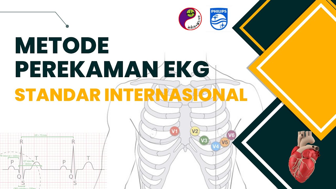
- 😀 Chest (precordial) electrodes are placed at specific intercostal spaces and positions: V1–V6, each with precise placement guidelines.
- 😀 Electrode placement accuracy is critical for obtaining a reliable and interpretable ECG recording.
- 😀 Gel is applied and electrodes are cleaned before placement to ensure good electrical contact.
- 😀 After recording, all electrodes should be removed carefully, and the ECG results reviewed with the patient.
- 😀 ECG installation is suitable for nursing education, patient monitoring, and assessing post-surgical heart conditions.
- 😀 Visual guidance and careful adherence to electrode positioning prevent errors and improve the quality of the ECG reading.
Q & A
Who is presenting the electrocardiography (ECG) procedure in the video?
-The procedure is presented by Nurse Rudy Kurniawan, who is also a lecturer at Muhammadiyah Ciamis Health College.
What is the purpose of performing an electrocardiography (ECG)?
-The purpose of an ECG is to check and record the electrical activity of the heart, providing information about heart rhythm and function.
Is the ECG procedure painful or dangerous for the patient?
-No, the ECG procedure is simple, painless, does not cause headaches, and does not electrocutethe patient.
What are the main components of the ECG machine mentioned in the video?
-The ECG machine consists of the main machine, cables, electrodes, ECG paper, display screen, conductive gel, alcohol swabs, and clamps for attaching electrodes.
How many limb electrodes are used and where are they placed?
-Four limb electrodes are used: right hand (red, R), left hand (yellow, L), left foot (green, F), and right foot (black, N for Neutral).
How many chest (precordial) electrodes are used and how are they positioned?
-Six chest electrodes (C1 to C6) are used. They are positioned in specific intercostal spaces: C1 (V1) right parasternal 4th intercostal, C2 (V2) left parasternal 4th intercostal, C3 (V3) between V2 and V4, C4 (V4) midclavicular 5th intercostal, C5 (V5) anterior axillary 5th intercostal, and C6 (V6) midaxillary 5th intercostal line.
What preparation steps are necessary before placing the electrodes?
-Before placement, the patient should remove any metallic objects on the chest, and all electrodes must be cleaned with alcohol swabs. Conductive gel is applied to improve signal transmission.
Why is it important not to rely solely on the color of the electrodes?
-The color may vary or some clamps may be colorless, so the letters on each electrode (e.g., R, L, F, C1-C6) are the critical identifiers for correct placement.
How does the presenter ensure proper placement of the chest electrodes?
-The presenter locates anatomical landmarks such as the clavicle and intercostal spaces to correctly position each chest electrode according to standard ECG placement guidelines.
What is the final step after recording the ECG?
-After recording, all electrodes are carefully removed from the patient, the skin is cleaned, and the results are reviewed to assess the heart's electrical activity.
What message does the presenter convey about the importance of the ECG procedure?
-The presenter emphasizes that the ECG procedure is easy, safe, and beneficial for both patients and healthcare professionals, contributing to the understanding of heart health.
Outlines

Esta sección está disponible solo para usuarios con suscripción. Por favor, mejora tu plan para acceder a esta parte.
Mejorar ahoraMindmap

Esta sección está disponible solo para usuarios con suscripción. Por favor, mejora tu plan para acceder a esta parte.
Mejorar ahoraKeywords

Esta sección está disponible solo para usuarios con suscripción. Por favor, mejora tu plan para acceder a esta parte.
Mejorar ahoraHighlights

Esta sección está disponible solo para usuarios con suscripción. Por favor, mejora tu plan para acceder a esta parte.
Mejorar ahoraTranscripts

Esta sección está disponible solo para usuarios con suscripción. Por favor, mejora tu plan para acceder a esta parte.
Mejorar ahoraVer Más Videos Relacionados

Pemeriksaan Elektrokardiografi (EKG)
Metode Perekaman EKG Standar Internasional | ECG Recording Technique

BAG TECHNIQUE - COMMUNITY HEALTH NURSING(CHN) l RETURN DEMONSTRATION (student nurse)

#pemeriksaanvisus #skilllab PEMERIKSAAN VISUS MATA. SKILL LAB

PERTEMUAN 7A - PROSEDUR PEMASANGAN EKG

Performing Oropharyngeal Suctioning
5.0 / 5 (0 votes)
