Diagnostic Imaging Explained (X-Ray / CT Scan / Ultrasound / MRI)
Summary
TLDRThis video explains four imaging modalities commonly used by physiotherapists to assess injuries: X-ray, CT scan, ultrasound, and MRI. X-rays are quick and cost-effective for identifying fractures and dislocations but provide only 2D images. CT scans create 3D cross-sectional images, useful for detecting small fractures and complex injuries but involve more radiation. Ultrasound is radiation-free and ideal for evaluating soft tissues during movement but cannot capture deep structures. MRI offers detailed imaging of soft tissues without radiation but is more expensive and slower. X-rays and CT scans assess bone injuries, while MRI and ultrasound focus on soft tissues.
Takeaways
- 🏥 X-ray is commonly used to evaluate possible fractures, dislocations, and bone infections.
- 📊 X-ray's main limitation is that it produces a 2D image of a 3D object, requiring multiple views for proper evaluation.
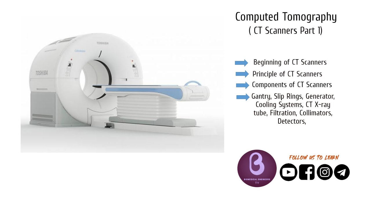
- 🧬 CT scan provides detailed cross-sectional images of bones, blood vessels, and soft tissues, useful for complex body regions.
- 💸 CT scan is more expensive and exposes patients to more radiation compared to X-ray.
- 🔊 Ultrasound uses high-frequency sound waves to image soft tissues like muscles, tendons, and ligaments.
- 🌡️ Ultrasound is radiation-free, cost-effective, and can be performed during movement, beneficial for dynamic evaluations.
- 🚫 Ultrasound's limitation is its inability to image deep structures like menisci and ACL.
- 🧲 MRI uses magnets and radiofrequency waves to provide detailed images of soft tissues without radiation exposure.
- 🕒 MRI is time-consuming, expensive, and not suitable for patients with certain metal implants.
- 🔍 X-ray and CT scan are typically used for bony injuries, while MRI and ultrasound are for soft tissue injuries.
- 📺 The video encourages viewers to subscribe for more informative content.
Q & A
What is the primary purpose of an X-ray in evaluating injuries?
-X-rays are used to evaluate possible fractures, dislocations, and bone infections.
What are the benefits of using X-rays for injury assessment?
-X-rays can be performed quickly, are readily available, and are cost-effective.
What is the main limitation of X-rays when assessing injuries?
-X-rays produce a 2-dimensional image of a 3-dimensional object, requiring at least two different views to evaluate the injury site.
How does a CT scan differ from an X-ray in terms of imaging?
-A CT scan uses multiple X-rays from different angles to produce detailed cross-sectional images of bones, blood vessels, and soft tissues.
What are the advantages of using a CT scan over an X-ray?
-CT scans can detect small fractures that may be missed on X-rays and can produce 3-dimensional images, aiding in locating the injury site.
What are the limitations of CT scans compared to plain X-rays?
-CT scans are relatively expensive and expose the person to a greater amount of radiation.
How does an ultrasound work and what is it used for?
-Ultrasound uses high-frequency sound waves to produce images of soft tissues such as muscles, tendons, and ligaments.
What are the benefits of using ultrasound for injury assessment?
-Ultrasound does not expose the person to radiation, is readily available, cost-effective, and can be performed during movement.
What is the main limitation of ultrasound imaging?
-Ultrasound cannot penetrate tissues to produce images of deep structures such as the meniscus and ACL.
How does MRI imaging work and what is it commonly used for?
-MRI uses strong magnets and radiofrequency waves to produce images of the meniscus, labrum, spinal disc, joint, and soft tissues.
What are the advantages of MRI over other imaging modalities?
-MRI provides detailed information about body structures without exposing the person to radiation, enabling accurate assessment of soft tissue injuries.
What are the limitations of MRI imaging?
-MRI takes longer to perform, is relatively expensive, and cannot be performed on people with certain metal implants.
How do X-ray and CT scan differ in their applications for injury assessment?
-X-rays are often used to evaluate bony injuries such as fractures, while CT scans are used for more complex regions of the body.
What types of injuries are MRI and ultrasound typically used to evaluate?
-MRI and ultrasound are often used to evaluate soft tissue injuries, including muscles, tendons, and ligaments.
Outlines
🔍 Understanding the Differences: Fractures, Ligament Tears, and Muscle Strains
This paragraph introduces the topic of differentiating between fractures, ligament tears, and muscle strains, and discusses the four common imaging modalities used to evaluate injuries. X-rays are introduced as a basic, cost-effective method primarily used by physiotherapists to assess fractures, dislocations, and bone infections. However, it has limitations such as producing only a 2D image, requiring multiple views to fully assess an injury.
🖼️ CT Scans: A Deeper Look into Complex Body Regions
The next section focuses on CT scans, which use multiple X-rays to generate detailed, cross-sectional images of bones, blood vessels, and soft tissues. Physiotherapists rely on CT scans for assessing more complex areas like the spine, pelvis, ankle, and foot. It is particularly useful for detecting small fractures missed by X-rays and can generate 3D images for more precise injury localization. However, it comes with higher costs and increased radiation exposure compared to standard X-rays.
🔊 Ultrasound: A Safe Option for Evaluating Soft Tissues
Ultrasound imaging, which uses high-frequency sound waves, is discussed as a non-invasive and radiation-free option for evaluating soft tissues, such as muscles, tendons, and ligaments. It is especially helpful in diagnosing conditions like shoulder impingement since it can be used during movement. Despite its advantages, ultrasound has limitations as it cannot capture deep structures like the meniscus or ACL, reducing its effectiveness in certain injury evaluations.
🧲 MRI: Detailed Imaging for Soft Tissue Injuries
MRI technology, which uses strong magnets and radiofrequency waves, is explained as a go-to method for evaluating soft tissues like the meniscus, labrum, spinal discs, and joints. Its primary advantage is that it offers detailed insights without the use of radiation, making it ideal for accurate assessment of soft tissue injuries. However, MRI scans are time-consuming, expensive, and pose risks for people with certain metal implants due to potential interactions with the magnetic field.
📊 Comparing Imaging Modalities for Bone and Soft Tissue Injuries
The conclusion summarizes the use cases for each imaging technique. X-rays and CT scans are favored for assessing bone injuries like fractures, while MRI and ultrasound are preferred for soft tissue injuries involving muscles, tendons, and ligaments. The video wraps up with a call to action, encouraging viewers to subscribe if they found the content useful.
Mindmap
Keywords
💡X-ray
💡CT scan
💡Ultrasound
💡MRI
💡Fracture
💡Soft tissue
💡Radiation
💡Dynamic imaging
💡Meniscus
💡Cost-effective
Highlights
Explains 4 imaging modalities used after an injury.
X-ray uses radiation to produce an image.
X-ray is used to evaluate possible fractures, dislocations, and bone infections.
X-ray benefits include speed, availability, and cost-effectiveness.
X-ray limitation is producing a 2D image of a 3D object.
CT scan uses X-rays from different angles to produce detailed cross-sectional images.
CT scan is used to evaluate complex regions like the spine, pelvis, ankle, and foot.
CT scan benefits include detecting small fractures and producing 3D images.
CT scan limitations include cost and higher radiation exposure.
Ultrasound uses high-frequency sound waves to produce images.
Ultrasound is used to evaluate soft tissues like muscles, tendons, and ligaments.
Ultrasound benefits include no radiation exposure, availability, cost-effectiveness, and movement during imaging.
Ultrasound limitation is inability to penetrate tissues for deep structures imaging.
MRI uses strong magnets and radiofrequency waves to produce images.
MRI is used to evaluate meniscus, labrum, spinal disc, joint, and soft tissues.
MRI benefits include no radiation exposure and detailed information about body structures.
MRI limitations include longer procedure time, cost, and incompatibility with certain metal implants.
X-ray and CT scan are often used for bony injuries.
MRI and ultrasound are often used for soft tissue injuries.
Encourages viewers to subscribe for more content.
Transcripts
Is this a fracture ligament tear or a simple muscle strain
today we will explain the 4 imaging modalities commonly used after an injury
X-ray is a form of radiation it penetrates the body to produce an image
physiotherapists commonly use X-Ray to evaluate a possible fracture
dislocation and bone infection X-ray has many benefit it can be performed quickly
they are readily available and cost-effective the main limitation of X-ray
is it produces a 2 dimensional image of a 3 dimensional object
so a minimum of 2 different views are often required to evaluate the site of injury
CT scan fires a number of X-rays from different angles
it can produce a detailed cross-sectional image of the bone blood vessel and soft tissues
physiotherapists commonly use CT scan to evaluate complex regions of the body
these include the spine pelvis ankle and foot CT scan has many benefit
it can detect small fractures that may have been missed on a X-ray
it can also produce a 3 dimensional image which makes it easier to locate the site of injury
CT scan has many limitations it is relatively expensive
and compared to the plain X-ray it can expose the person to a greater amount of radiation
Ultrasound uses high-frequency sound waves to produce an image
physiotherapists commonly use ultrasound to evaluate soft tissues
such as the muscle tendon and ligament Ultrasound has many benefit
it does not expose the person to any radiation they are readily available cost-effective
and most importantly they can be performed during movement
this is particularly helpful in the evaluation of shoulder impingement
the main limitation of Ultrasound is it cannot penetrate the tissues
and produce image of deep structures such as the meniscus and the ACL
MRI uses strong magnets and radiofrequency waves to produce an image
physiotherapists commonly use MRI to evaluate the meniscus labrum spinal disc
joint and soft tissues MRI has many benefit it does not expose the person to any radiation
it provides detailed information about the body structures
this enables us to make accurate assessment of soft tissue injuries
MRI has many limitations it takes a lot longer to perform it is relatively expensive
and it cannot be performed in people with certain metal implants
as some metals can heat up or attract to the magnet
In summary X-ray and CT scan are often used to evaluate bony injuries such as fracture
MRI and ultrasound are often used to evaluate soft tissue injuries
including the muscle tendon and ligament
If you liked the content from today’s video don’t forget to subscribe
thank you for watching we will see you next time
Ver Más Videos Relacionados
5.0 / 5 (0 votes)