Male Genital System (Internal & External) - Anatomy
Summary
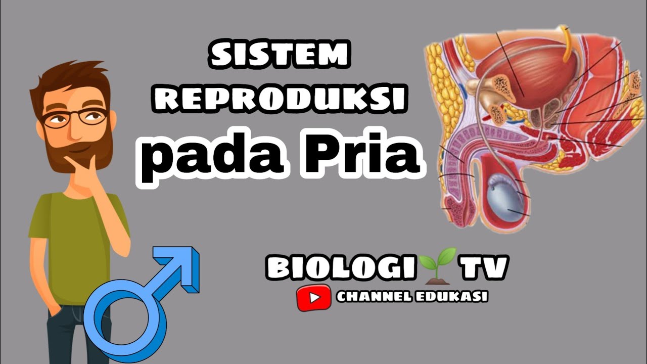
TLDRThis educational video script offers a comprehensive overview of the male genital system, detailing both internal and external structures. It covers the testes, epididymis, ductus deferens, seminal glands, prostate, bulbourethral glands, and the urethra. The script also discusses the anatomy of the penis and scrotum, emphasizing their functions and significance in male reproduction, such as sperm maturation and the role of testosterone in development.
Takeaways
- 🔬 The male genital system is divided into internal and external genital organs, each playing a crucial role in male reproduction.
- 🥚 The testes are the primary male reproductive glands, responsible for producing sperm and testosterone, and are located in the scrotum outside the body.
- 🧬 Leydig cells in the testes produce testosterone, which is vital for male development, while Sertoli cells provide structural support and nourishment for developing sperm.
- 📍 The testes descend from the lumbar region during prenatal development, taking with them the testicular arteries and lymphatic drainage to the lumbar nodes.
- 🌀 The epididymis is not an organ with functional cells but serves as a tightly coiled tube reservoir for maturing sperm, providing them with nutrition.
- 💧 The seminal glands produce 50-80% of the ejaculate fluid, while the prostate gland contributes about 30%, both being essential for semen composition.
- 🚦 The ductus deferens is a tubular organ that transports sperm from the epididymis to the prostatic urethra through peristaltic contractions.
- 💉 The bulbourethral glands are accessory glands that produce a viscous fluid to lubricate the urethra during ejaculation.
- 🍌 The penis consists of three erectile bodies: two corpora cavernosa and one corpus spongiosum, which fill with blood during an erection.
- 🌡 The scrotum's temperature is maintained 2-4 degrees lower than body temperature to ensure optimal sperm production.
- 🔄 The cremaster muscle controls the scrotum's position based on external temperature, pulling it closer to the body in cold and relaxing in heat.
Q & A
What are the primary functional cells in the male reproductive system?
-The primary functional cells in the male reproductive system are Leydig and Sertoli cells, both of which are found in the testes.
What is the role of testosterone in male development?
-Testosterone influences male development and maturation by developing muscles, deepening the voice, growing body hair, and promoting the production of red blood cells. It also maintains spermatogenesis and male fertility.
Where are the testes located in the body?
-The testes are located outside the body in a pouch called the scrotum. They originate in the lumbar region near the kidneys and descend into the scrotum during prenatal development.
What is the function of the Tunica Albuginea?
-The Tunica Albuginea is a dense membrane of connective tissue that covers the testis and provides structural support.
How does the blood-testis barrier function?
-The blood-testis barrier is formed by tight junctions between Sertoli cells, which prevents sperm from entering the bloodstream and protects against an immune response against sperm cells.
What is the role of the epididymis in the male reproductive system?
-The epididymis serves as a reservoir for spermatozoa, allowing them to mature and gain the capacity to move and fertilize the ovum.
How does the temperature of the scrotum affect sperm production?
-The temperature inside the scrotum is 2–4 degrees lower than the core body temperature, which ensures optimal spermatogenesis as sperm production is sensitive to temperature.
What is the function of the cremaster muscle in the scrotum?
-The cremaster muscle contracts to pull the scrotum towards the abdominal wall in cold conditions and relaxes when it's hot, helping to regulate the temperature of the testes.
What are the parts of the ductus deferens and their locations?
-The ductus deferens has a scrotal part in the scrotum, a funicular part within the spermatic cord, an inguinal part as it passes through the inguinal canal, and a pelvic part as it enters the pelvis.
What percentage of the ejaculate fluid does the seminal gland produce?
-The seminal gland produces 50–80% of the ejaculate fluid, which is a significant portion of the semen released during ejaculation.
How is the urethra divided in the male reproductive system?
-The male urethra is divided into the prostatic urethra in the prostate, the membranous urethra in the perineum, and the spongy urethra in the penis.
Outlines
🌟 Overview of the Male Genital System
The paragraph introduces the male genital system, emphasizing its internal and external components. It highlights the importance of the testes, which house the Leydig and Sertoli cells crucial for male reproduction. Leydig cells produce testosterone influencing male characteristics, while Sertoli cells support sperm development. The testes' descent from the lumbar region to the scrotum is described, along with the testes' external and internal anatomy, including the Tunica Albuginea and seminiferous tubules.
🔬 Internal Structures and Functions of the Testes
This section delves deeper into the testes' internal structures, explaining the role of Sertoli cells in forming the blood-testis barrier and Leydig cells in testosterone production. It also discusses the mediastinum of the testis, rete testis, and the epididymis' role in sperm maturation. The epididymis is described as a reservoir for sperm, providing nutrition and facilitating sperm motility and fertilization capacity.
🧵 The Path of the Spermatic Cord and Descent of the Testes
The paragraph traces the layers that form during the descent of the testes from the lumbar region, which include the Tunica Vaginalis, internal and external spermatic fasciae, and the cremaster muscle. It also describes the spermatic cord, containing the ductus deferens, blood vessels, and nerves, and how these structures connect the testes to the abdominal wall and scrotum.
💧 The Ductus Deferens and Seminal Glands
This part covers the ductus deferens, detailing its role in transporting sperm from the epididymis to the prostatic urethra. It also describes the seminal glands' function in producing a significant portion of the ejaculate fluid. The structure and composition of the prostate and its importance in male reproductive health are also discussed, including the different zones within the prostate and their functions.
💦 The Urethra and Bulbourethral Glands
The paragraph discusses the male urethra, its parts, and its journey through the prostate, perineum, and penis. It also describes the bulbourethral glands, which contribute to the lubrication of the urethra during ejaculation. The structures and functions of the penis, including its erectile bodies and external features, are also outlined.
🌡 The Scrotum: Temperature Regulation and Anatomy
The final paragraph focuses on the scrotum, explaining its role in temperature regulation for optimal sperm production. It describes the scrotum's layers, including the dartos fascia and skin, and how they contribute to the scrotum's function. The paragraph concludes with a summary of the entire male genital system's anatomy.
Mindmap
Keywords
💡Male genital system
💡Testes
💡Epididymis
💡Ductus deferens
💡Seminal gland
💡Prostate
💡Male urethra
💡Bulbourethral glands
💡Scrotum
💡Penis
💡Spermatogenesis
Highlights
Introduction to the male genital system, covering both internal and external genital organs.
Description of the internal genital organs, including the testes, epididymis, ductus deferens, seminal gland, ejaculatory duct, prostate, male urethra, and bulbourethral glands.
Explanation of the primary functional cells in the male reproductive system: Leydig and Sertoli cells.
Role of testosterone in male development, maturation, and spermatogenesis.
The descent of the testes from the lumbar region to the scrotum during prenatal development.
External features of the testes, including the upper pole, lower pole, anterior border, posterior border, lateral surface, and medial surface.
Internal anatomy of the testes, including the Tunica Albuginea, Tunica Vasculosa, and lobules of testes.
Function of the blood-testis barrier in preventing sperm from entering the bloodstream and avoiding immune responses.
Description of the epididymis as a reservoir for spermatozoa and its role in sperm maturation.
Anatomy of the spermatic cord, including the ductus deferens, vessels, and nerves.
Transport of spermatozoa by the ductus deferens through peristaltic contractions.
Function of the seminal gland in producing 50–80% of the ejaculate fluid.
Details of the prostate's role in producing seminal fluid and its histological zones.
Description of the bulbourethral glands as accessory genital glands that lubricate the urethra during ejaculation.
Division of the male urethra into prostatic, membranous, and spongious sections.
Structure of the penis, including the corpora cavernosa, corpus spongiosum, and the glans penis.
Function of the cremaster muscle in regulating scrotal temperature to optimize spermatogenesis.
Layers of the scrotum, including the tunica vaginalis, internal spermatic fascia, cremaster muscle, external spermatic fascia, dartos fascia, and skin.
Transcripts
What's up.
Let's go ahead and cover the male genital system.
We will cover the most important aspect of the male genitals, and hopefully, in the end,
you'll have a pretty good understanding about this topic
Alright.
The male genital organs are divided into the internal genital organs and the external genital
organs.
We'll cover both of these in detail, And we'll start with the internal genital
organs So the internal genital organs consist of
the testes, epididymis, ductus deferens, seminal gland, Ejaculatory Duct, Prostate, Male urethra,
and the bulbourethral glands.
Alright, let's follow this arrangement, starting with the testis.
Now, of the whole male reproductive system, the primary functional cells the male has
are Leydig and Sertoli cells.
And both of them are found in the testes.
So the testicle or testis is the male reproductive gland or gonad in all animals, not just humans.
Leydig cells produce testosterone, which is a steroid hormone that binds to intracellular
receptors and regulates protein synthesis.
And what that does is it influences the male development and maturation for developing
muscles, deepening the voice, growing body hair, and promoting the production of red
blood cells.
Testosterone also stimulates the Sertoli cells to provide structural support and secrete
fluid to nourish and support developing sperm cells.
So that means that testosterone is also essential to maintain spermatogenesis and male fertility.
And In the absence of testosterone stimulation, spermatogenesis does not proceed.
It doesn't happen, basically.
The testes are located outside the body, right?
In a pouch called the scrotum.
During prenatal development, they originate in the lumbar region, near the kidneys, and
then they descend through the retroperitoneal space and inguinal canal to reach the scrotum.
As they descend, the layers of the abdominal wall form the layers of the scrotum as the
testes pass through them.
And here's also a fun fact, the testicular vessels descend together with the testes,
and that is why the testicular arteries, or gonadal arteries, come from the lumbar region
of the abdomen as well.
So same goes for females and the ovaries.
Lymphatic drainage is similarly directed to the lumbar nodes.
Ok.
So we're first going to cover the testes external structures,
Then we'll open it up and cover its internal anatomy.
After that, we're going to talk a little bit about the epididymis and finally go through
their coverings and external fixation.
Awesome.
Let's do the external structures first.
Externally, the testicles have an upper pole and a lower pole.
It has an anterior border and a posterior border.
And if you look at the testes from an anterior perspective, you'll also see that it has a
lateral surface and a medial surface.
So the external features of the testes are very simple.
Let's now go ahead and look at the internal structures of it,
And we'll start by removing all other structures we're not interested in for now.
Now first off, we need to identify the most external layer.
And that is a capsule called Tunica Albuginea.
Tunica Albuginea is a dense membrane of connective tissue covering the testis.
And if we remove the lateral wall of the testis, you'll see that Tunica albuginea covers the
external part of the testis.
Right underneath tunica albuginea, you'll find the vascular layer, or tunica vasculosa,
containing primarily blood vessels.
Within the actual testis, you'll find septae.
Or septa of the testis.
They are connective tissue that separates the inner structures of the testes into lobules,
called lobules of testes Now the lobules of the testis are regions
that contain single tubules.
Right?
So each lobule has a single tubule.
And each of these single tubules consists of convoluted seminiferous tubules, which
are coiled.
And once the tubule leaves the lobule, it'll become straight, so straight tubules.
Now, what do these tubules look like underneath the microscope?
Here you can see lobules.
They literally look like pyramids, separated by septae of connective tissue that extend
inwards from the capsule, right?
Seminiferous tubules have a convoluted part that is within the lobule itself and a straight
part that concentrates towards where all the lobules meet.
These seminiferous tubules have Sertoli cells, which are larhe columnar cells as you see
here.
These cells have a very tight junction between them that they form the blood-testis barrier.
This barrier prevents the sperm from entering the bloodstream, which also prevents our body's
immune system from mounting an immune response against the sperm cells.
So this barrier is very, very important.
You can also see the process of spermatogenesis in this slide, which, remember is the process
by which spermatogonia develop into sperm.
You can see some sperm cells located in the lumen of the tubule.
Leydig Cells are also called Interstitial Cells, and they're called interstitial cells
because they're found in the connective tissue, or interstitium, between seminiferous tubules.
Now the Leydig cells are round cells, as you see here, with vesicular nuclei and very eosinophilic
cytoplasm.
Remember earlier I told you that the male reproductive system has two really important
functional cells?
One of them is the Sertoli cells.
The other one is the Leydig cells.
Leydig cells secrete testosterone, the male sex hormone.
Alright.
So that is the lobules and the septae of the testicles.
More posteriorly, we have the mediastinum of the testis.
The mediastinum of the testis has a network of tubules called rete testis.
This network of tubules will give off efferent ductules, which continue into the epididymis.
And this happens in the head of the epididymis, the upper part of it.
So let's go ahead a talk about the epididymis a little bit.
Now I want you to understand something about the epididymis.
It's not really an organ of its own.
It doesn't really have any functional cells.
The epididymis is actually just tubes that are coiled together so tightly that they form
a structure called the epididymis.
Now even though it doesn't really have any functional cells on its own, the epididymis
does something really important for the testes.
And that is that it serves as a reservoir for the spermatozoa.
So the sperm cells mature within the epididymis.
And is provided with nutrition by the epididymis, so that they gain the capacity to move and
fertilise the ovum.
So, in reality, Spermatozoa spend 10–12 of their total 72 days of maturation within
the epididymis.
Awesome.
So externally, the epididymis has a head or caput, and as I mentioned earlier, the cranial
part of the head is formed by the efferent ductules that come from the mediastinum of
the testes; right?
Then we have the body of epididymis, which is formed by the convoluted ducts of the epididymis.
We got the tail of the epididymis, which connects to the ductus deferens.
Another thing we can mention here is that anatomically, there's going to be a pouch
located at the lateral surface between the testis and the epididymis.
That pouch is called the sinus of the epididymis.
And here's an anterior view just to make it easier to visualize it.
Alright, let's go ahead and recap the ducts of the testes and the epididymis.
First, we got the convoluted seminiferous tubules within the lobules of the testes.
Then, we got the straight tubules that form the rete tests.
They continue into the epididymis as the efferent ductules.
Within the epididymis, we got the duct of epididymis, which continues as the ductus
deferens.
So that's really how the tubules are arranged.
Let's now go ahead and look at the coverings and the external fixation of the testes and
the epididymis.
Alright, let's zoom in first.
The layer that is closest to the testes and the epididymis is the Tunica Vaginalis.
Tunica Vaginalis is originally a part of the peritoneum that became one of the layers of
the scrotum during the descent of the testis.
And notice that the tunica vaginalis is composed of two layers with a little space between
them.
We got the visceral layer covering the testis and the epididymis from the anterior and the
lateral aspects.
It fuses with the tunica albuginea, and then posteriorly the visceral layer continues into
the parietal layer.
Between the visceral and the parietal later, there's a little space which contains a small
amount of serous fluid, just like the peritoneum does.
Notice how the tunica vaginalis covers the whole thing as a pouch, except the posterior
ends of the epididymis.
And that is because the visceral layer envelops all but the posterior aspect of the testis,
while the parietal layer lies against the scrotal wall.
Alright.
Remember I told you that the testes descend from the lumbar region in the retroperitoneal
space?
And as they descend, they take with them all layers of the abdominal wall to form the scrotum?
The inner layer comes from the peritoneum, called tunica vaginalis.
Externally to that, there's the internal spermatic fascia, which is a continuation of the transveralis
fascia of the abdominal wall.
Then externally to the internal spermatic fascia, we have a muscle called the cremaster
muscle, which is a skeletal muscle that pulls the scrotum towards the abdominal wall.
So when it gets cold, these muscle fibers contract to pull the testes closer to the
body, and if it gets hot, they relax.
These muscle fibers, the cremaster muscle, consists of muscle bundles from the internal
oblique and transversus abdominis muscles, and it's covered by the cremasteric fascia.
Externally to this muscle, we have the external spermatic fascia, which is a continuation
of the superficial abdominal fascia Now just to complete the picture here.
Externally to this layer, we're going to have the dartos fascia, which is a layer of connective
tissue found in the scrotum and foreskin in males, and the vaginal lips in females.
And then externally to that, we have the skin.
These are basically all the layers of the scrotum.
But now, these layers.
The external spermatic fascia, the cremaster muscle, and the internal spermatic fascia.
They're going to continue upwards and form the spermatic cord.
Or Funiculus Spermaticus.
So here you see a naked testicle covered by tunica vaginalis.
Externally to that, we can see the internal spermatic fascia.
Externally to that, we see the cremaster muscle.
External to that, again, we see the external spermatic fascia.
And again, here we see the spermatic cord.
So let's expose the spermatic cord a little.
Let's cut the external spermatic fascia here.
Let's cut the cremaster muscle here.
Let's cut the internal spermatic fascia here.
And let's cut through the anterior part of the tunica vaginalis, and then look at the
whole thing from this perspective.
We'll see this.
I know I'm repeating myself many times here but I really want you to remember this.
So here we see the external spermatic fascia.
Cremaster muscle, Internal Spermatic fascia, and down here, we can see the tunica vaginalis.
We can also see the testes and the epididymis.
Alright.
So up here, we can see the inguinal canal.
And down here, we can see the scrotum.
Right?
Between the inguinal canal and the scrotum, we can see the spermatic cord.
So the spermatic cord is a tough rope-like structure that goes from the scrotum to the
inguinal canal.
Within the spermatic cord.
We can find the ductus deferens coming from the epididymis.
But we can also find vessels.
We can find the artery to the ductus deferens, which is a branch of the internal iliac artery
that supplies the ductus deferens.
We can find the Testicular artery, which is a branch of the abdominal aorta supplying
the testis and the epididymis.
We have lymph vessels from the testis and the epididymis that flow to the lumbar lymph
nodes.
We have the Pampiniform plexus, which is a venous plexus conveying blood from the testis
and epididymis to the inferior vena cava on the right and to the left renal vein on the
left We got the Testicular plexus which is basically
the nervous plexus of the testis, and the Deferential plexus, which are visceral sensory
nervous plexus of the ductus deferens.
So that was everything I had regarding the spermatic cord and the testes and epididymis
as well.
Let's now go ahead and talk a little bit about the ductus deferens
The ductus deferens is a tubular organ that transports spermatozoa from the epididymis
to the prostatic urethra by peristaltic contractions.
Right?
So It passes from the scrotum through the spermatic cord and inguinal canal into the
lesser pelvis, where it crosses the ureter and enters the Prostate.
Alright.
Now the ductus deferens have different parts according to their location.
If it's in the scrotum, it's called the scrotal part.
After the scrotal part, it goes within the spermatic cord.
Called the funicular part.
Then it enters the inguinal canal through the superficial inguinal ring, getting the
name inguinal part.
Then it leaves the inguinal canal through the deep inguinal ring and enters the pelvis,
called the pelvic part.
The pelvic part of the ductus deferens is going to go through the lesser pelvis, and
pass anterior to the external iliac vessels.
It'll run close to the urinary bladder And dilate to form the ampulla of the ductus
deferens, which is the most distal segment of the ductus deferens.
Then ductus deferens will fuse with the duct of the seminal gland to form the ejaculatory
duct, which will enter the Prostate and join the urethra—the prostatic urethra at the
seminal colliculus.
Alright.
That was all for the ductus deferens.
Let's go through the next segment of the male genital system, which is this one.
The Seminal gland.
The seminal gland is very, very important as well.
The seminal glands are located behind the urinary bladder above the Prostate.
Each seminal gland is a simple tubular gland composed of a single duct with multiple convolutions.
The seminal glands produce 50–80 % of the ejaculate fluid.
So it produces a fluid that makes up semen, which is released during ejaculation.
So the seminal gland has an excretory duct, which joint joins the ductus deferens, as
you see here, to form the ejaculatory duct that empties into the urethra.
Awesome.
Let's continue to the next segment and talk about the Prostate.
Now the Prostate is the largest male genital gland.
And it produces roughly about 30% of the ejaculate fluid.
It is located sub peritoneally, so under the peritoneum.
And also inferior to the urinary bladder.
The urethra goes through the Prostate along with the paired ejaculatory ducts.
Alright.
Externally, there's a base called the base of the Prostate, which is the broad cranial
part of the Prostate located around the neck of the urinary bladder.
We have an apex of the Prostate, which is the tip of the Prostate pointing towards the
pelvic floor There's a posterior surface facing the rectum
and an anterior surface facing the pubic symphysis.
And then, within the Prostate, we got the urethra.
Or, to be specific, the prostate part of the urethra.
Alright, let's zoom in a little bit.
Now the Prostate is divided into zones.
Histological zones, not anatomical.
But I wanna mention them anyway because the Prostate is built differently across the inner
surface of it.
So the outer part of the Prostate, we got the peripheral zone.
This zone is rich in glands that produce seminal fluid.
Around the ejaculatory ducts and the prostatic utricle, there's the central zone.
There's also a transitional zone around the proximal part of the urethra.
The transitional zone is where benign prostatic hyperplasia occurs and can lead to bladder
outlet obstruction when an adenoma grows to a significant size.
Then the rest is usually a non-glandular zone composed of fibromuscular stroma
Again, these are microscopic zones, so histological zones not anatomical.
Just keep in mind that the Prostate has zones.
Some parts have more glands; some parts have more ducts.
Some parts have more muscles and fibers.
Now, if we make a vertical cut like this, then look at the Prostate from this perspective.
We'll see this.
So on the posterior surface of the Prostate.
We can see prostatic ductules, which secrete the prostatic secretions.
We ca see the urethral crest, which is an elevated crest on the posterior surface of
the urethra There's an elevated area on the urethral crest
called Seminal colliculus And on the seminal colliculus, you can find
the prostatic utricle, which is an unpaired remnant of the paramesonephric duct.
And we can find the openings of the ejaculatory ducts on both sides of the seminal colliculus
for the sperma.
So that was all I had for the Prostate.
Let's now do the bulbourethral glands.
The bulbourethral glands are a pair of accessory genital glands.
They lie closely above the bulb of the Penis, and empty their fluid into the beginning part
of the spongious urethra.
These pea-sized glands are located on the inferior surface of the deep, transverse perineal
muscle.
They produce a viscous liquid that lubricates the internal surface of the urethra during
ejaculation.
Awesome.
Let's quickly go through the male urethra.
So here we see the urinary bladder.
The Prostate.
The Perineal muscles and the Penis.
The male urethra is here, going through all of these structures.
So it goes from the internal urethral orifice to the urinary bladder.
To the external urethral orifice, exiting the glans penis.
Now.
The urethra is divided into several parts based on the region they're located in.
In the Prostate It's called the prostatic urethra.
In the perineum, it's called the membranous urethra.
And in the Penis, it goes through the corpus spongiosum so it's called the spongious urethra.
Alright!
That was all the internal make genital organs that we needed to cover.
We went through the testes and the epididymis.
We went through the ductus deferens.
The seminal gland and the ejaculatory duct We went through the Prostate, the male urethra,
and the bulbourethral glands Let's now go through the external genital
organs, which consist of the Penis and the Scrotum.
We'll start with the Penis.
And similar to most things, it has external structures and internal structures.
So let's start by talking about the external structures we'll see looking at the Penis.
So here's a lateral view of the Penis.
But to really be able to cover all external structures, we need to look at it from an
inferior surface as well, so let's go ahead and do that.
So essentially, the Penis consists of 3 pipes, or three erectile bodies, that courses throughout
its entire length.
We have 2 Corpora cavernosa penis, which are erectile bodies that erect the Penis when
engorged with blood, and 1 Corpus spongiosum penis, which is the unpaired erectile body
containing the urethra.
So within the corpus spongiosum, that's where we have the spongy part of the male urethra.
The Penis can be divided as the root of the Penis, which is the inner part covered by
skin and muscles.
We have the body of the Penis, which is the external movable part of the Penis beneath
the pubic symphysis.
And we have the glans penis, which is the anterior end.
Now the root of the Penis, consists mainly of the bulb of Penis, which is the dilated
part of the corpus spongiosum underneath the perineum.
And two crura of Penis, which are the paired internal part of the corpora cavernosa.
Now, the body of Penis is also composed of corpora cavernosa and the corpus spongiosum.
But anatomically, the body of the Penis has surfaces as well.
It has the Dorsum of Penis, which is the "anterior" surface in a flaccid penis in the anatomical
position, And when erect, you know, it becomes the upper to posterior surface.
It has a Urethral surface, which is the surface facing the scrotum
Then we can see Fundiform ligament of Penis, which originates at the linea alba of the
anterior abdominal wall, and makes a loop around the Penis, attaching the Penis to the
pubic symphysis.
And we can see the Suspensory ligament of Penis, which attaches the dorsum of the Penis
to the anterior surface of the pubic symphysis.
Now, there's one more structure that I wanna mention in the body of Penis.
And that is, if we look at the Penis from an inferior perspective.
On the inferior side, there's going to be a line, or a raphe called the raphe of Penis.
So that was the body of Penis.
Now let's do the glans penis.
The glans penis is the most distal segment of the corpus spongiosum.
It's also referred to as the head of the Penis.
Similar to glans clitoris, the glans penis is the most sensitive erogenous zone and primary
anatomical source of male sexual pleasure, due to the number of nerve endings.
Alright, let's zoom in and look at the structures.
The proximal dilated part of the glans that faces the body of the Penis, is called the
corona of glans.
It also has a neck, which is a groove that separates the glans from the body of the Penis.
Around the glans penis, we can find the foreskin or the prepuce.
The foreskin is a fold of skin that originate from the body of the Penis and cover the glans.
It can be pulled over the glans to a variable extent.
Then we can find a frenulum, which is ventrally located.
It's the transition between the skin of glans and the skin of the prepuce.
And lastly, we can also find some preputial glands, which are small smegma-producing glands
at the inner margin of the foreskin.
Smegma is a thick, white, cheesy substance composed of dead skin cells and oil and other
fluids, that collects under the foreskin of the Penis.
It's more common in uncircumcised men who don't clean that area well enough.
Women can also get smegma in their vaginal area
So that was the external surfaces of the Penis.
Let's now make a transverse cut, like this.
Remove some parts of the Penis.
And look at it from this perspective We'll see this.
This image illustrated the penis structures very well,
We can see the Corpus spongiosum down here, containing the urethra.
We can see corpora cavernosa Around the corpora cavernosa, we can see tunica
albuginea, which is a dense layer of connective tissue that from a septum, called septum penis
between the corpora cavernosa.
We can also see the deep fascia of the Penis, which is a layer of connective tissue covering
the erectile bodies.
And we have the superficial fascia of Penis, which enables free sliding of the skin.
So it's located right underneath the skin.
Alright.
Other structures we can see, which are very very important, is the superficial dorsal
vein of the Penis.
This is an unpaired vein that drains through the external pudendal veins into the common
femoral vein.
We also have the deep dorsal vein of the Penis, which is an unpaired vein that bifurcates
into the internal pudendal vein, which converges into the internal iliac vein
On the sides of it, we can see the dorsal artery of the Penis, which supplies the skin
and glans of the Penis and the foreskin.
Then within the corpora cavernosa, we can see cavernous spaces and trabeculae of corpora
cavernosa.
These are spaces that fill with venous blood during an erection.
So they're small cavities inside the erectile bodies lined with endothelium
And in the middle, we can find the Deep artery of Penis, which is inside the corpus cavernosum.
And in corpus spongiosum, we can see the urethral artery.
So that was everything I had for the Penis.
Now lastly, let's talk a little bit about the scrotum.
The scrotum is a sac located on the outside of the body.
Right?
So if we go ahead and look at the scrotum from this perspective, we'll see this.
So again, as we talked about earlier, the scrotum contains the testis and epididymis.
And also the ductus deference on the posterior aspect.
The temperature inside the scrotum is 2–4 degrees lower than the core body temperature.
This ensures optimal spermatogenesis because the optimal temperature for spermatogenesis
is a little colder than our core temperature.
If if it gets too cold outside, the cremaster muscle contracts and pulls the scrotum upwards,
towards the perineum.
Similarl if it gets too hot, the cremaster muscle relaxes.
Alright.
So the layers are as follows.
First, we have the tunica vaginalis.
Which remember comes from the peritoneum.
Externally to that, there's the internal spermatic fascia, which is a continuation of the transveralis
fascia of the abdominal wall.
Then externally to the internal spermatic fascia, we have a muscle called the cremaster
muscle, which consists of muscle bundles from the internal oblique and transversus abdominis
muscles.
Externally to this muscle, we have the external spermatic fascia, which is a continuation
of the superficial abdominal fascia Externally to this layer, we're going to have
the dartos fascia.
This is a 1 – 2 mm thick layer of connective tissue with elastic fibers and smooth muscles.
And it's going to form a septum, or scrotal septum, which divides the scrotal pouch into
two cavities for each testicle.
Outside the dartos fascia is the skin, as you see here.
All of these layers form the scrotum.
So that was everything I had for the anatomy of the male genital system.
I really hope you found this video helpful.
If you did, don't forget to like, comment, and subscribe!
See you next time.
Ver Más Videos Relacionados
Anatomy of Male Reproductive System | Model
Anatomy of the male reproductive system | Reproductive system physiology | NCLEX-RN | Khan Academy
Male Reproductive System
Sistema reprodutor masculino – Ciências – 8º ano – Ensino Fundamental
Anatomy Series, Male Urethra, Vas Deferens and Ejaculatory Duct by Dr. Shakti Chandra
The Journey of Sperm: How the Male Reproductive System Works
5.0 / 5 (0 votes)