Sliding Filament Theory Of Muscle Contraction Explained
Summary
TLDRThe Sliding Filament Theory explains muscle contraction at a cellular level. Muscles consist of fibers with myofibrils, containing actin and myosin proteins. Nerve impulses trigger the release of acetylcholine, leading to calcium release from the sarcoplasmic reticulum. This calcium binds to troponin, allowing myosin to form cross-bridges with actin, pulling the filaments and contracting the muscle. The process continues as long as ATP and calcium are available, and muscles relax when calcium is pumped back.
Takeaways
- 💪 The sliding filament theory explains muscle contraction at a cellular level.
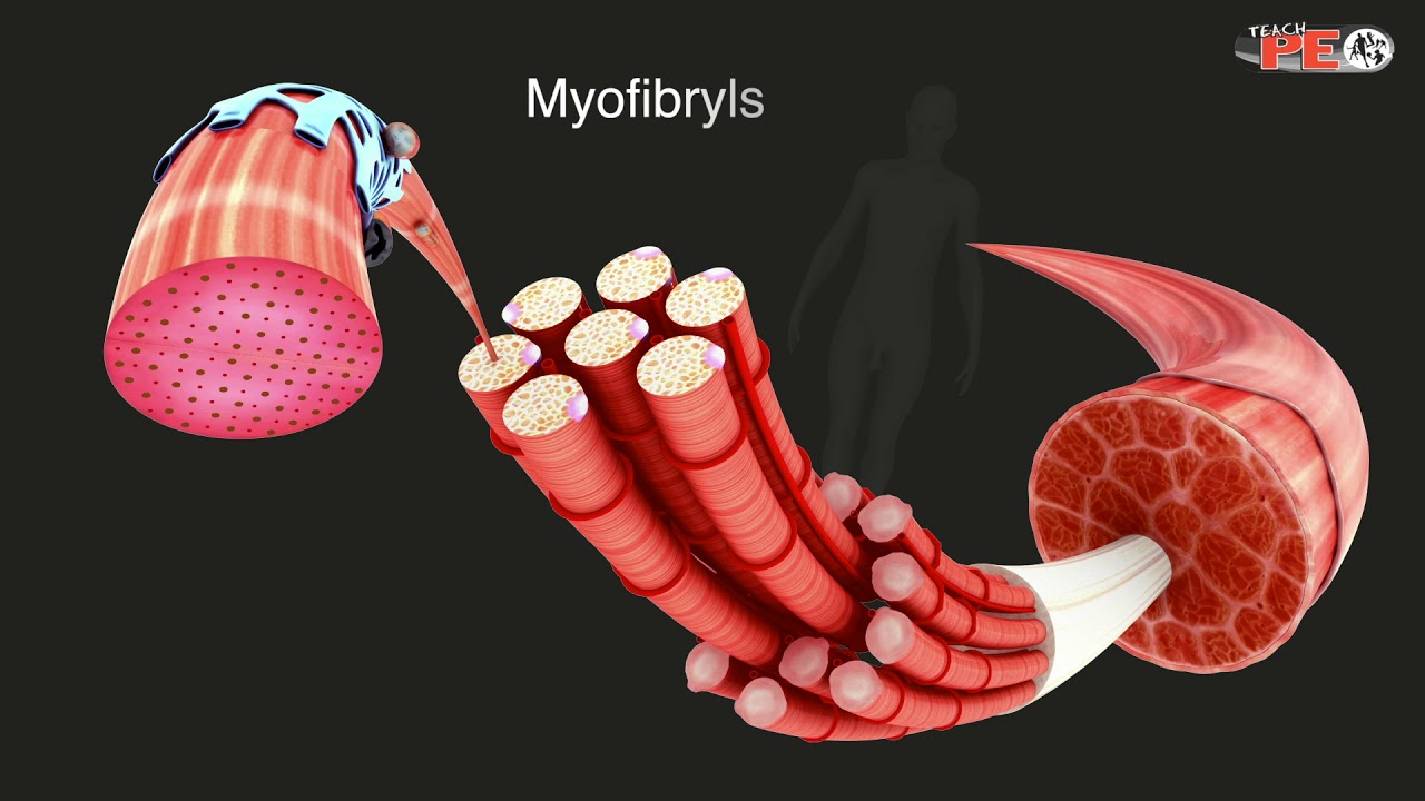
- 🏋️♂️ Skeletal muscles are composed of bundles of muscle fibers, each containing many individual fibers.
- 🧬 Each muscle fiber contains myofibrils, which are bundles of actin and myosin proteins.
- 🔬 The sarcoplasmic reticulum is a network of tubules that stores calcium within the muscle fiber.
- 🔄 Sarcomeres are the functional repeating units within myofibrils, consisting of actin and myosin filaments.
- ⚡️ A nerve impulse triggers the release of acetylcholine, leading to calcium release from the sarcoplasmic reticulum.
- 🔗 Calcium binding to troponin changes its shape, allowing myosin to attach to actin and form cross bridges.
- 🔋 The breakdown of ATP provides energy for myosin to pull actin filaments, causing muscle contraction.
- 🔄 The ratchet mechanism refers to the repeated pulling of actin over myosin, powered by ATP cycling.
- ⏸ When the nerve impulse stops, calcium is pumped back, and muscles relax by returning actin to its resting position.
Q & A
What is the sliding filament theory?
-The sliding filament theory is the mechanism by which muscles contract at a cellular level. It describes how muscle fibers shorten by the sliding of actin and myosin filaments past each other.
What are the basic components of a muscle?
-A muscle is made up of bundles of muscle fibers, which in turn contain individual fibers. Each muscle fiber is composed of cylindrical organelles called myofibrils.
What are myofibrils and what are they made of?
-Myofibrils are cylindrical organelles within muscle fibers and are composed of proteins called actin and myosin, which are arranged in a repeating pattern.
What is the function of the sarcoplasmic reticulum in muscle contraction?
-The sarcoplasmic reticulum is a network of tubules and channels that store calcium. During muscle contraction, it releases calcium, which is essential for the activation of the contractile process.
What are sarcomeres and how do they relate to muscle contraction?
-Sarcomeres are the functional repeating segments of a myofibril and are the basic units of muscle contraction. They consist of overlapping actin and myosin filaments, and their shortening leads to muscle contraction.
How does a nerve impulse trigger muscle contraction?
-A nerve impulse causes the release of acetylcholine, which leads to depolarization and the subsequent release of calcium from the sarcoplasmic reticulum, initiating muscle contraction.
What is the role of calcium in the muscle contraction process?
-Calcium binds to troponin, changing its shape and causing tropomyosin to move away from the active site on actin, allowing myosin to attach and form cross bridges, which leads to muscle contraction.
What is the role of ATP in muscle contraction?
-ATP (adenosine triphosphate) provides the energy required for the myosin head to detach from actin and reset for another contraction cycle. The breakdown of ATP powers the ratchet mechanism of muscle contraction.
What is the ratchet mechanism in muscle contraction?
-The ratchet mechanism refers to the repeated pulling of the actin filaments over the myosin filaments. This process is powered by the release and reattachment of myosin heads to actin, driven by ATP.
How does the muscle return to its resting state?
-When the nerve impulse stops, calcium is pumped back into the sarcoplasmic reticulum, and the troponin and tropomyosin return to their original positions, blocking the active sites on actin and causing the muscle to relax and lengthen.
What happens when the muscle contraction process is sustained?
-Muscle contraction can be sustained as long as there are adequate ATP and calcium stores. The continuous cycling of ATP binding, myosin head attachment and detachment, and calcium release and reuptake allows for prolonged muscle contraction.
Outlines

Esta sección está disponible solo para usuarios con suscripción. Por favor, mejora tu plan para acceder a esta parte.
Mejorar ahoraMindmap

Esta sección está disponible solo para usuarios con suscripción. Por favor, mejora tu plan para acceder a esta parte.
Mejorar ahoraKeywords

Esta sección está disponible solo para usuarios con suscripción. Por favor, mejora tu plan para acceder a esta parte.
Mejorar ahoraHighlights

Esta sección está disponible solo para usuarios con suscripción. Por favor, mejora tu plan para acceder a esta parte.
Mejorar ahoraTranscripts

Esta sección está disponible solo para usuarios con suscripción. Por favor, mejora tu plan para acceder a esta parte.
Mejorar ahoraVer Más Videos Relacionados

Skeletal muscle contraction : Muscle physiology Animations

Skeletal Muscle Contraction and Relaxation Physiology Animation / Excitation Contraction Coupling 💪
Structure of Skeletal Muscle Explained in simple terms

Muscular System Sliding Filament Theory

Mekanisme Kontraksi Otot

Mekanisme Kontraksi Otot Rangka
5.0 / 5 (0 votes)
