Pulmonary 2 Lung Volumes and Ventilation
Summary
TLDRThis lecture delves into the mechanics of a spirometer, a device measuring lung volumes and capacities. It explains the traditional water displacement method and modern alternatives, and covers both static and dynamic lung function assessments. The importance of understanding lung volumes, such as vital capacity and total lung capacity, is highlighted, along with dynamic measures like forced vital capacity and maximum voluntary ventilation, crucial for diagnosing respiratory conditions. The lecture also touches on concepts like dead space and alveolar ventilation, essential for gas exchange efficiency.
Takeaways
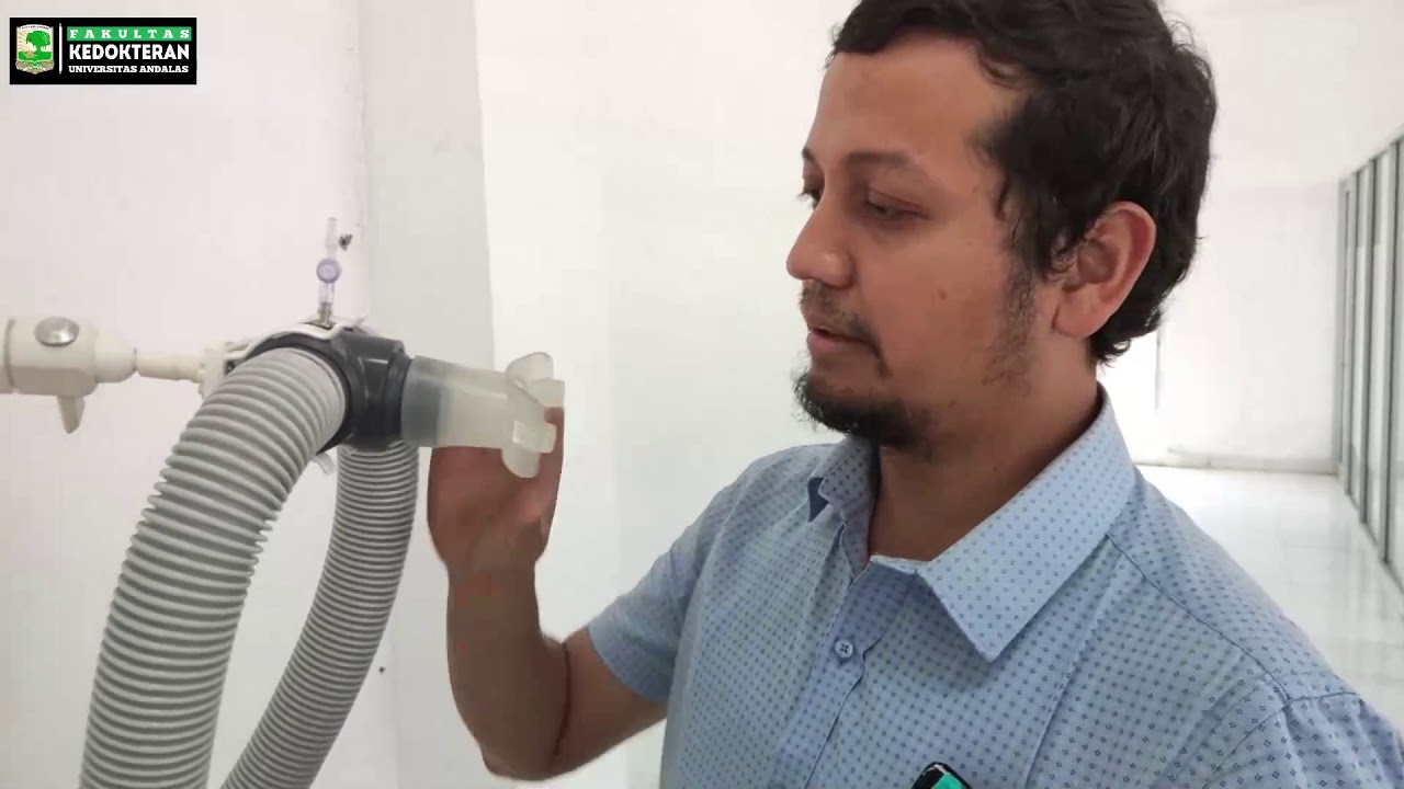
- 🌊 A spirometer is a device used to measure lung volumes and capacities, traditionally involving a water-filled chamber and a tube for breathing.
- 📏 The movement of air due to breathing causes the water level in the spirometer to rise and fall, which is directly proportional to the volume of air displaced.
- 📊 Lung volume measurements are calibrated using markers or scales on the spirometer for precise readings.
- 🚀 Modern spirometers have evolved to be smaller, with examples like turbine models and clinical ear displacement boxes.
- 🔍 Lung volumes can vary significantly based on factors such as age, gender, body size, and height.
- 🏗️ Static lung volume tests measure the dimensional aspects of air movement without time limitations, providing a snapshot of lung volume.
- 🌪️ Dynamic lung volume tests assess air movement over time, focusing on expiratory power and resistance to air movement in the lungs.
- 📉 The ratio of forced expired volume in 1 second to forced vital capacity (FEV1/FVC) is a key dynamic measure, with lower ratios indicating potential lung obstruction.
- 🏋️♂️ Maximum voluntary ventilation (MVV) measures the maximum volume of air a person can breathe in one minute, with elite athletes showing remarkably high values.
- 🌀 Dead space refers to the portion of the respiratory tract where gas exchange does not occur, including both anatomic and physiological dead spaces.
- 🔄 Alveolar ventilation is the portion of minute ventilation that reaches the alveoli for gas exchange, affected by the presence of dead space.
Q & A
What is a spirometer and what is its primary function?
-A spirometer is a device used to measure lung volumes and capacities in respiratory physiology. It operates by having a subject breathe in and out through a tube connected to a chamber filled with water. The fluctuation in water level, which is proportional to the volume of air displaced, is used to measure lung function.
How are the markers or scales on a spirometer used?
-The markers or scales on a spirometer are calibrated to specific volume measurements, allowing for precise readings of the water level changes. This enables researchers and clinicians to quantify various lung parameters with accuracy.
What are the two types of measurements used to assess lung volumes and capacities?
-The two types of measurements used to assess lung volumes and capacities are static and dynamic measurements. Static lung volume tests focus on the dimensional aspects of air movement within the pulmonary tract without time limitation, while dynamic tests assess expiratory power and resistance to air movement in the lungs.
What is the significance of the forced vital capacity (FVC) in lung function testing?
-The forced vital capacity (FVC) represents the total lung volume moved in one breath from full inspiration to maximum expiration. It is a crucial measure in lung function testing as it provides insights into the overall lung capacity and can indicate the presence of respiratory conditions.
How does the forced expiratory volume in 1 second (FEV1) relate to FVC and why is it important?
-The FEV1 is the volume of air expired during the first second of a forced vital capacity maneuver. The ratio of FEV1 to FVC is important as it provides diagnostic insights into expiratory power and resistance in the lungs. A decreased ratio can indicate conditions like severe obstructive pulmonary disease or asthma.
What is the maximum voluntary ventilation (MVV) and how is it measured?
-The maximum voluntary ventilation (MVV) is the volume of air breathed during rapid and deep breathing for 15 seconds, multiplied by four to represent the volume breathed for 1 minute. It is an assessment of ventilatory capacity and can vary significantly among individuals, including elite athletes.
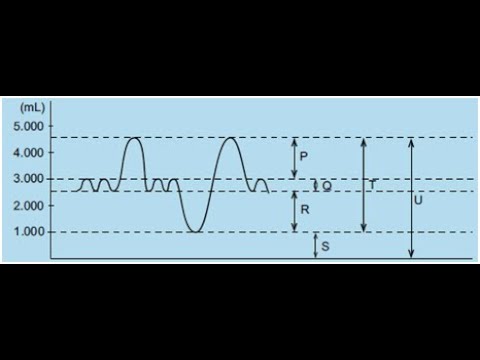
What is the role of the tidal volume in respiratory physiology?
-Tidal volume refers to the volume of air moved during either the inspiratory or expiratory phase of each breathing cycle under resting conditions. It is a key parameter in understanding the efficiency of the respiratory system and gas exchange.
What is the significance of the residual lung volume (RLV) in lung function?
-The residual lung volume (RLV) is the volume of air that remains in the lungs after maximal exhalation. It contributes to the total lung capacity and can be affected by various factors, including the individual's height and potential genetic influences.
What is meant by 'anatomic dead space' in the respiratory tract?
-Anatomic dead space refers to the portion of the respiratory tract, such as the nose, mouth, and trachea, where air remains and does not participate in gas exchange. It is a part of the total volume of air inhaled but does not contribute to alveolar ventilation.
How does physiological dead space differ from anatomic dead space?
-Physiological dead space refers to the portion of alveolar volume with poor tissue perfusion or inadequate ventilation, which does not participate in gas exchange. Unlike anatomic dead space, physiological dead space can increase significantly in certain conditions due to inadequate perfusion or ventilation.
What factors can affect the efficiency of alveolar ventilation?
-Factors that can affect the efficiency of alveolar ventilation include the presence of dead space, the depth of breathing, and the individual's overall lung function. Efficient alveolar ventilation is crucial for maintaining consistent alveolar air composition and stable arterial blood gases.
Outlines
🌡️ Understanding the Spirometer and Lung Volumes
This paragraph introduces the concept of measuring lung volumes and capacities using a spirometer, a device traditionally involving a water-filled chamber. The movement of air by the subject's breathing causes the water level to fluctuate, which is proportional to the air volume displaced. Markers or scales on the spirometer allow for precise volume readings. The paragraph also explains the evolution of spirometers and the importance of factors like age, gender, body size, and height in lung volume variations. It distinguishes between static and dynamic lung measurements, with static measurements providing a snapshot of lung volume at a specific time without time constraints, and dynamic measurements assessing the power and resistance of air movement in the lungs.
🏃♂️ Dynamic Lung Function Measurements and Ventilation
This section delves into dynamic lung function measurements, focusing on the forced vital capacity (FVC) and the speed of air movement, which is influenced by the resistance of the pulmonary airways and lung compliance. The forced expiratory volume in one second (FEV1) to FVC ratio is highlighted as a key diagnostic tool for conditions like obstructive pulmonary disease and asthma. The maximum voluntary ventilation (MVV) test, which measures the volume of air breathed during rapid and deep breathing, is also discussed, with variations in MVV among healthy individuals, athletes, and those with lung diseases. The paragraph concludes with an explanation of minute ventilation, anatomical dead space, and the physiological significance of alveolar ventilation in maintaining stable arterial blood gases.
🌬️ Alveolar Ventilation and Dead Space
The final paragraph discusses the intricacies of alveolar ventilation and the concept of dead space in the respiratory system. It explains the difference between anatomical dead space, which is the volume of air that does not reach the alveoli, and physiological dead space, which refers to alveolar regions with poor tissue perfusion or inadequate ventilation. The paragraph illustrates how changes in breathing patterns, such as shallow or deep breathing, affect minute ventilation and alveolar ventilation. It emphasizes the importance of understanding the relationship between these two types of ventilation for assessing lung function and gas exchange efficiency.
Mindmap
Keywords
💡Sphygmomanometer
💡Lung Volumes
💡Tidal Volume
💡Inspiratory Reserve Volume (IRV)
💡Expiratory Reserve Volume (ERV)
💡Forced Vital Capacity (FVC)
💡Residual Lung Volume (RLV)
💡Total Lung Capacity (TLC)
💡Forced Expiratory Volume in 1 Second (FEV1)
💡Maximum Voluntary Ventilation (MVV)
💡Dead Space
Highlights
Introduction to the water-filled spirometer, a device for measuring lung volumes and capacities in respiratory physiology.
Explanation of how the spirometer works, with the water level fluctuating in proportion to the volume of air displaced by breathing.
Description of the markers or scales on the spirometer for precise readings of lung volume changes.
Evolution of spirometers over the last century, with modern devices being smaller and more portable.
Importance of considering factors like age, gender, body size, and height when measuring lung volumes.
Introduction to static and dynamic measurements for assessing lung volumes and capacities.
Static lung volume tests provide a snapshot of lung dimensions without time limitations.
Dynamic lung function tests assess expiratory power and resistance to air movement in the lungs.
Definition and explanation of various lung volumes, including tidal volume, inspiratory reserve volume, and expiratory reserve volume.
Forced vital capacity (FVC) represents the total lung volume moved in one breath from full inspiration to maximum expiration.
Residual lung volume (RLV) is the air remaining in the lungs after maximal exhalation.
Total lung capacity is calculated by adding various lung volume measurements together.
Forced expiratory volume in 1 second (FEV1) and its ratio to FVC are important dynamic lung function measures.
Decreased FEV1/FVC ratio indicates compromised lung function, such as in obstructive pulmonary disease.
Maximum voluntary ventilation (MVV) test assesses the volume of air breathed during rapid and deep breathing.
Elite athletes can achieve remarkably high MVV values, exceeding 240 liters per minute.
Minute ventilation is calculated by multiplying the breathing rate by the tidal volume.
Alveolar ventilation refers to the portion of minute ventilation that reaches the alveoli for gas exchange.
Anatomic dead space is the volume of air that does not reach the alveoli and is fully saturated with water vapor.
Physiological dead space refers to alveolar volume with poor tissue perfusion or inadequate ventilation.
The relationship between minute ventilation and alveolar ventilation is crucial for understanding gas exchange efficiency.
Different breathing patterns, such as shallow or deep breathing, impact alveolar ventilation and gas concentrations at the alveolar-capillary membrane.
Transcripts
welcome to the second of the pulmonary
physiology lectures this short video
will provide you with the information to
be able to address the following
learning
objectives let's uncover the inner
workings of a water field spherometer a
device used to measure lung volumes and
capacities in respiratory
physiology picture this at the heart of
the spherometer is a chamber filled with
water typically contained within a
sealed container
connected to this chamber is a tube or
mouthpiece through which the subject
breathes as the subject breathes in and
out the movement of air causes the water
level within the chamber to rise and
fall and it's this fluctuation in water
level which is directly proportional to
the volume of air displaced by the
subject's
breathing now to measure lung volumes
accurately the spirometer is equipped
with a series of markers or scales that
allow for precise readings of the water
level changes now these markers are
calibrated to specific volume
measurements enabling researchers and
clinicians to quantify various lung
parameters with
Precision overall the water field
barometer provides a simple yet
effective means of assessing lung
function now while this is the
traditional me method to measure lung
volumes spirometers have evolved over
the last century with more common
devices now being much smaller such as a
turbine model pictured at the top right
or the clinical ear displacement box
seen at the bottom
right let's take a closer look at aung
volume tracing now it's important to
note that these lung volumes can and
will vary significantly based on factors
such as age gender body size and
composition with the person's height
playing a particularly significant
role let's explore the two types of
measurements used to assess lung volumes
and capacities these are static and
dynamic measurements now a static lung
volume tests focus on the dimensional
aspects of air movement within the
pulmonary tract unlike Dynamic tests
static measurements impose no time
limitation on the individual undergoing
the test instead they provide a snapshot
of the volume dimensions of the lungs at
a specific point in time offering
valuable insights into lung function
let's dive into the intricacies of
static lung function measurement where
the spirometer Bell serves as our guide
falling and Rising with each inhalation
and exhalation to record ventilatory
volume and breathing rate total volume
is our starting point point and it
describes the air move during e either
the inspiratory or the expiratory phase
of each breathing cycle under resting
conditions tital volume ranges between
point4 and 1 L of air per breath for
healthy
individuals moving on WE encounter the
inspect Reserve volume or the
Irv an additional volume of 2.5 to 3.5 L
above tital volume reserved for deep
inhalation to measure inpat Reserve
volume individuals Inspire normally and
then maximally after recording several
representative title
volumes now let's explore the expert
Reserve volume or Erv and this ranges
between 1 and 1 and 1/2 L for men and
it's about 10 to 20% lower for women
following a normal exhalation
individuals continue to Exhale
forcefully to determine their expert
Reserve
volume now the force votal capacity or
fvc represents the total ear volume
moved in one breath from Full
inspiration to maximum expiration or
vice versa and values usually aage
between 4 to 5 l in healthy young men
and 3 to 4 L in healthy young
women interestingly large lung volumes
such as Force votal capacities of 6 to 7
l or even higher are not uncommon for
tall individuals and some professional
athletes now these variations likely
reflect genetic factors rather than
training
effects and finally we encounter the
residual lung volume or the rlv the
volume of air that remains in the lungs
after maximal
exhalation residual lung volume averages
between .9 and 1.2 L for young adult
women and 1.1 and 1.7 L for men now this
gives a total lung capacity of 6 L on
average for males and 4.2 L on average
for
females note that capacities are
produced by adding one or more volume
measurements together
let's unravel the dynamic measures of
pulmonary ventilation which hinge on two
crucial factors firstly we have the
maximum ear volume expired known as the
force vital capacity or fvc and secondly
we consider the speed of moving a volume
of air which relies on two key factors
the resistance of the pulmonary Airways
to smooth air flow and the resistance or
stiffness presented by chest and lung
tissue to changes in their shapee during
breathing termed lung
compliance now let's delve into the
ratio of forced expired volume in 1
second to force vital
capacity now unlike static lung function
measures Dynamic measures like the F1
provide valuable diagnostic insights as
they assess expiratory power and overall
resistance to ear movement in the
lungs under normal conditions the fv1 to
fvc ratio averages around
85% however in conditions like severe
obstructive pulmonary osma and bronchial
asthma this ratio typically decreases
below 40% of vital
capacity a clinical demarcation for
Airway obstruction is established when
an individual expels less than 70% of
the force vital capacity in 1 second
indicating a compromised lung function
another Dynamic assessment of
ventilatory capacity is the maximum
voluntary ventilation test during this
test individuals engage in Rapid and
deep breathing for 15 seconds the volume
of air breathe during this time is then
multiplied by four to represent the
volume breathe for 1 minute giving us
the maximum voluntary ventilation or
mvv in healthy young men mvv typically
ranges between 140 and 180 L per minute
while women generally have an average
mvv ranging from 80 to 120 L per minute
it's fascinating to note that Elite
athletes can achieve remarkable mvv
values for instance male members of the
United States Nordic ski team have been
known to average 192 lers per minute
with individuals reaching values in
excess of 240 L per
minute at the other end of the Continuum
is individuals with obstructive lung
disease who face significant challenges
in achieving mvv values comparable to
those predicted for their normal age and
height match controls typically they
only achieve about 40% of what is
predicted during quiet breathing at rest
adults typically breathe at a rate of
around 12 breaths per minute with each
breath moving around half a liter of air
this means we move 6 l or 6,000 M of air
into and out of our lung each minute to
calculate our minute ventilation we
simply multiply the breathing rate by
the tital
volume the alola ventilation refers to
the portion of minute ventilation that
actually reaches the alola chambers
where gas exchange with the bloodstream
occurs however not all inspired air
makes it to the alvioli
a portion remains in the non- diffusible
conducting portions of the respiratory
tract such as the nose mouth and trache
this portion is known as the anatomic
dead space in healthy individuals the
anatomic Dead Space volume equals
approximately 150 to 200 mL or about 30%
of a resting total volume despite its
composition being almost equivalent to
ambient air Dead Space air is fully
saturated with water
vapor now due to the presence of Dead
Space only a portion of the ambient ear
inspired in each breath about 350 of the
500 mL of the title volume mixes with
existing lvol air however it's important
to note that this doesn't mean only 350
mL of air enter and leave the LVL with
each breath in reality the full total
volume of 500 m enters the
Alvi but only 350 ml represents fresh
air which is about 17th of the total air
in the
alvolo this seemingly inefficient
alviola ventilation is actually an
evolutionary protective mechanism to
maintain consistency in alveola air
composition and ensure stable arterial
blood gases throughout the breathing
cycle
now the only remaining component of the
tital volume is the physiological Dead
Space a term used to describe the
portion of alveola volume with poor
tissue Regional profusion or inadequate
ventilation as Illustrated in the figure
healthy lungs typically have only a
negligible amount of physiological Dead
Space however in certain conditions
physiological Dead Space can increase
significantly reaching up to 50% of the
resting title volume
this increase can occur due to two main
factors firstly inadequate perfusion may
occur during condition such as
Hemorrhage or an embolism which is a
blood clot that blocks pulmonary
circulation in such cases blood flow to
certain regions of the lungs may be
compromised leading to an increase in
physiological Dead
Space secondly inadequate lvol
ventilation may result from chronic
pulmonary diseases further contributing
to an increase in physiological Dead
Space now then let's look at this table
which sheds light on the complexity of
minute ventilation and its relationship
to real alola
ventilation in the first example of
shallow breathing where total volume
decreases to 150 ml minute ventilation
remains at 6 L per minute due to an
increase in breathing rate to 40 breaths
per
minute conversely the same minute
ventilation of 6 L per minute can be
achieved by decreasing breathing rate to
12 breaths and increasing tidal volume
to 500
mL now let's consider the example of
deep breathing where tidal volume is
doubled and ventilatory rate is
halfed again we achieve a minute
ventilation of 6 L per minute however
each ventilatory adjustment drastically
impact and impacts lvol
ventilation in the case of shallow
breathing dead SP space ear represents
the entire ear volume moved as no alola
ventilation
occurs conversely in the other examples
involving deeper breathing a larger
portion of the a breath of it sorry of
each breath mixes with existing alveola
air leading to effective alviola
ventilation it's crucial to note that
alveola ventilation and not dead space
ventilation determines the gaseous
concentrations at the elola capillary
membrane highlighting the significance
of understanding the relationship
between minute ventilation and Lola
ventilation but now you should feel well
prepared to tackle the learning
objectives
Weitere ähnliche Videos ansehen
IMAT Biology Lesson 6.6 | Anatomy and Physiology | Respiratory System
How to Interpret a Chest X-Ray (Lesson 7 - Diffuse Lung Processes)
Het ademhalingsstelsel | Samenvatting
How to Breathe in Pilates
Respiratory System of the Human Body - How the Lungs Work! (Animation)
Simple Linear Regression in R | R Tutorial 5.1 | MarinStatsLectures
5.0 / 5 (0 votes)