9. Transport in Animals (Part 1) (Cambridge IGCSE Biology 0610 for exams in 2023, 2024 and 2025)
Summary
TLDRThis IGCSE study video explores the transport systems in animals, focusing on the circulatory system's role in nutrient and gas transport. It explains the single circulatory system of fish and the double circulation found in mammals, highlighting the heart's structure and function. The video also delves into the effects of physical activity on heart rate and discusses coronary heart disease, its causes, and prevention methods. Aimed at providing a clear understanding of these biological concepts, the video invites viewers to subscribe for more educational content.
Takeaways
- 🌊 Fish have a single circulatory system with a two-chambered heart consisting of an atrium and a ventricle, which allows blood to pass through the heart only once for a full circuit.
- 🐟 In fish, oxygenation occurs at the gills, and the circulatory system includes gill circulation and systemic circulation, with oxygenated blood moving to body capillaries after gill exchange.
- 💓 Mammals possess a double circulatory system with a four-chambered heart, enabling blood to pass through the heart twice for one complete body circuit.
- ❤️ The right side of the mammalian heart handles pulmonary circulation, pumping deoxygenated blood to the lungs for oxygenation.
- 💚 The left side is responsible for systemic circulation, delivering oxygenated blood from the lungs to the rest of the body.
- 🔄 Double circulation in mammals is advantageous for efficient oxygen and glucose delivery due to higher blood pressure compared to single circulation systems.
- 🦴 The heart's structure includes the right atrium, right ventricle, left atrium, and left ventricle, with valves ensuring one-way blood flow.
- 🔍 Coronary arteries are crucial for supplying oxygen-rich blood to the heart muscle itself, wrapping around the exterior of the heart.
- 💪 The ventricles have thicker muscular walls than the atria, with the left ventricle being the thickest to handle higher pressure requirements for systemic circulation.
- 🏃♂️ Physical activity increases heart rate as muscles require more oxygen and glucose for respiration, and waste products need to be removed more rapidly.
- 🚑 Coronary heart disease occurs when coronary arteries are blocked, leading to a heart attack due to oxygen starvation in heart muscles, often caused by cholesterol buildup.
- 🍏 Risk factors for coronary heart disease include diet, lack of exercise, diabetes, obesity, stress, smoking, genetic predisposition, age, and gender, with males being at higher risk.
Q & A
What is the main function of the circulatory system in animals?
-The main function of the circulatory system in animals is to transport nutrients and gases, such as oxygen, throughout the body via the blood.
How does the single circulatory system in fish differ from the double circulatory system in mammals?
-In fish, which have a single circulatory system, blood passes through the heart only once to complete a full circuit through the body, whereas in mammals, with a double circulatory system, blood passes through the heart twice for every one circuit of the body.
What are the two main types of blood flow in the circulatory system of fish?
-The two main types of blood flow in fish are oxygenated blood flow (pink arrows), which is rich in oxygen, and deoxygenated blood flow (blue arrows).
How does a fish's heart structure support its single circulatory system?
-A fish's heart supports its single circulatory system with two chambers: an Atrium and a Ventricle, which allow blood to pass through the heart only once for a complete circuit.
What is the advantage of a double circulatory system in mammals over a single circulatory system in fish?
-A double circulatory system in mammals allows for faster and more efficient delivery of oxygen and glucose throughout the body due to higher blood pressure, which is essential for larger organisms with greater oxygen demands.
How many chambers does the heart of a mammal have, and what is the significance of this?
-A mammalian heart has four chambers, which allows for the separation of pulmonary and systemic circulation, enhancing the efficiency of oxygen and nutrient delivery to the body.
What are the functions of the valves in the heart?
-The valves in the heart, such as the tricuspid and bicuspid valves, ensure one-way flow of blood by preventing it from flowing back into the atria. Semilunar valves prevent blood from flowing back into the heart from the arteries.
What is the role of the coronary arteries in the heart?
-The coronary arteries supply oxygen-rich blood to the heart muscle itself, ensuring that the heart can function and pump blood efficiently.
Why do the ventricles have thicker muscle walls than the atria?
-The ventricles have thicker muscle walls than the atria because they are responsible for pumping blood out of the heart at higher pressures, whereas the atria only need to move blood into the ventricles.
How does physical activity affect the heart rate, and why?
-Physical activity increases the heart rate because the body's muscle cells require more energy, oxygen, and glucose for respiration, and the waste products need to be removed more quickly. The heart rate increases to meet these demands.
What is coronary heart disease, and what are its main causes?
-Coronary heart disease occurs when the coronary arteries become blocked, leading to a lack of blood and oxygen supply to the heart muscles, which can result in a heart attack. It is mainly caused by the buildup of cholesterol and fatty substances within the coronary arteries.
Outlines
🐟 Single and Double Circulatory Systems
This paragraph introduces the concepts of single and double circulatory systems, focusing on the transport of nutrients and gases in animals. It explains that the circulatory system is a network of blood vessels with a pump and valves ensuring one-way blood flow. Fish have a single circulatory system with two-chambered hearts, where blood is oxygenated at the gills and completes a circuit through the body. The paragraph also contrasts this with the double circulatory system of mammals, which has four-chambered hearts and two distinct circulations: pulmonary (deoxygenated blood to the lungs) and systemic (oxygenated blood to the body). Diagrams are used to illustrate these processes.
💓 Detailed Anatomy of the Mammalian Heart
This section delves into the anatomy of the mammalian heart, highlighting its four chambers: the right and left atria and ventricles. It describes the flow of deoxygenated blood from the body to the lungs via the right side of the heart and the oxygenated blood from the lungs to the body via the left side. The paragraph explains the function of various valves, such as the tricuspid and bicuspid valves, and the semilunar valves in the pulmonary artery and aorta. It also touches on the importance of the septum in preventing the mixing of oxygenated and deoxygenated blood. A simplified diagram aids in visualizing the double circulation process.
🚀 Impact of Physical Activity on Heart Rate
This paragraph explores the relationship between physical activity and heart rate, detailing an experiment to measure pulse rate at rest and during exercise. It explains that during exercise, the body's muscle cells require more energy, oxygen, and glucose for respiration, leading to an increased heart rate to meet these demands. The paragraph also discusses the concept of oxygen debt and how the heart rate increases to repay it. Additionally, it covers the monitoring of heart activity through ECG, pulse rate, and stethoscope sounds, emphasizing the need for a healthy lifestyle to maintain heart health.
💔 Coronary Heart Disease and Its Risk Factors
The final paragraph discusses coronary heart disease, a condition where the coronary arteries become blocked, leading to a lack of oxygen in the heart muscles and potentially causing a heart attack. It explains the causes of this disease, which include the buildup of cholesterol and fatty substances in the arteries. The paragraph outlines the risk factors for coronary heart disease, such as diet, lack of exercise, diabetes, obesity, stress, smoking, genetic predisposition, age, and gender. It concludes with ways to reduce the risk of coronary heart disease, including quitting smoking, maintaining a healthy diet, and regular exercise.
Mindmap
Keywords
💡Circulatory System
💡Single Circulatory System
💡Double Circulatory System
💡Heart Chambers
💡Oxygenated and Deoxygenated Blood
💡Gill Circulation
💡Systemic Circulation
💡Pulmonary Circulation
💡Coronary Arteries
💡ECG
💡Coronary Heart Disease
Highlights
Introduction to IGCSE study buddy for biology revision.
Explanation of the circulatory system's role in transporting nutrients and gases in animals.
Description of the single circulatory system in fish with two-chambered hearts.
Illustration of oxygenated and deoxygenated blood flow in fish.
Clarification on fish respiration through gills instead of lungs.
Overview of the double circulatory system in mammals with four-chambered hearts.
Diagrammatic representation of mammalian double circulation.
Differentiation between pulmonary and systemic circulation in mammals.
Advantage of double circulation for efficient oxygen and glucose delivery in mammals.
Structural details of the mammalian heart, including atria and ventricles.
Function of valves in preventing backflow of blood in the heart.
Role of the septum in separating oxygenated and deoxygenated blood.
Importance of the aorta in distributing oxygenated blood throughout the body.
Simplified diagram for understanding the double circulation process.
Memory aid for differentiating arteries and veins based on blood flow direction.
Discussion on coronary arteries supplying oxygen-rich blood to the heart muscle.
Comparison of muscular walls' thickness between atria and ventricles.
Explanation of how physical activity affects heart rate and its recovery.
Investigation of the impact of exercise on heart rate through an experiment.
Understanding coronary heart disease and its causes.
Risk factors and prevention methods for coronary heart disease.
Conclusion of chapter 9 part 1 on transport in animals.
Transcripts
hi everyone welcome to IGCSE study buddy
where you can revise biology topics from
the Cambridge IGCSE syllabus
if you are enjoying this video so far
please don't forget to hit the like
button and subscribe to my channel
this video summarizes part 1 of chapter
9 transport in animals
in most animals the circulatory system
is the primary method used to transport
nutrients and gases through the body in
the blood
the circulatory system is a system of
blood vessels with a pump and valves to
ensure one-way flow of blood
fish have a single circulatory system
the heart of a fish has two Chambers an
Atrium and a ventricle
in a single circulatory system blood
passes through the heart only once to
complete a full circuit through the body
of the fish
this diagram illustrates the single
circulation of a fish
the pink arrows represent the oxygenated
blood flow or blood rich in oxygen
the blue arrows represent deoxygenated
blood flow as you may notice fish do not
have lungs since oxygen is absorbed when
the blood passes the gills
so deoxygenated blood from the body
capillaries move through the two
chambers of a fisher's Heart The Atrium
and The ventricle and then reaches the
Gill capillaries where oxygen is
absorbed into the blood
thereafter the blood becomes oxygenated
and moves from the Gill capillaries to
the body capillaries completing one
circuit
so when the heart pumps the blood to the
gills to be oxygenated it's called Gill
circulation
the blood then continues through the
rest of the body before arriving back at
the atrium and this is called systemic
circulation
moving on to the double circulation of a
mammal
so mammals have a double circulatory
system
the mammalian heart has four chambers
this means that blood passes through the
heart twice for every one circuit of the
body
so here's a diagram to explain the
double circulation of a mammal
the Red Arrows represent the oxygenated
blood flow and the blue arrows represent
deoxygenated blood flow
the heart is labeled as if it was in the
chest so what is on your left on a
diagram is actually the right hand side
and vice versa
so deoxygenated blood enters the right
atrium labeled as r a on the diagram via
the vena cava
then it moves into the right ventricle
next the deoxygenated blood is pumped by
the right ventricle to the lungs to
become oxygenated
the pulmonary artery carries this blood
away from the heart
gas exchange happens at the lungs and
the deoxygenated blood becomes
oxygenated blood
then oxygenated blood enters the left
atrium via the pulmonary vein
then it moves to the left ventricle
oxygenated blood is pumped by the left
ventricle to the wrist of the body
the aorta carries this oxygenated blood
away from the heart
body cells use the oxygen and cause the
blood to become deoxygenated
the deoxygenated blood returns to the
heart and the cycle repeats
the right side of the heart receives
deoxygenated blood from the body and
pumps it to the lungs that is called
pulmonary circulation
the left side of the heart receives
oxygenated blood from the lungs and
pumps it to the body this is called
systemic circulation
what is the advantage of a double
circulation
mammals are larger than fish so they
have a greater requirement for oxygen
and glucose for respiration
double circulation allows this oxygen
and glucose to be delivered faster and
more efficiently through the blood since
a double circulation maintains a higher
blood pressure than a single circulation
system
let's learn the structures of the
mammalian heart now
this is the right atrium one of the
heart's four chambers
this is the vena cava deoxygenated blood
coming from the body flows into the
right atrium through this vein
this is the tricuspid valve it is an
atrioventricular valve which separates
the Atria from the ventricles on each
side of the heart and prevents the blood
from flowing back into the Atria
once the right atrium is filled with
blood the heart muscles of the right
atrium contracts and the blood is pushed
through the tricuspid valve into the
right ventricle
this is the right ventricle the next
chamber of the heart
this is the semilunar valve it prevents
blood flowing backwards into the heart
the walls of The ventricle contract and
the blood is pushed through the
semilunar valve into the pulmonary
artery
this is the pulmonary artery it is a
blood vessel that carries deoxygenated
blood away from the heart to the lungs
to be oxygenated
this is the septum it is a muscle wall
that separates the two sides of the
heart therefore it prevents the mixing
of oxygenated and deoxygenated blood
this is the left atrium another one of
the heart's four chambers
this is the pulmonary vein oxygenated or
oxygen rich blood returning from the
lungs enters the left atrium through
this vein
this is the bicuspid valve it is an
atrioventricular valve which separates
the left atrium from the left ventricle
and prevents the blood from flowing back
into the lift atrium
once the left atrium is filled with
blood the heart muscles of the left
atrium contracts and the blood is pushed
through the bicuspid valve into the left
ventricle
this is the left ventricle a chamber of
the heart
this is the semilunar valve in the aorta
it prevents blood flowing backwards into
the heart
the thicker muscle walls of The
ventricle contracts strongly to push the
blood forcefully through the semilunar
valve into the aorta
this is the aorta it is a blood vessel
that carries oxygenated blood away from
the heart all the way around the body
here's a simpler diagram that will guide
you through double circulation
deoxygenated blood enters the right
atrium through the vena cava
then it moves into the right ventricle
through the atrioventricular valve the
special name for the one on this side is
called the tricuspid valve
the right ventricle pumps the
deoxygenated blood out to the pulmonary
artery via the semilunar valve
the blood travels to the lungs and moves
through the capillaries past the alveoli
where gas exchange takes place
the deoxygenated blood becomes
oxygenated blood and returns to the
heart via the pulmonary vein into the
left atrium
it then moves into the left ventricle
through the atrioventricular valve the
special name for the one on this side is
called the bicuspid valve
the left ventricle pumps oxygenated
blood out to the aorta via the semilunar
valve
the aorta takes the oxygenated blood
away from the heart to the wrist of the
body
here's a small tip to remember which
vessels take blood away from the heart
remember a
pulmonary artery and aorta are arteries
and they carry blood away from the heart
so artery begins with a and so does away
blood this pump towards the heart in
veins and away from the heart in
arteries
next what are coronary arteries
the heart is made of muscle tissues
like all other tissues in the body the
heart muscle needs oxygen rich blood to
function
coronary arteries Supply blood to the
heart muscle
the coronary arteries wrap around the
outside of the heart
moving on to muscular walls the
ventricles have thicker muscle walls
than the Atria since they pump blood out
of the heart so they need to do so at a
higher pressure
so you will notice that the lower two
Chambers the ventricles have thicker
muscle walls than the upper two Chambers
the Atria
also comparing both ventricles the left
ventricles muscle wall is thicker than
the right ventricles muscle wall
this is because it has to pump blood at
a higher pressure to the whole body
whereas the right ventricle has to pump
blood just to the lungs
the activity of the heart may be
monitored by
using an ECG
measuring pulse rate
or listening to the sounds of valves
closing using a stethoscope
let's investigate the effect of physical
activity on the heart rate
first record the pulse rate at rest for
a minute
next exercise for a while
then record the pulse rate every minute
until it returns to the resting rate
this experiment will show that during
exercise the heart rate increases and
may take several minutes to return to
normal
so why does the physical activity have
this effect on the heart rate this is
because during exercise the muscle cells
of the body need more energy
therefore they need more oxygen and
glucose for respiration
the waste products of respiration also
needs to be removed faster from the
cells
moreover some of the muscle cells may
have respired anaerobically without
oxygen so the oxygen debt needs to be
repaid
the heart rate therefore increases to
meet these demands
finally let's learn about coronary heart
disease
so far we know that the heart functions
as a pump which delivers blood to the
rest of the body
however the heart muscles themselves
also need a blood supply because they
too are respiring muscles
coronary arteries Supply blood to the
heart muscles in order for them to
respire and perform their function
coronary heart disease is when the
coronary artery becomes blocked leading
to blood and therefore oxygen starvation
in the heart muscles
this leads to a heart attack
coronary heart disease is caused by the
buildup of cholesterol and other fatty
substances within the coronary arteries
so in this diagram you can see a
coronary artery with normal blood flow
and a coronary artery with a cholesterol
buildup which is restricting the blood
flow
the possible risk factors for coronary
heart disease include
diet eating unhealthy amounts of
saturated fats increases cholesterol
levels in the body lack of exercise
physical activity can help control blood
cholesterol diabetes and obesity so an
inactive person is at a higher risk of
getting this disease
stress increases the blood pressure
increasing the chance of coronary
arteries getting blocked
smoking increases blood pressure and
increases the likelihood of clots
forming in the arteries
genetic predisposition children of
parents with coronary heart disease are
more likely to develop coronary heart
disease
age with increasing age the risk of
getting coronary heart disease increases
and gender males have a higher chance of
developing coronary heart disease than
females
the risk of coronary heart disease may
be reduced in the following ways
quit smoking
maintain a healthy diet with reduced
animal fats
and exercise regularly
so that concludes part 1 of chapter 9
transport in animals
hope you found this video useful thank
you for watching and please don't forget
to subscribe to IGCSE study buddy for
mobile larger revision videos bye bye
Weitere ähnliche Videos ansehen
Module 2 Revision | HSC Year 11 Biology
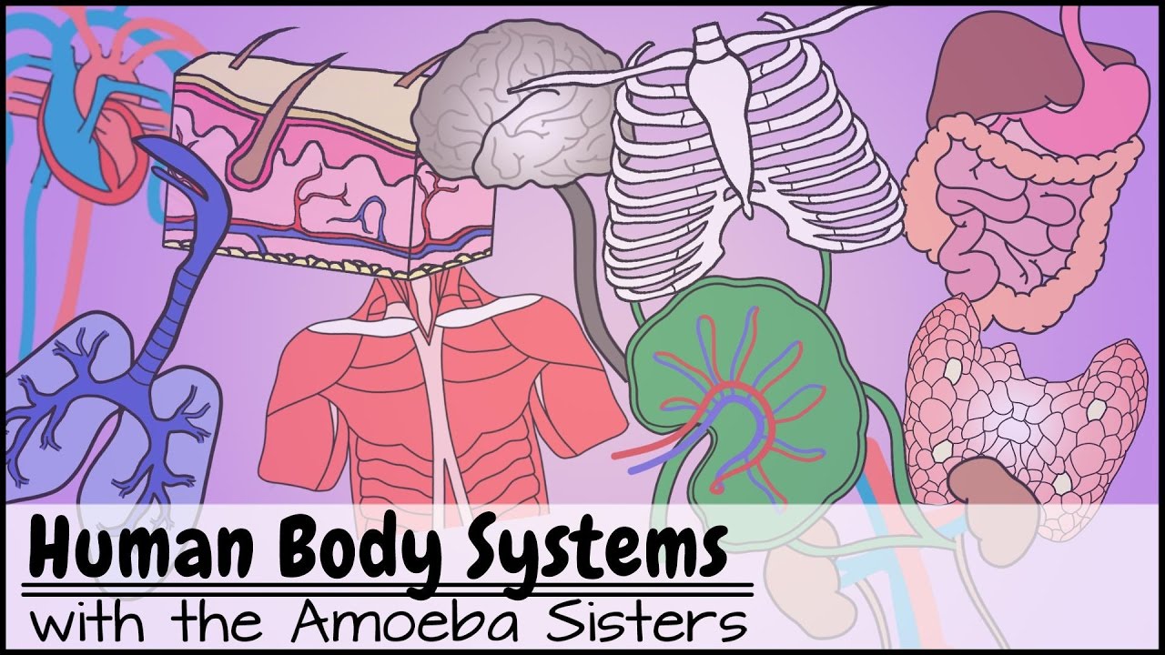
Struktur dan Fungsi Jantung dan Pembuluh Darah Pada Sistem Peredaran Darah Manusia
B3.2 HL Transport in Animals [IB Biology HL]
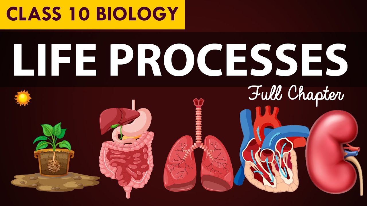
Q1W1 | Respiratory & Circulatory System
CVS 3 The Heart
Life process class 10 Animated video | 10th BIOLOGY | ncert #science | Chapter 7
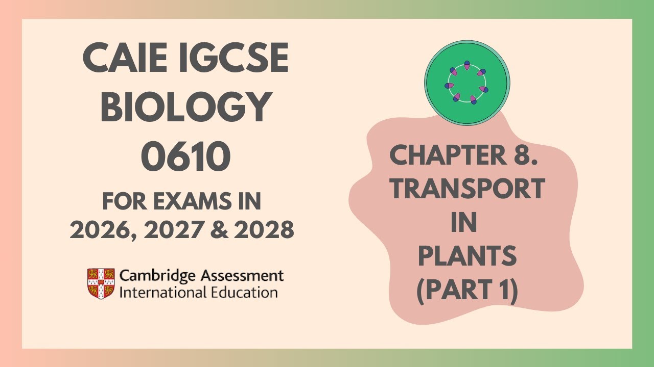
 #transportation
5.0 / 5 (0 votes)