Neurology | Optic Nerve | Cranial Nerve II: Visual Pathway and Lesions
Summary
TLDRThis educational video script delves into the intricacies of the human visual pathway, starting from the retina's interaction with light to the processing in the brain. It explains the journey of visual information through the optic nerve, optic chiasma, and lateral geniculate nucleus, highlighting how lesions at various stages can lead to different visual field defects. The script aims to clarify complex concepts like nasal and temporal hemiretina, homonymous hemianopia, and the impact of damage to the optic radiations and occipital cortex, using visual aids to illustrate the effects on visual fields.
Takeaways
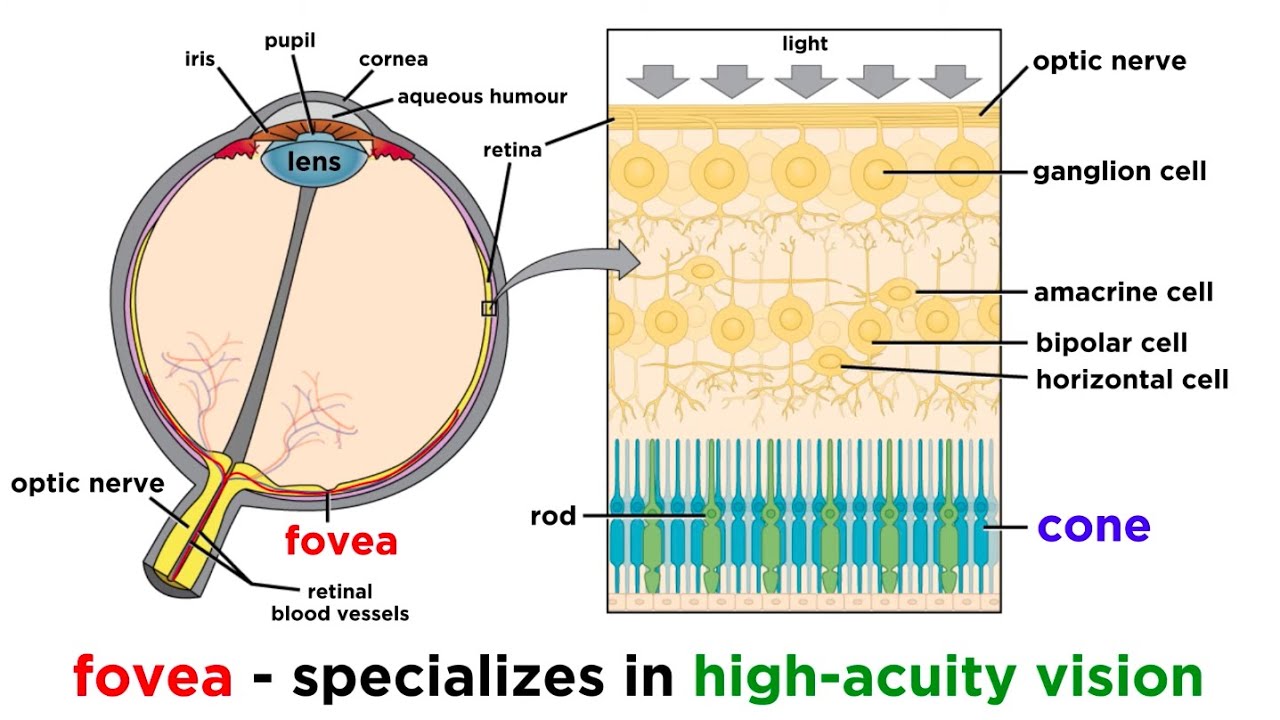
- 👁 The video discusses the visual pathway, detailing how light hits the retina and is processed through the optic nerve and other components.
- 🌟 It emphasizes the importance of understanding the phototransduction cascade for those who have seen the related video, as it explains the conversion of light into chemical and electrical changes.
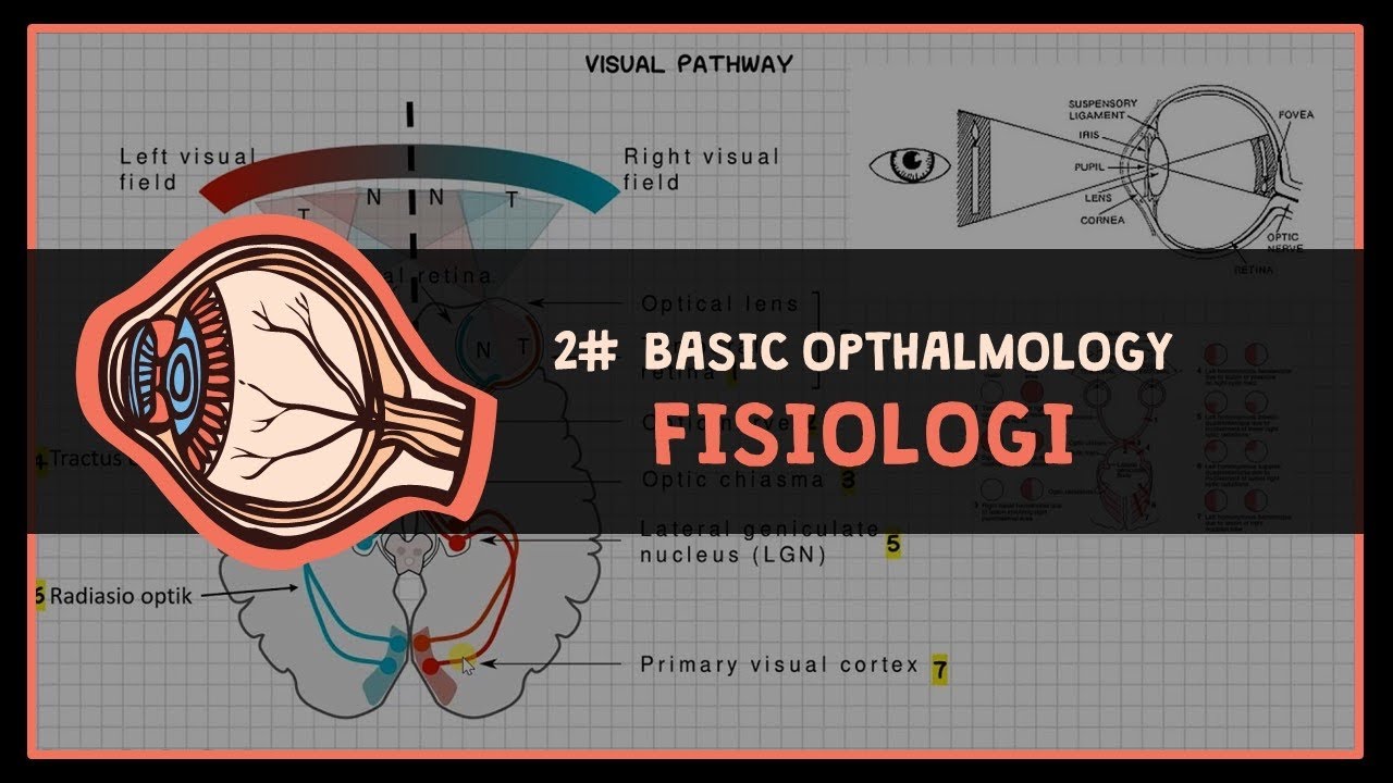
- 🔍 The script explains the concept of visual fields, distinguishing between the nasal and temporal components and how they relate to the right and left visual fields of each eye.
- 🤔 It clarifies potential confusion by redefining the terms for the visual fields, ensuring that the audience understands which part of the brain processes which visual field.
- 💡 The video describes how the retina's temporal hemiretina receives light from the opposite visual field, and how this information is transmitted to the brain without crossing over.
- 🔄 At the optic chiasm, fibers from the nasal hemiretina cross over to the opposite side of the brain, while fibers from the temporal hemiretina stay on the same side.
- 🧠 The optic tract carries visual information to the lateral geniculate nucleus (LGN) in the thalamus, which is a crucial part of the visual pathway.
- 📏 The LGN is structured in six layers, with ipsilateral fibers connecting to layers two, three, and five, and contralateral fibers to layers one, four, and six.
- 🌐 Some fibers from the LGN project to the midbrain, affecting the pupillary light reflex and involving structures like the superior colliculus and pretectal nucleus.
- 🧬 The majority of fibers from the LGN continue to the occipital lobe, specifically to the striate cortex, which is the primary visual cortex responsible for visual perception.
- 🛑 The script also covers the implications of various lesions in different parts of the visual pathway, explaining the resulting visual field deficits such as monocular blindness, homonymous hemianopia, and quadrantanopia.
Q & A
What is the primary focus of the video script?
-The video script primarily focuses on explaining the visual pathway, detailing how light hits the retina and is processed through the optic nerve and other components of the visual system.
What is the significance of the phototransduction cascade in the visual pathway?
-The phototransduction cascade is significant because it is the process by which light is converted into chemical and then electrical changes, which are essential for the visual pathway to function properly.
How does the script define the visual field and its components?
-The script defines the visual field as the area that can be seen at any given moment. It is divided into components such as the nasal and temporal components, which are related to the proximity to the nose and temple, respectively.
What is the role of the optic nerve in the visual pathway?
-The optic nerve plays a crucial role in the visual pathway by transmitting the visual information from the retina to the brain. It is composed of the ganglion cell axons that carry the information after phototransduction.
Can you explain the concept of the optic chiasma in the visual pathway?
-The optic chiasma is a structure where the optic nerves from both eyes meet. Here, the fibers from the temporal hemiretina cross over to the opposite side, while the fibers from the nasal hemiretina remain on the same side, allowing for the integration of visual information from both eyes.
What is the optic tract and where does it lead?
-The optic tract is a long tube-like structure that extends from the optic chiasma. It carries the visual information from the retina to the lateral geniculate nucleus in the thalamus.
How does the script describe the organization of the lateral geniculate nucleus (LGN)?
-The script describes the LGN as having six layers, with ipsilateral fibers (from the temporal hemiretina) going to layers two, three, and five, and contralateral fibers (from the nasal hemiretina) going to layers one, four, and six.
What are the two types of fibers mentioned in the script that contribute to the visual pathway, and how do they differ?
-The two types of fibers mentioned are the ipsilateral fibers and the contralateral fibers. Ipsilateral fibers stay on the same side of the brain and come from the temporal hemiretina, while contralateral fibers cross to the opposite side of the brain and come from the nasal hemiretina.
What is the striate cortex and its relevance to the visual pathway?
-The striate cortex, located in the occipital lobe, is the primary visual cortex. It is the area where the perception of visual stimuli occurs, processing the information received from the optic radiations.
How does the script explain the impact of different types of lesions on the visual pathway?
-The script explains that lesions in different parts of the visual pathway can result in various types of visual field loss, such as monocular blindness, homonymous hemianopia, and quadrantanopia. The specific type of visual impairment depends on the location and extent of the lesion.
Outlines
👀 Introduction to the Visual Pathway
The script begins by introducing the visual pathway, focusing on how light hits the retina and travels through the optic nerve. It emphasizes the importance of understanding the phototransduction cascade, which converts light into chemical and electrical changes. The video explains the concept of visual fields, distinguishing between the nasal and temporal components for both the right and left eyes. It clarifies potential confusion by redefining the terms for the visual fields as they relate to the right and left eyes, ensuring the viewer grasps the basics before delving into more complex topics.
🔄 Optic Nerve Crossing and Pathways
This paragraph delves into the mechanics of the optic nerves, explaining how the fibers from the temporal hemiretina cross over at the optic chiasm, while those from the nasal hemiretina do not. It describes the optic tracts and how the fibers from each eye are directed to the opposite side of the brain. The script uses the mnemonic 'anything from the right goes to the left, and anything from the left goes to the right' to help viewers remember the pathway of the visual information. It also introduces the concept of ipsilateral and contralateral fibers, which are crucial for understanding the effects of various lesions on vision.
🌐 Lateral Geniculate Nucleus and Visual Processing
The script moves on to the lateral geniculate nucleus (LGN) within the thalamus, highlighting its six layers and explaining the distribution of ipsilateral and contralateral fibers to different layers. It discusses the importance of the LGN in processing visual information before it is sent to other parts of the brain. The paragraph also touches on the connections between the LGN and the midbrain, specifically mentioning the superior colliculus and the pretectal nucleus, which are involved in the pupillary light reflex and other visual responses.
🧠 Optic Radiations and the Visual Cortex
The discussion continues with the optic radiations, which are the fibers that carry visual information from the LGN to the occipital lobe. The script differentiates between the superior and inferior retinal fibers, also known as the Bärum's loop and Meyer's loop, respectively. It explains how these fibers reach the striate cortex, also known as the primary visual cortex, where the perception of visual stimuli occurs. The paragraph also introduces the concept of lesions and how they can affect the visual field, providing a foundation for understanding the clinical implications of damage to different parts of the visual pathway.
🛑 Lesions and Their Impact on Vision
This paragraph explores various hypothetical lesions that could affect the visual pathway, from damage to the optic nerve causing monocular blindness to lesions in the optic chiasm leading to bitemporal hemianopia. The script describes the visual field deficits associated with each type of lesion, such as right homonymous hemianopia resulting from damage to the optic tract. It uses visual aids to illustrate the effects of these lesions on the visual fields, helping viewers to understand the clinical manifestations of different types of damage to the visual pathway.
🔍 Superior and Inferior Retinal Fibers Lesions
The script focuses on the impact of lesions affecting the superior and inferior retinal fibers, also known as Bärum's loop and Meyer's loop. It explains how damage to these fibers can lead to specific types of visual field loss, such as left inferior quadrant hemianopia or right superior quadrant hemianopia. The paragraph uses detailed visual representations to show how the superior and inferior visual fields are affected by lesions in different parts of the visual pathway, providing a clear understanding of the consequences of such damage.
🌀 Optic Radiations Damage and its Effects
This paragraph discusses the effects of damage to the optic radiations, which can result in a loss of vision in both the superior and inferior planes of the visual field. The script describes the concept of homonymous hemianopia, which occurs when there is damage to the optic radiations on one side, leading to a loss of the same visual field in both eyes. It also explains how damage to the right optic radiations can result in left homonymous hemianopia, affecting the viewer's understanding of the comprehensive impact of such lesions.
🏥 Clinical Implications of Occipital Lobe Damage
The final paragraph addresses the clinical implications of damage to the occipital lobe, particularly the macular region, which is responsible for acute vision. The script explains how occlusion of the posterior cerebral artery can affect blood flow to the occipital lobe but spares the macula due to the blood supply from the middle cerebral artery. This can result in a unique condition where the peripheral vision is lost, but the central vision remains intact, a phenomenon referred to as hemianopia with macular sparing. The paragraph concludes by summarizing the key points discussed in the video and encourages viewers to engage with the content.
Mindmap
Keywords
💡Visual Pathway
💡Retina
💡Phototransduction Cascade
💡Optic Nerve
💡Optic Chiasma
💡Visual Field
💡Nasal and Temporal Hemi-retina
💡Lateral Geniculate Nucleus
💡Optic Tract
💡Hemianopia
💡Striate Cortex
Highlights
Introduction to the visual pathway, explaining how light hits the retina and its journey through the optic nerve and other processes.
Importance of understanding the phototransduction cascade for the visual pathway discussion.
Description of the right and left visual fields, and the nasal and temporal components of these fields.
Clarification of the confusion between the right eye's left visual field and the right visual field.
Explanation of how the temporal hemiretina receives light rays from the opposite visual field.
Crossing of fibers at the optic chiasm, where fibers from the right eye's nasal hemiretina cross to the left side.
Identification of ipsilateral and contralateral fibers and their respective paths in the visual pathway.
Role of the optic tract in transmitting visual information from the optic chiasm to the brain.
Function of the lateral geniculate nucleus in the thalamus and its six layers' involvement in processing visual information.
Distribution of ipsilateral and contralateral fibers to specific layers in the lateral geniculate nucleus.
Connection of fibers to the midbrain, specifically the superior colliculus and pretectal nucleus, for reflex actions.
Path of fibers through the temporal and parietal lobes to the occipital lobe, highlighting the difference between superior and inferior retinal fibers.
Importance of the striate cortex in the occipital lobe as the primary visual cortex for visual perception.
Clinical implications of lesions in different parts of the visual pathway and their effects on visual fields.
Description of specific types of visual field loss due to lesions in the optic nerve, optic chiasm, and optic tract.
Impact of damage to the optic radiations and its similarity to homonymous hemianopia.
Special case of occipital lobe damage sparing the macula, resulting in hemianopia with macular sparing.
Conclusion summarizing the visual pathway processing, associated lesions, and their clinical relevance.
Transcripts
I'm in Aries in this video we're going
to talk about specifically the visual
pathway so we're going to take how the
light hits the retina and we're going to
take it through the optic nerve and all
the other different process if you guys
have already watched our video on the
phototransduction cascade that's going
to be very very important that you do
that before we get into this visual
pathway because we already talked about
exactly how those light rates got
converted into chemical changes and then
electrical changes and how they went
down the axon to the ganglion cells
which basically made up the optic nerve
all right so now we're going to have two
eyeballs right this is the we're going
to say this the left eyeball and this is
the right eyeball okay now first things
first
what I want us to try to imagine if
we're gonna put a lot of different
concepts together when you guys are
looking you guys are like looking at
anything around you we our eyes are so
amazing that they can pick up different
types of visual fields so for example
this is the right visual field or the
right visual field and this is the left
eyes visual field
okay so what is this one right here this
is the right eyes visual field and this
one over here is the left eyes visual
field okay now with in the visual field
you have two different components of
this visual field we're going to say
this is the nasal so since your you'd
have your honker right there pretend
this is your nose okay so pretend this
structure here is your nose okay since
this is the nose these two structures
that are near the nasal region this is
the nasal component of the visual field
alright so this is the nasal component
of the visual field and then this one
over here would be near the temple so
this is the temporal component of the
visual field and this over here is going
to be the
temporal component of the visual field
okay now to make it easy though now that
we know that this is what it is it's
nasal and the temporals these words are
going to get interchanged a lot so I do
not want to confuse you guys so what I'm
going to do is for the sake of it is I'm
going to switch nasal and since this is
the right visual field this is what gets
confusing the right visual field has a
left visual field and a right visual
field okay so the right eye has a left
visual field and a right visual field so
we're going to say this is the left
visual field and this is the right
visual field okay so I just want to make
it as simple as I possibly can here this
whole right eye is going to have two
fields it's going to have the right
visual field and last visual field same
thing this left eye is going to have two
visual fields it's going to have a left
visual field and a right visual field
okay simple as that now we're going to
be able to kind of do this a lot easier
now so instead of using the words nasal
and tempore but understand those words
because it's going to be important
lesions okay let's say first off when
we're looking out let's say that this
part of the retina there's two parts of
the retina so this is the right eye this
part of the retina is closest to the
temple or the right temple right so
we're going to call this part of the
retina we're going to call this the
temporal Hemi retina okay that's a
temporal Hemi retina now here's what
gets really funky the temporal Hemi
retina is receiving light rays from the
left visual field okay so from here this
part here this left visual field is
hitting this part of the retina okay so
if light rays from the left visual field
are hitting this part of the hit retina
that temporal heavy right now
but then the what is this one this one
is close to the actual nose so since
it's close to the nose this is called
the nasal right Hemi retina so it's
called the nasal hemorrhage now and it's
called the temporal Hemi retina the
nasal hemorrhage now is receiving light
from the right visual field so the nasal
Henry retina is getting hit with light
from the right visual field all right so
just to make sure we're clear left
visual field hits the temporal hit me
right now and the right visual field
hits the nasal heavy retina sweet deal
same thing would happen up here what
would this one be this one right here
would be the temporal Hemi retina want
to put a char here and this right up
here is going to be the nasal Hemi
retina okay and same thing nasal heavy
rain is going to be receiving visual
field from left visual field so it's
going to be getting it from the left
visual field and then the temporal Hemi
retina on the left eyeball is going to
be receiving it from the right visual
field okay now that we're done with that
what happens with these fibers here's
what I want you to remember anything
from the right goes to the left anything
with the left goes to the right that's
it anything that's coming from the left
okay well this temporal Hemi retina this
is on the right eyeball okay so this is
the right eyeball it's picking up
information from the left visual field
well anything from the left has to go to
the right oh so these should stay on the
same side then so these fibers right
here they should not go and cross over
to the other side they should stay on
this same side so they're going to come
down the optic nerve and you know what
happens the two optic nerves actually
come together they come together and
form this big old structure that you
kind of see right here I'm actually not
going to do it in brown here because I'm
going to use this brown a lot
I'm just going to say here at this point
here where the two optic nerves are
connected all of this part right here
this whole thing is called the optic
chiasma and the optic chiasma is where
the right optic nerve and the left optic
nerve come together right near the
pituitary gland so this is right here
the pituitary gland okay now what
happens here the fibers from the
temporal Hemi retina which is receiving
it from the left visual field we said
anything from the left visual field will
go to the right eye okay so this is
going to cross means lock of the cross
it's going to stay on the same side so
is going to come down the right optic
nerve where the optic chiasma is it's
not going to cross it's going to stand
the same side and it's going to move
down this structure right here which is
called the optic tract and we're going
to stop right there okay so what is this
structure right here this long tube-like
structure here this whole tube like
structure here is called the optic tract
so this would be the right optic tract
what do you think this one is this is
the left optic tract and this is the
optic chiasma
okay now let's follow this nasal
hemorrhage huh well the nasal henry
retina is receiving information from the
right visual field well anything from
the right goes to the left so this is
got a cross so these fibers from the
nasal Heming retina it's going to come
through the optic nerve the right optic
nerve and it's going to cross at the
optic chiasma and go over here to the
opposite side it's going to come over to
what optic tract it's going to come over
here to the left optic tract and it's
going to come right to there and that's
what we'll stop at that point okay now
we understand this this should be a lot
easier now let's do this one
nasal hit me right now it's receiving
what it's getting hit from the last
visual field well if it's receiving
information for the left visual field
anything from the left has to go to the
right okay
well this is on the left that means it
has to cross over here so it'll come
down the left optic nerve but then
instead of going on the same side it's
going to cross over it's going to become
contralateral right and these fibers are
going to come over here
and they're going to move all the way
over here on this side and come right
there
okay so all the things that are coming
from the left visual field on the left
nasal Hemi ratney is going to be coming
from the left visual field it's going to
be taking that information on the right
optic tract and into this area that
we're going to talk about in a second
let's keep going temporal Hemi retina
it's going to be receiving information
from the right visual field right of the
left eye so it's going to be receiving
from the right visual field one new
thing from the right goes to the left
where are they on the left side of the
eye let's just keep it up see lateral so
these fibers here are going to come down
the left optic nerve it's not going to
cross at the optic chiasm it's going to
stay on the same side and move down the
left optic tract and then to this area
right here okay so these are your fibers
that are crossing and these are the
ipsilateral fibers so what do these
fibers right here called these fiber
gears that are staying on the same side
are your itsy lateral fibers these are
the ipsilateral fibers and these fibers
here that are crossing are called your
contralateral fibers contra lateral
fibers okay all right sweet deal now
that we know that let's keep it bring or
keep bringing it to where we're going to
go next so we already went from these
hmmm your retinues from the visual
fields we followed it down their optic
nerves the optic chiasm we folder down
the optic tract we see the FC lateral
fibers and we know the contralateral
fibers sweet deal what happens is from
this optic tract it's going to come to a
special area in the thalamus I'm
actually zooming in on the specific
nucleus on that ballast but let's say
pretend here that I have I draw kind of
like a little view of the thalamus it
has kind of like this like piece like
symbol a little bit and it kind of looks
like this and what happens is it has a
special nucleus coming off of it right
it has a special nucleus coming off of
it like this it's called the lateral
geniculate nucleus what I'm doing is I'm
taking the lateral geniculate nucleus
and just zooming it on it okay so this
right here is the thalamus
but a special nucleus of the thalamus is
called the lateral geniculate nucleus
I'm taking a large Nikolov nucleus and
zooming in on it okay so that we're
clear so this is lateral geniculate
nucleus it has a total of six layers six
layers so let's layer this one two three
four five six and the same thing over
here one two three four five six why am
I telling you this
because any epsy lateral fibers go to
two three and five layers so FC lateral
fibers within the lateral geniculate
nucleus are going to go to layers two
layers three and layers five so what
does that leave for the contralateral
the contralateral is left with going to
one four and six so then the
contralateral fibers will go to one four
and six okay so now it comes in to this
area right so I'm going to kind of make
these different because obviously the
same color but look here from this one
this is the contralateral so this one is
the contralateral fiber the
contralateral fiber goes to what one
four and six so if I were to do it let's
actually just make it a different color
from this point so that we're not
confusing this so now I'm going to come
from this contralateral fiber it's going
to be maroon it's going to give
connections to what for it's going to
give connections to six and it's going
to give connections to one then the
ipsilateral fibers are going to go to
two three and five okay and the same
thing is going to happen over here let's
say that I just for the sake of it I do
the same thing over here at the FC
lateral fibers and the contralateral
fibers over here so this one here is the
contralateral I'm going to keep the
contralateral maroon contralateral goes
to what it goes to six it goes to four
and it goes to one if see lateral fibers
are going to go to two three and five
now from this they're going to come out
of lateral geniculate nucleus so these
fibers are going to be coming out of the
lateral geniculate nucleus so the
terminate in this area some of the
fibers we'll talk about when we talk
about the pupillary light reflex some of
these fibers can actually go from the
lateral geniculate nucleus and into the
specific area of the midbrain there go
into the midbrain and there's two
structures in the midbrain that's a very
important one is going to be right here
called the superior colliculus it's
going to give off some branches that can
go there so some of the fibers can go
here to the superior colliculus so some
of the fibers can come to the superior
colliculus but a really really important
one is going to be just anterior to the
superior colliculus there's a very
important nucleus here called the pre
tectal nucleus so really important
nucleus here called the pre tectal
nucleus and we'll talk about this one
how it's connected with your basically
the constriction of the pupil okay so
that's the protective nuclei we're not
gonna talk about that now we're going to
talk about the rest of this pathway okay
I suppose you realize that some of these
fibers can go into the midbrain however
most of the fibers are going to continue
let's do this in pink now let's bring it
all nice and nice and pretty alright so
coming out of the lateral geniculate
nucleus there's going to be two types of
fibers one is going to run through the
temporal lobe and come to this area of
the occipital lobe so you see this right
here
all this structure that we have right
here this is representing the occipital
lobe so this is the occipital lobe and
this would technically be on the left
side and this is the occipital lobe and
this would be on the right side and
what's it separating them is what's
called the calcarine fissure right so
it's called the calcarine fissure right
here I'm not going to do that I was
remember it's the calcarine fissure all
right now some of the fibers from
ladder's nickel a nucleus move through
what's called the temporal lobe so some
of them move through an area of the
brain called the temporal lobe to go to
the occipital lobe some of the fibers
actually move through the parietal lobe
to come to
the occipital lobe so what's the other
one right here this would be the
parietal lobe and then the same thing
would happen over here so if we just
continuously do this because it's going
to be important that we make this very
very consistent here one's going to
remove through the temporal lobe to come
to the occipital lobe and one's going to
move through the parietal lobe to come
to the occipital lobe okay sweet
stinking deal this is the temporal and
this one right here would be the trial
the reason why I'm being so picky is
because these ones have different
they're different types of fibers these
are actually the ones that are moving to
the parietal lobe these are called the
superior retinal fibers they also call
it baram's loop so some bærum do musta
came up with it right so these superior
retinal fibers are actually also called
barrels and they're moving to the
parietal lobe these ones that are moving
to this temporal lobe are the inferior
retinal fibers so these are the inferior
retinal fibers and this is actually
called meyers loop so again some some
dude named meyers came up with that
right so again
inferior retinal fibers and this is a
part of meyers loop and it's important
because it talks about and when we get
to lesions right okay now these are
moving to deprive the lobe they'll come
to the occipital lobe and they'll go to
a specific area specific like part of
the occipital lobe and that specific
area of the occipital lobe is actually
called the striate cortex and the
striate cortex is basically the primary
visual cortex so the striate cortex is
basically the primary visual cortex this
is where the perception of these visual
stimuli become what we see okay so how
we perceive these different types of
images okay so that's an important thing
to know these fibers are coming to the
striate cortex of the occipital lobe
which is the primary visual cortex
where their perception of these visual
sensations are actually occurring all
right now that we've done that I need us
to apply some visual like correlations
that there are certain types of lesions
and different fibers here okay now the
only reason I'm saying that is to know
this is great but why do we need to
notice because we need to apply it to
clinical settings so let's say here that
I have a couple different lesions I'm
going to make it as simple as simple as
possible let's keep all the colors here
let's do it in like this maybe we'll do
in purple here will represent the lesion
by purple I'll say there's a lesion here
so that's leaves you're number one okay
then let's say that there's a lesion
right here so there's a lesion right
here this is leisure number two then
let's say that there's actually going to
be a lesion on the outside here and
outside here at the same time so let's
say that this is lesion number three
then let's say that we have a lesion
that's occurring right here leisure
number four and then we'll have a lesion
here like lesion number five lesion six
and then what happens if we had damaged
the whole thing lesion seven and the
last thing is what if we develop some
type of damage to the occipital lobe and
that will be lesion eight so eight total
lesions is what we're going to talk
about here okay here we go if the right
optic nerve is damaged let's do our
visual fields here let's get our little
visual fields here keep it super super
simple here here's our visual field for
the right eye so we're gonna have two
visual fields here one right there for
the right eye one right there for the
left eye and we'll separate into the
left visual field and right visual
fields so again this is for the left eye
this is for the right eye if this right
here is damaged
this right here is damaged this optic
nerve then I'm losing what I'm losing
the certain types of visual sensations
from the right visual field of the right
eye
okay so right visual field of the right
eye it's gone I have no vision in this
area so let's go ahead and shade that in
this whole thing go on
I got no vision there I developed some
scotoma right there I lose all my vision
in that that visual field
then any of the information all the
visual sensations that are coming from
the left visual field of the right eye
that's gone - alright see ya
see you later that's it I lose all the
information from these two visual fields
of the right eye
what do I develop I develop mono ocular
blindness which side right sided mono
ocular blindness so what is this called
right here mono ocular blindness or an
obeah so we can say that this is right
side mono ocular blindness or an opium
okay simple of that and this would be
lesion number one so Louise you're
number one if you damage the optic nerve
you're going to have complete right side
of the eye mono ocular blindness no
vision there second one let's say that
you develop a pituitary tumor and it
compresses the medial part here of the
optic chiasma it's compressing the
contralateral fibers here so let me make
two visual fields here okay so this one
is going to be again this is for your
left eye and this is for your right eye
well it's going to be affected here if I
damage the contralateral fibers what
does that mean okay let's follow this
one over your nasal Hemi ratney I'm
losing the visual sensations from the
left visual field of the left eye that's
gone
see you later they're like hey let's do
this in a different color here like this
this is gone okay so I lose that sight I
lose my temporal visual field or the
left visual field of that left eye right
then what I'm also damaging these fibers
and if I damage these fibers this is
picking up information from the right
visual field of the right eye so now
this is gone so I'm losing my temporal
vision on the right eye because I'm
losing my vision and both the visual
fields two temporal fields right the
temporal fields this is called they got
a heck of a name for this one they call
it by temporal Hemi a no Pia or enough
and they not hemianopsia you can even
say like that
so bitemporal hemianopia
okay that's done what about three okay
let's say that there's some type of you
know the internal carotid arteries kind
of run nearby here
so there's actually internal carotid
arteries that runs very very near this
optic chiasma right here let's say that
for some reason these guys develop like
an aneurysm right and they compress
these outer parts here be careful you
have an aneurysm of both of them and
it's actually compressing both these
fibers what fibers the ipsilateral
fibers so now let's draw your two visual
fields here this is four three okay this
is your left eye this is your right eye
okay if it's damaging the ipsilateral
fibers you're going to lose information
from the nasal field of the left eye and
if you're damaging this FC lateral fiber
you're going to lose the nasal field of
the right eye
well that's by temporal this is going to
be by nasal by nasal hemianopia will
turn me over and tickle me twisted look
at that that's called by nasal Hemi a
Nokia or in nobs synopses right so by
nasal hemianopia is the third type of
lesion
so I'd be number three so this is number
three here this was number two and this
was number one
okay now off the number four if you lose
number four let's make our visual fields
here and I'll do it like this again left
eye and right eye left and right visual
fields
bye damage these bad boys all this right
optic tract let's follow it the right
optic tract let's follow this one first
okay come over here come over here come
over here
okay Mazal heavy right knees not going
to work so I'm gonna loop my would lose
the left visual field of my left eye so
last visual field of the left eye gone
that's gone okay follow this one if we
follow this one follow this one for this
one we go over to the temporal Hemi
retina which I'm losing it from the
nasal side of my right eye that socks
and if I loose it from the nasal side of
my right eye what would I call this one
they got a heck of a name for this one
all right
it's a little tight it's a tiny bit
confusing but I want you guys to
remember remember we said that this was
the temporal and the nasal nasal
temporal right we also said that we
could say that this is the left visual
field in the right visual field and left
eye left visual field of the right eye
and right visual field of the right eye
what did I lose I lose both of my left
visual fields of both eyes so because of
that they call it left homo
nyam is Hemi and Opia why not
so that is the fourth type of lesion and
you would have the same thing at that
hashing on the left optic tract except
to be right homonymous hemianopia so
again this is lesion number four
it's called left homonymous hemianopia
now let's say that we get back here we
get back here now these are the these
are the frackers these are the one that
can be a little bit confusing here let's
say that I do number five here right so
now I damage this part the actual what
loop the baram's loop which is called
the superior retinal fibers before I
move on let me make a mini little
diagram here so that I can really really
make sense of this we can split the
visual fields up like we did over there
but we can actually if we really want to
be very very specific let's say I make
here this is my left visual field my
right eye left eye sorry left eye right
eye
remember how we split it into a left
visual field and a right visual field
for both of them guess what we can split
it into a upper left upper right lower
left lower right okay now let's say
everything up above this part that I
have in blue this is all the superior
part right so this is the superior part
of the retina all this down below is the
in fear red now when we're talking about
the superior etta no fibers okay now I'm
looking at the eye like from my eyes
like imagine here I have my eyeball like
this I'm actually fixing my eye and
looking straight down like this into the
eyeball okay let's pretend for a second
though that I look at it like this now I
take this eyeball here and let's say up
here I have my superior part of the
retina and then down here I have the
inferior part of the retina the superior
part of the retina is getting hit with
light rains that's coming from the lower
visual fields and then the inferior
retina is getting hit with light range
that's coming from the superior visual
field it's frickin crazy I know but I'm
sorry that's how it works
so now when we make this one we have to
make it like this here's left eye right
eye we have to split it like this but
then into fours okay superior quadrant
in
your quadrant okay okay
now let's let's think about this if this
suit we damaged this right side the
right superior retinal fibers if the
right superior retinal fibers are
damaged remember anything from the right
side is coming from the left eye or the
left visual field so again anything
that's coming from this is coming from
the left visual field anything that's on
the right eye is coming from the left
visual fields so want to make sure that
we get that completely clear if this is
damaged the superior edge no fibers then
I'm losing the vision within the
inferior visual field on the left side
okay so we already know that I'm losing
the actual left visual field that's
already clear does anything from the
right eye is picking up information from
the left visual fields so all of this is
gone the left side
but remember superior at no fibers only
pick up the information from the
inferior visual field so really we're
only losing this part here and we're
only losing this part here so because
I'm losing vision within the lower
quadrant of the left visual field we
call this what this one's a heck of 1 to
left inferior quadrant Hemi and Opia
okay so left inferior quadrant
hemianopia and it's because we're losing
our vision within the left visual field
and in the inferior plane okay then if
we did a lesion here in the sixth one
right so we said that we damage this six
one here so let's actually do this one
up here so physically we're running out
of room up there so let's do the six one
up here so the sixth one we're going to
damage right here okay so let's make our
visual fields same thing we got to make
this into four parts we're going to cut
it up like this cut them up like this in
fear retinal fibers and this is from
this is actually on the left side so
this is the left inferior retinal fibers
so that means that it's receiving
information from the right visual field
okay so we already know it's right
visual field that are being affected
done okay and again this is left eye and
this is right eye we know that the right
visual fields are going to be affected
but we know it's the inferior part of
the retina that's being affected if the
inferior part of the retina is being
affected we're losing vision in the
superior plane so now what's going to
happen we're losing the right visual
fields but on the inferior the superior
plane so now you're losing this part
here what is this one called this is
called right superior quadrant Hemi and
Opia
okay that's that one so right superior
quadrant hemianopia and if you get this
one then you should be able to get the
same thing if I damaged this other side
okay it would just be the exact opposite
maybe left superior quadrant hemianopia
if I damage this soupy retinal fibers of
this side on the left side then I'd have
right inferior quadrant hemianopia the
same thing okay
holy sweet goodness now we have this
seventh one here seventh one if I damage
this whole thing you know I never even
told you what the heck these pink
structures are I'm so sorry about that
these pink structures here all of these
this one here this one here from both
sides
they're called optic radiations they're
called optic radiations so these
superior retinal fibers and in
theoretical fibers are make up what's
called the optic radiations or you can
call this the Janicki low calcarine
tract and the reason why it's coming
from the Janicki body to the calcarine
fissure okay so these optic radiations
let's say that I damage the right optic
radiation so I damage my right optic
radiations okay right optic radiations
bricking going okay so if that's damaged
let me get rid of this right here so I
can make room here same thing let me
write draw this right here this right
here but because not affecting it's a
losing if you're losing both the quadric
so we're just going to make it to the
boxes here if I damage this one and I
damage this one what do I remember
anything that's on the right side I'm
losing it from the left visual fields so
if I'm losing it from the left visual
fields then what must be happening here
I'm losing it from both the superior and
for your planes so that means I'm losing
all of the information from the left
visual field of the left eye and the
left visual field on B right eye what do
we call this one if you remember doesn't
it seem very very familiar it looks just
like this one right over here what is
this one over here called
this one was called left homonymous
hemianopia it's the exact height
vaccine-type here so
is called left homonymous hemianopia
one and we're thankfully done here let's
say that you damage a specific part of
the occipital cortex you know there's
two arteries that are supplying the
occipital lobe here there's what's
called the posterior cerebral artery
which is a part of the Circle of Willis
and there's another one here called the
middle cerebral artery which is actually
a branch of the Circle of Willis let's
say that you develop some type of most
common cause here some type of lesion
with the unlocks epital lobe is you
develop a lesion here within the
posterior cerebral artery you still have
blood that can supply the occipital lobe
from the middle cerebral this is the
middle cerebral artery you still can get
blood to the occipital lobe now there's
a specific area in occipital lobe that
picks up very very acute vision that is
within the macula of the eye okay so
there's the macular region of the
occipital lobe if the post a cerebral
artery is occluded you might affect the
blood flow to the macular region of the
occipital lobe well because we have
these middle cerebral arteries they're
still going to give blood to the macula
so what happens here something really
freaking weird is because you spared the
macula by giving a blood flow from the
middle cerebral artery you spared the
macular region the occipital lobe then
whenever you do their visual fields so
again here's the left here's the right
eye split up their visual fields here
into left and right there's something
really thing a weird here that we can
actually include into this I can put a
circle here and this circle here is
representing the center of that part in
the eye you know how we have the macula
lutea where the fovea centralis is the
highest concentration of cones what
happens is if you can actually spare
that macula because the posterior artery
is occluded but the middle cerebral
artery is still getting blood flow to
this area this area of the macula is the
this area of the occipital lobe where
the macular impulses are coming to that
will be spared but again everything is
on this right side so if the right part
except a little bit of damage anything
from the left visual field will be lost
so what am I going to lose
I'll lose this part of the left visual
field and I'll lose this part of the
left visual field but guess what I will
not lose I won't lose this specific area
right there which is actually called the
macula region so this is called it looks
like oh isn't this left homonymous
hemianopia yeah this right here is
called left homo 9ms hemianopia but with
sparing of the macula with sparing of
macula
okay that's the whole purpose of this
all right so in this video we talked
about the visual field processing we
talked about the visual field processing
we talked about the actual visual
pathway and we talked about associated
lesions that could develop within the
different parts of this visual pathway
guys I really hope all of this made
sense I really really really do hope
that you guys did enjoy it and I hope it
helped if it did please hit the like
button comment down the comment section
and please subscribe
alright engineers as always until next
time
Weitere verwandte Videos ansehen
Fisiologi Melihat (Refraksi & Visual Pathway) #1 BASIC OPTHALMOLOGY
Vision: Crash Course Anatomy & Physiology #18
Human Eye and Colourful World Full chapter (Animation) | Class 10 Science Chapter 11 | CBSE
Divisão Funcional Proposta por Brodmann - Divisão Funcional do Córtex #neuropsicologia
Matrix multiplication as composition | Chapter 4, Essence of linear algebra
The Reading Brain | How We Learn To Read
5.0 / 5 (0 votes)