10-Minute Neuroscience: Visual Pathways

Neuroscientifically Challenged
26 Aug 202309:52

Summary

TLDRThis video script offers a comprehensive overview of the visual information pathway in the brain, starting from the eye's retina to the visual cortex and associated areas. It explains the roles of the cornea, lens, and pupil in focusing light, the function of photoreceptors like rods and cones, and the neural processing involving bipolar, ganglion, horizontal, and amacrine cells. The script delves into the optic nerve, chiasm, and tract, leading to the primary visual cortex (V1) and other visual areas responsible for higher-level visual processing, such as object recognition.

Takeaways

  • 👀 Vision starts in the eye, with the retina being the neural structure that detects light and produces signals for the nervous system.
  • 🔍 The cornea and lens work together to focus light onto the retina, with the cornea refracting light and the lens adjusting its shape for different distances.
  • 👓 As people age, their lenses become less flexible, leading to a need for reading glasses due to difficulty focusing on close objects.
  • 🌑 The pupil size adjusts to regulate light intake, dilating in low light and constricting in bright conditions.
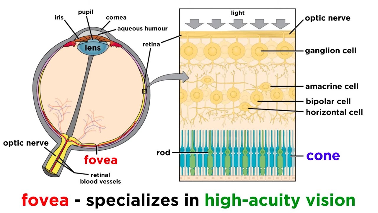
  • 🧠 The retina contains five basic types of neurons organized in layers, making it relatively simple and aiding in our understanding of vision.
  • 🌟 Photoreceptors, located at the back of the retina, convert light into electrochemical signals through a process called phototransduction.
  • 🌈 Rods and cones are the two types of photoreceptors, with cones responsible for color vision and high spatial resolution, while rods are sensitive to light but have low spatial resolution.
  • 🌙 Rods are dominant in low light conditions, making it difficult to see details or colors, as cones are less sensitive and provide higher acuity in brighter light.
  • 👁️ The fovea, especially the foveola, is rich in cones and is responsible for our highest visual acuity, prompting the eyes to move to focus important details there.
  • 🛣️ The optic nerve carries visual information from the retina to the brain, with a blind spot at the optic disc where no photoreceptors are present.
  • 🔄 At the optic chiasm, most axons from the nasal retina cross to the opposite side of the brain, while those from the temporal retina do not, integrating visual fields.
  • 🧬 The optic tract leads to various brain areas, including the lateral geniculate nucleus in the thalamus, which projects to the primary visual cortex (V1).
  • 🌆 The primary visual cortex and surrounding areas process visual information, with specialized neurons detecting orientation, movement, contrast, and depth.
  • 🧠 Higher-level visual processing involves additional brain areas that build upon basic visual aspects to enable complex tasks like object recognition.

Q & A

  • What is the starting point of the neural aspect of vision?

    -The neural aspect of vision starts with the retina, which is the neural structure of the eye.

  • How does the cornea contribute to vision?

    -The cornea is a transparent layer at the front of the eye that lets light in and bends or refracts the light rays to direct them onto the retina.

  • What role do the ciliary muscles play in vision?

    -The ciliary muscles modify the shape of the lens to maintain focus on objects that are closer or farther away.

  • Why do people need reading glasses as they age?

    -As people age, their lenses become less flexible and less capable of changing shape to focus on nearby objects.

  • How does the size of the pupil regulate the amount of light reaching the retina?

    -The size of the pupil can be adjusted by muscles in the iris, dilating in low-light situations and constricting in brighter environments to regulate the amount of light.

  • What is the main function of the retina?

    -The retina's main function is to detect light and produce electrical and chemical signals that the rest of the nervous system can understand.

  • Why is the location of photoreceptors at the back of the retina considered strategic?

    -The location is strategic because photoreceptors are next to the pigment epithelium, which helps maintain the cells and keep them functioning properly.

  • What are the two main types of photoreceptor cells and their respective functions?

    -The two main types are rods and cones. Cones enable color vision and high spatial resolution, while rods are sensitive to light and have low spatial resolution but do not provide color perception.

  • What is the significance of the fovea in the retina?

    -The fovea, especially the foveola, has a high concentration of cones and is responsible for our highest acuity vision, allowing us to discern important details.

  • How do horizontal and amacrine cells contribute to early visual processing?

    -Horizontal cells modulate the function of photoreceptor cells to enhance contrast and adapt to lighting conditions. Amacrine cells refine the visual signal by modifying the functions of other retinal cells.

  • What is the consequence of the optic disc having no photoreceptors?

    -The absence of photoreceptors at the optic disc creates a blind spot in our visual field, which the brain fills in with information from other photoreceptors.

  • How does the optic chiasm contribute to the processing of visual information?

    -At the optic chiasm, about 60% of the axons from the optic nerve cross over to the other side of the brain, ensuring that information from the right visual field is processed by the left side of the brain and vice versa.

  • What is the primary function of the primary visual cortex (V1)?

    -The primary visual cortex (V1) helps to create a visual image from the information received by the retina, with neurons activated preferentially by different characteristics of a visual stimulus.

  • How do higher-level visual areas contribute to the processing of visual information?

    -Higher-level visual areas, such as V2, V3, V4, V5, and V6, are specialized for detecting specific aspects of a visual scene, such as movement, and work in conjunction with V1 for more complex visual processing.

Outlines

00:00

👀 Visual Pathway Introduction

This paragraph introduces the journey of visual information from the eye to the brain, focusing on the pathway rather than the processing. It explains the role of the eye's components, such as the cornea, lens, and pupil, in creating a focused image on the retina. The retina's function in detecting light and producing signals is highlighted, along with the strategic placement of photoreceptors near the pigment epithelium for maintenance. The paragraph also distinguishes between rods and cones, detailing their roles in low light and color perception, and notes the high concentration of cones in the fovea for high acuity vision.

05:02

🌐 Retinal Neurons and Visual Processing

This paragraph delves into the types of neurons in the retina, including photoreceptors, bipolar, ganglion, horizontal, and amacrine cells, and their functions in early visual processing. It discusses the transmission of visual information from the retina to the brain via the optic nerve and the phenomenon of the blind spot due to the absence of photoreceptors at the optic disc. The optic chiasm's role in directing visual information to the appropriate hemisphere is explained, along with the distribution of fibers to various brain regions for different visual functions. The paragraph concludes with the description of the primary visual cortex (V1) and its surrounding areas, which are involved in higher-level visual processing and object recognition.

Mindmap

Keywords

💡Visual Information

Visual information refers to the data that the brain receives and interprets from the eyes. It is the core theme of the video, which discusses how this information travels from the eye to the brain. The script explains that vision begins with the eye but the neural aspect starts with the retina, which is responsible for detecting light and producing signals that the nervous system can interpret.

💡Retina

The retina is the neural structure at the back of the eye that plays a crucial role in vision. It is outlined in the script as the starting point of the neural pathway of visual information. The retina contains photoreceptors that convert light energy into electrochemical signals, initiating the process of vision.

💡Cornea

The cornea is the transparent layer at the front of the eye that allows light to enter and is responsible for bending or refracting light rays onto the retina. It is essential for creating a focused image on the retina and is mentioned in the script as a key component in the process of vision.

💡Lens

The lens is another component of the eye that helps direct light onto the retina. It is unique because its shape can be modified by the ciliary muscles to maintain focus on objects at varying distances. The script explains how the flexibility of the lens decreases with age, leading to the need for reading glasses.

💡Pupil

The pupil is the opening in the middle of the iris that can be adjusted to regulate the amount of light reaching the retina. The script describes how the size of the pupil changes in response to different light levels, with muscles in the iris causing it to dilate in low light and constrict in bright light.

💡Photoreceptors

Photoreceptors, specifically rods and cones, are cells in the retina responsible for converting light energy into electrochemical signals through a process called phototransduction. The script highlights their strategic location and specialization, with rods being sensitive to light and cones being involved in color perception.

💡Rods and Cones

Rods and cones are the two main types of photoreceptor cells in the retina. Rods are sensitive to light and provide low spatial resolution, useful in low light conditions but do not perceive color. Cones, on the other hand, are less sensitive to light, provide high spatial resolution, and are responsible for color vision. The script explains their different functional specializations and how they contribute to vision under varying light conditions.

💡Fovea

The fovea is a part of the retina with a high concentration of cones, which allows for high visual acuity. The script mentions that the center of the fovea, known as the foveola, contains no rods and is the area of the retina responsible for our sharpest vision, important for discerning details.

💡Ganglion Cells

Ganglion cells are neurons in the retina that carry visual information to the brain. Their axons form the optic nerve, and the script explains that these cells are crucial for transmitting the signals generated by photoreceptors to the brain for further processing.

💡Optic Chiasm

The optic chiasm is the point where the optic nerves from both eyes meet and about 60% of the axons cross to the opposite side of the brain. This decussation ensures that the visual information from the right visual field is processed by the left side of the brain and vice versa, as detailed in the script.

💡Primary Visual Cortex (V1)

The primary visual cortex, also known as V1 or the striate cortex, is the region of the brain that processes visual information received from the retina. The script describes how this area is involved in creating a visual image and how it communicates with other visual areas to detect specific aspects of a visual scene.

Highlights

The pathway of visual information in the brain is covered from the eye to the visual cortex and surrounding areas.

Vision starts with the retina, which is the neural structure of the eye.

The cornea and lens play crucial roles in focusing light onto the retina.

Ciliary muscles adjust the lens shape for focus on objects at varying distances.

Aging affects lens flexibility, leading to the need for reading glasses.

The iris controls the size of the pupil to regulate light entering the eye.

The retina is part of the central nervous system and detects light to produce signals.

There are only 5 basic types of neurons in the retina, making its anatomy relatively simple.

Photoreceptors, located at the back of the retina, convert light energy into electrochemical signals.

Rods and cones are the two main types of photoreceptor cells with different functional specializations.

Cones enable color vision, while rods are sensitive to light and provide low spatial resolution.

The fovea, with its high cone concentration, is responsible for our highest acuity vision.

Horizontal and amacrine cells refine the visual signal in the retina through their interactions with other cells.

The optic nerve carries information from the retina to the brain, creating a blind spot at the optic disc.

At the optic chiasm, about 60% of the axons from the optic nerve cross to the opposite side of the brain.

The optic tract extends to various areas, including the pretectum, suprachiasmatic nucleus, and superior colliculus.

The lateral geniculate nucleus in the thalamus is a key relay point for visual information.

The primary visual cortex, or V1, is responsible for creating a visual image from the received information.

Neurons in the primary visual cortex are activated by different visual stimulus characteristics.

Additional visual areas surrounding V1 are specialized for detecting specific aspects of a visual scene.

The brain's higher-level processing of visual information enables object recognition and meaning assignment.

Transcripts

play00:00

Hi everyone, welcome to 10 minute neuroscience.

play00:03

In this installment, I’ll be covering the pathway of visual information through the

play00:06

brain starting with the eye and ending with the visual cortex and surrounding areas.

play00:10

This’ll be a very general overview, and I’ll be focusing more on the pathway of

play00:14

visual information than on the processing of that information, but this should serve

play00:18

as a good introduction to the way visual information travels from the eye through the brain.

play00:23

Of course, vision begins with the eye, but the neural aspect of vision really starts

play00:26

with the retina, which is the neural structure of the eye and is outlined in blue here.

play00:32

Other components of the eye, however, help to create a focused image on the retina.

play00:35

This is accomplished to a large degree through the actions of the cornea and the lens.

play00:40

The cornea is a transparent layer at the front of the eye that lets light into the eye.

play00:45

It also bends, or refracts, those light rays to direct the light onto the retina.

play00:51

The lens also helps to direct light on the retina.

play00:53

It has less refractive power than the cornea, but it has an advantage in that its shape

play00:57

can be modified by muscles in the eye called ciliary muscles, and changing the shape of

play01:02

the lens can help to maintain focus for objects that are closer or farther away.

play01:07

As people get older, their lenses become less flexible and less capable of changing shape

play01:10

to focus on nearby objects; this is why people tend to need reading glasses when they get

play01:15

older.

play01:16

The size of the pupil, the opening in the middle of the iris, which is the colored part

play01:19

of your eye, can be adjusted to regulate the amount of light that reaches the retina.

play01:22

For example, there are muscles in the iris that cause the pupil to dilate (or open up

play01:26

more) in low-light situations, and constrict in an environment with higher levels of illumination

play01:31

since it doesn’t need to let in as much light in that type of environment.

play01:35

The retina is a neural structure, and it’s actually considered part of the central nervous

play01:38

system.

play01:39

Its main function is to detect light and use that light to produce electrical and chemical

play01:44

signals that the rest of the nervous system can understand.

play01:47

You can see here a close-up of a section of the retina, and although there are over 1,000

play01:52

different types of neurons in the nervous system, there’s only 5 basic types of neurons

play01:56

in the retina, and those neurons are situated in distinct layers.

play02:00

So, even though there are hundreds of millions of neurons in the retina, compared to the

play02:04

rest of the nervous system it’s relatively simple anatomically.

play02:07

This has helped us to develop a better understanding of vision than of any other sensory system.

play02:12

Surprisingly, the layer of the retina that’s at the very back of the eye is the layer that

play02:16

contains photoreceptors, the cells responsible for converting light energy into electrochemical

play02:21

signals, a process known as phototransduction.

play02:25

The location of the photoreceptors is surprising because it seems counterintuitive to have

play02:29

the light detecting cells at the very back of the retina, so light has to travel through

play02:32

the eye and then through several layers of cells to reach the photoreceptors, but it’s

play02:36

thought that their location is strategic in the sense that they’re next to a layer of

play02:40

the retina called the pigment epithelium, and the cells of the pigment epithelium help

play02:44

to maintain photoreceptor cells and keep them functioning properly.

play02:47

There are two main types of photoreceptor cells: rods and cones, which are named for

play02:52

their shape as you can see here.

play02:55

These photoreceptor cells are the site where vision really begins; they each contain hundreds

play02:58

of disks that are capable of absorbing photons of light and absorbing photons causes the

play03:04

photoreceptors to change levels of neurotransmitter release in order to convey information about

play03:09

a visual scene.

play03:11

Rods and cones have different functional specializations; they’re involved in distinct aspects of

play03:15

vision.

play03:16

First off, cones enable us to see color, while rods don’t provide for color perception.

play03:21

Rods are also very sensitive to light and have low spatial resolution, meaning they’re

play03:24

not good at seeing details.

play03:26

Cones, on the other hand, are not very sensitive to light and have high spatial resolution,

play03:31

so they provide us with higher visual acuity.

play03:34

In low light conditions, only rods are activated.

play03:37

This makes it more difficult for us to make out details when there’s very little light

play03:40

due to the poor spatial resolution of rods, and it also means that in dim light we can’t

play03:45

perceive color.

play03:46

When levels of illumination increase, eventually rods stop responding and fail to convey information.

play03:51

Essentially their high sensitivity to light causes them to become overstimulated, or saturated

play03:56

as its often called, in normal lighting situations like sunlight or even typical indoor lighting.

play04:01

So in those conditions, cones are the dominant photoreceptor in determining how we see.

play04:05

Surprisingly, even though cones mediate perception in typical light situations, we have far more

play04:10

rods than cones in the retina.

play04:12

There’s somewhere around 90 million rods and only about four and a half million cones.

play04:16

However, there is one part of the retina, an area called the fovea, where there are

play04:21

many more cones than rods.

play04:23

In fact, at the very center of the fovea, which is called the foveola, there are no

play04:26

rods at all.

play04:28

Because of its high cone content the fovea is the part of our retina that has the capacity

play04:32

for our highest acuity vision.

play04:34

This causes us to unconsciously move our eyes so that important visual information lands

play04:38

on our fovea, since this is the part of our eye most capable of discerning important details.

play04:44

When photoreceptors absorb photons, it causes changes in the amount of neurotransmitters

play04:48

these cells release, and this affects the activity of the next layer of cells: bipolar

play04:52

cells.

play04:54

Bipolar cells pass on signals about perceived light to the next layer of cells, which are

play04:58

called ganglion cells, and these cells will carry the visual information to the brain.

play05:02

We’ll talk more about that in a moment but I want to mention the other two major cell

play05:05

types in the retina: horizontal cells and amacrine cells.

play05:09

Horizontal cells have dendrites that spread horizontally and make contact with multiple

play05:13

photoreceptor cells.

play05:16

Horizontal cells modulate the function of photoreceptor cells to do things like enhance

play05:19

contrast and adapt to changes in lighting conditions, among other things.

play05:24

Amacrine cells make contact with bipolar cells, ganglion cells, and other amacrine cells,

play05:29

and they also have a number of functions, but like horizontal cells they’re generally

play05:32

thought to be involved with refining the visual signal through their ability to modify the

play05:36

functions of other retinal cells.

play05:39

So horizontal and amacrine cells are involved with very early processing of visual information.

play05:43

But most of the visual processing occurs in the brain, so the information from the retina

play05:47

has to be carried out of the eye and to the brain.

play05:49

This is accomplished by the ganglion cells, whose axons leave the eye in a bundle at a

play05:55

region called the optic disc.

play05:58

Because the optic disc is the area where the ganglion cells leave the eye, and essentially

play06:01

they need a place where they can exit the eye, there are no photoreceptors there.

play06:05

So, this creates a small region where we don’t receive any visual information, or a blind

play06:09

spot.

play06:10

Amazingly, we don’t notice this blind spot in our visual scene because the brain fills

play06:13

it in with information from other photoreceptors.

play06:16

If you find it hard to believe that you’ve got a blind spot always present in your field

play06:19

of vision, click on this link above for a quick and simple experiment that proves the

play06:23

existence of your blind spot.

play06:25

The ganglion cells leaving the eye form the optic nerve, which is one of our cranial nerves.

play06:30

The optic nerve extends back to this region just below the hypothalamus called the optic

play06:34

chiasm.

play06:35

At the optic chiasm, about 60% of the axons from the optic nerve cross over to the other

play06:40

side of the brain, while the rest stay on the side they originated on.

play06:44

The fibers that are coming from the nasal part of the retina—the part closer to the

play06:48

nose—cross over to the other side, or decussate, while the fibers from the temporal part of

play06:52

the retina—the part closer to the temple–do not cross over.

play06:56

The result of this is that all of the information from the right visual field ends up traveling

play07:00

to the left side of the brain, and vice versa.

play07:04

After the optic chiasm, the visual fibers are no longer called the optic nerve—they’re

play07:07

now called the optic tract.

play07:10

The optic tracts extend to multiple areas.

play07:12

For example, some of the fibers go to an area in the brainstem called the pretectum, which

play07:15

is involved in a number of visual functions such as the pupillary light reflex, which

play07:19

causes your pupils to constrict when there’s greater illumination in your environment.

play07:24

Other fibers go to a region of the hypothalamus called the suprachiasmatic nucleus; this nucleus

play07:28

helps to maintain circadian or daily rhythms and uses information about light in the environment

play07:32

to help to do that.

play07:34

And still other fibers go to the superior colliculus, a region in the brainstem that

play07:38

among other things helps to coordinate head and eye movements to focus on objects of interest

play07:42

in the visual field.

play07:44

Most of the fibers in the optic tracts, however, end in a nucleus in the thalamus called the

play07:49

lateral geniculate nucleus, there’s one of these on each side of the brain here.

play07:52

Here, the optic tracts synapse on neurons that leave the lateral geniculate nucleus

play07:57

and extend toward the back of the brain as bundles of fibers called the optic radiations.

play08:04

The optic radiations travel back to a region of cortex that surrounds a fissure called

play08:09

the calcarine sulcus.

play08:12

This small area of cortex that surrounds the calcarine sulcus is called the primary visual

play08:17

cortex, or V1, it's represented by this striped area here.

play08:21

There’s a collection of myelinated fibers that forms a white stripe here that can be

play08:25

seen with the naked eye in anatomical brain sections of this region and because of this

play08:30

striation or stripe, sometimes the primary visual cortex is called the striate cortex.

play08:35

The primary visual cortex helps to make a visual image out of the information that has

play08:39

been received by the retina.

play08:40

To do that, there are neurons in the primary visual cortex that are activated preferentially

play08:44

by different characteristics of a visual stimulus, such as orientation, movement, contrast, depth,

play08:50

etc.

play08:51

The primary visual cortex also communicates with a multitude of other visual areas that

play08:55

surround it, including visual area 2, or V2, visual area 3 or V3, V4, V5, and V6.

play09:03

Neurons in these other areas seem to be specialized to some degree for detecting specific aspects

play09:08

of a visual scene.

play09:09

Neurons in V5, for example, seem to be specialized for detecting movement.

play09:13

These additional visual areas are recruited by V1 and they also begin to recruit other

play09:18

areas of the brain to accomplish higher-level processing of visual information.

play09:22

This enables our brain to move from identifying the basic aspects of a visual scene, such

play09:26

as shape and contrast, to more complex tasks like object recognition, which can provide

play09:32

the image in our brain with meaning based on previous experience.

play09:36

So we just scratched the surface there but I hope that gave you a sense of the major

play09:39

pathway that visual information travels in the brain.

play09:41

Thanks for watching.

Rate This

5.0 / 5 (0 votes)

Related Tags
NeuroscienceVision PathwayEye AnatomyCornea LensPhotoreceptorsRods ConesVisual CortexRetinal LayersOptic ChiasmOptic TractVisual Processing