X-Ray Radiography
Summary
TLDRThis video script outlines the process of radiography testing, a method using ionizing radiation like X-rays to create images of objects. An examiner marks the weld area, sets up the X-ray equipment, and operates it remotely to avoid exposure. The X-rays penetrate the material, creating an image on film or a plate, which is then processed and analyzed to assess the object's quality and compliance with standards.
Takeaways
- 📸 Radiography testing uses ionizing radiation to create images of objects.
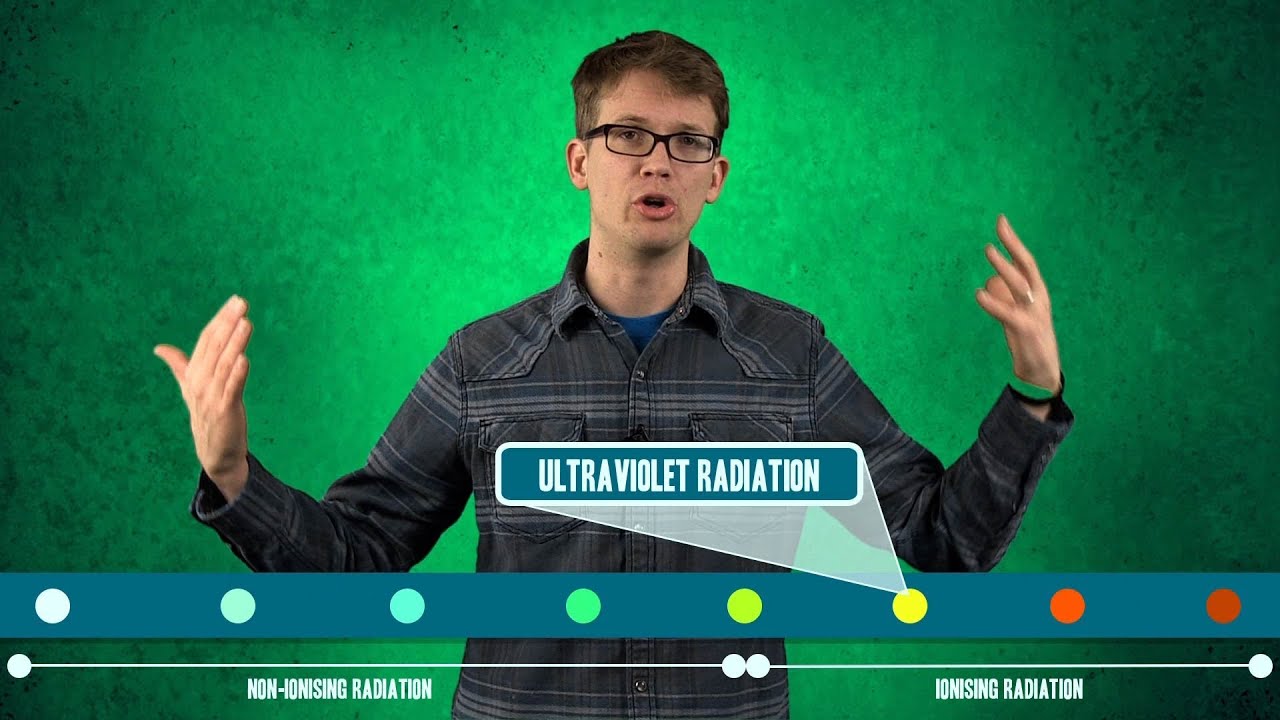
- 🔎 X-rays are a common type of radiation used for radiography.
- 📏 A measuring tape is used to mark the weld for inspection, making it visible on the x-ray.
- 🎞️ Film or imaging plates are mounted on the object to capture the radiographic image.
- 🚫 The examiner leaves the area and operates the x-ray device remotely to avoid radiation exposure.
- ⚙️ The x-ray machine's voltage, current, and duration are adjusted based on the material and wall thickness.
- 🔬 X-rays penetrate the material, creating an image on the film or imaging plate.
- 🔍 The image is processed to reveal details that the examiner can interpret.
- 👀 The examiner's experience is crucial for interpreting the image and assessing the object's quality.
- 🏗️ The process helps determine if the inspected object meets the required standards.
Q & A
What is radiography testing?
-Radiography testing is a non-destructive testing method that uses ionizing radiation, such as gamma rays or X-rays, to create images of an object's internal structure on various viewing media.
How is the weld marked for inspection in radiography testing?
-The examiner marks the weld to be inspected with a measuring tape, making it visible on the X-ray so that the exact location of possible irregularities can be determined.
What is the purpose of mounting the film on the object?
-The film is mounted on the object to capture the radiographic image produced by the X-rays passing through the material.
Why does the examiner leave the area during the irradiation process?
-The examiner leaves the area and operates the device remotely to avoid exposure to the ionizing radiation.
What safety measures are taken during radiography testing?
-The examiner closes the lead door and operates the device remotely to protect themselves from radiation exposure.
How does the examiner adjust the X-ray machine settings?
-The examiner adjusts the voltage, current, and duration of the X-ray exposure depending on the material being investigated and the wall thickness of the object.
What happens during the irradiation process?
-During irradiation, X-rays pass through the material and generate a radiographic image on the film or imaging plate.
What is the role of the imaging plate in radiography testing?
-The imaging plate captures the radiographic image produced by the X-rays, similar to how film does, and is used for digital radiography.
How is the quality of the inspected object determined after processing the image?
-The processed image is interpreted by the examiner, who uses their experience to determine the quality of the inspected object and whether it meets the required standards.
What are the potential irregularities that radiography testing can reveal?
-Radiography testing can reveal internal irregularities such as cracks, voids, inclusions, and other defects that are not visible from the surface.
Why is it important for the examiner to have a trained eye when interpreting the radiographic images?
-A trained eye is crucial for accurately interpreting radiographic images to identify and assess the severity of any defects, ensuring the safety and integrity of the inspected object.
Outlines

Dieser Bereich ist nur für Premium-Benutzer verfügbar. Bitte führen Sie ein Upgrade durch, um auf diesen Abschnitt zuzugreifen.
Upgrade durchführenMindmap

Dieser Bereich ist nur für Premium-Benutzer verfügbar. Bitte führen Sie ein Upgrade durch, um auf diesen Abschnitt zuzugreifen.
Upgrade durchführenKeywords

Dieser Bereich ist nur für Premium-Benutzer verfügbar. Bitte führen Sie ein Upgrade durch, um auf diesen Abschnitt zuzugreifen.
Upgrade durchführenHighlights

Dieser Bereich ist nur für Premium-Benutzer verfügbar. Bitte führen Sie ein Upgrade durch, um auf diesen Abschnitt zuzugreifen.
Upgrade durchführenTranscripts

Dieser Bereich ist nur für Premium-Benutzer verfügbar. Bitte führen Sie ein Upgrade durch, um auf diesen Abschnitt zuzugreifen.
Upgrade durchführen


5.0 / 5 (0 votes)
