Assessment of CXR Positioning & Views - How to Read a Chest X-Ray (Part 4) - MEDZCOOL
Summary
TLDRThis educational video script discusses the fundamentals of interpreting chest X-rays, emphasizing the importance of assessing X-ray quality before analysis. It differentiates between anterior posterior (AP), posterior anterior (PA), and lateral views, highlighting the PA view as superior due to increased patient mobility and reduced external distractions. The script also explains how the position of the X-ray affects the appearance of structures like the heart, crucial for diagnosing conditions like pericardial effusion or cardiomegaly.
Takeaways
- 🔍 Always assess the quality of an X-ray before interpretation to avoid making conclusions based on poor data.
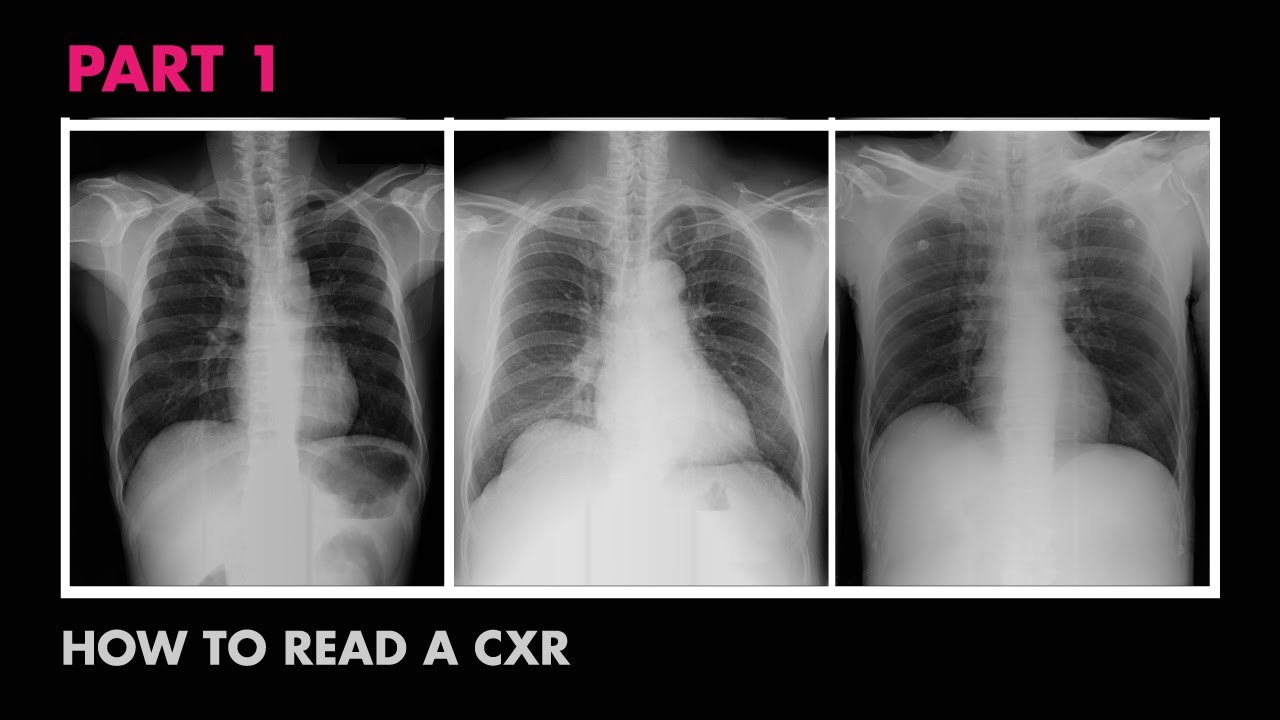
- 📚 Start with identifying the type of chest X-ray: anterior posterior (AP), posterior anterior (PA), or lateral.
- 👀 Recognize the patient's position and ensure you are viewing the correct patient's X-ray to avoid misdiagnosis.
- 📐 PA view is generally superior in quality to AP view, especially when obtained on ambulatory patients.
- 🛏️ AP X-rays are often obtained on bed-bound patients in the ICU, which can be of lower quality due to limited patient mobility and external distractions.
- 🏥 Portable X-ray machines are used for patients who cannot move, which can result in a less optimal study due to positioning challenges.
- 💡 The direction of the X-ray beam affects the appearance of organs; anterior structures appear larger on PA films compared to AP films.
- ❤️ Heart size is most accurately assessed on a PA film due to the way the X-ray beam casts shadows from the posterior to anterior.
- 📈 Understanding the type of X-ray film is crucial for accurate diagnosis, especially in conditions like pericardial effusion or cardiomegaly.
- 👨⚕️ The PA and lateral views are preferred for chest X-rays when possible, due to the increased mobility and positioning options available for ambulatory patients.
- 📹 Subscribe to the channel for updates on educational videos and support the creators on Patreon for more content.
Q & A
What is the first step in analyzing an x-ray according to the video?
-The first step is to assess the quality of the x-ray, determining if it is of good enough quality for interpretation.
What does the letter 'A' in the mnemonic stand for?
-The letter 'A' stands for 'Assessment of Quality', which is the first aspect to consider when reading x-rays.
Why is it important to recognize the type of chest x-ray being viewed?
-Recognizing the type of chest x-ray helps in making accurate assessments, as different types have different positional and quality implications.
What are the three types of chest x-ray films mentioned in the video?
-The three types of chest x-ray films are anterior posterior (AP), posterior anterior (PA), and lateral x-rays.
Why are posterior anterior (PA) views generally superior in quality to anterior posterior (AP) views?
-PA views are superior because they are usually obtained on ambulatory patients who can position themselves more flexibly, allowing the x-ray technician more freedom to maneuver without external distractions.
What challenges do bed-bound patients present when taking an anterior posterior (AP) chest x-ray?
-Bed-bound patients present challenges such as limited space and mobility, and the presence of equipment like IV poles and monitors, which can affect the quality of the AP x-ray.
How does the position of the x-ray machine affect the appearance of the heart on the film?
-If the x-ray is shot from the front to the back (PA view), the cardiac silhouette appears smaller because the heart is more anterior in the body. Conversely, if shot from the back to the front (AP view), the heart appears larger.
Why is the heart size most accurately assessed on a PA film?
-The heart size is most accurate on a PA film because the x-ray captures a smaller shadow of the heart due to the position of the film in relation to the heart's location in the body.
What are some factors that can affect the quality of a portable AP chest x-ray taken in the ICU?
-Factors affecting the quality of a portable AP chest x-ray in the ICU include the patient's inability to move, the presence of medical equipment, and the limited space for positioning the patient.
What is the significance of knowing the correct patient when assessing an x-ray?
-Knowing the correct patient ensures that the interpretation is based on the right individual, preventing misdiagnosis or incorrect treatment based on another patient's x-ray.
How can a mnemonic help in the process of reading x-rays?
-A mnemonic helps in systematically remembering and applying the key steps in reading x-rays, such as assessing quality, which can improve accuracy and efficiency in interpretation.
Outlines
📚 X-ray Assessment Basics
This paragraph introduces the fundamental concepts of x-ray interpretation, focusing on the importance of quality assessment. It contrasts an ideal x-ray with one of poor quality, highlighting issues such as patient rotation, obstructions in the field, and poor inspiration. The speaker emphasizes the necessity of evaluating the quality before making any interpretations to avoid incorrect conclusions based on substandard data. The paragraph also outlines the first step in x-ray quality assessment: recognizing the type of chest x-ray, noting the positioning and ensuring the correct patient is being examined.
🔍 Understanding Chest X-ray Types
The speaker elaborates on the three types of chest x-rays: anterior posterior (AP), posterior anterior (PA), and lateral. The paragraph explains how to distinguish between them, particularly noting the lateral x-ray's side shot. It also discusses the superiority of the PA view in terms of quality, especially when obtained from ambulatory patients, due to the flexibility it allows in patient positioning without external distractions. The paragraph further explains the technical aspects of how the position of the x-ray machine and detector affect the quality and appearance of the image, using the example of a sick patient in the ICU to illustrate the challenges of obtaining high-quality AP films in such settings.
📏 Significance of X-ray Positioning
This section delves into the importance of knowing the type of x-ray film being viewed, especially when assessing structures like the heart. It explains how the position of the x-ray source affects the size and appearance of internal organs on the film, using the analogy of casting a shadow. The paragraph clarifies that a PA film provides the most accurate representation of heart size, which is crucial for diagnosing conditions like pericardial effusion or cardiomegaly. The speaker concludes by summarizing the preference for PA and lateral views when possible, and the fallback to AP chest x-rays when not.
Mindmap
Keywords
💡Anatomy
💡Mnemonic
💡X-ray
💡Assessment of Quality
💡Chest X-ray
💡Anterior Posterior (AP)
💡Posterior Anterior (PA)
💡Lateral X-ray
💡Positioning
💡Inspiration
💡Cardiac Silhouette
Highlights
Introduction to the basic anatomy and mnemonics for reading x-rays.
Explanation of the importance of assessing x-ray quality before interpretation.
Comparison between ideal and poor quality x-rays with examples.
The necessity to determine the quality of the x-ray before making conclusions.
Differentiating between anterior posterior (AP), posterior anterior (PA), and lateral chest x-rays.
Visual distinction between lateral and AP/PA x-rays.
The superiority of PA view over AP view in terms of quality.
Explanation of how the position of the patient affects the x-ray quality.
The flexibility in positioning patients for PA x-rays compared to AP.
Challenges in obtaining high-quality AP x-rays with bed-bound patients in the ICU.
The impact of patient position on the appearance of the cardiac silhouette.
Why PA films are preferred for assessing heart size in cases of pericardial effusion or cardiomegaly.
The practicality of obtaining a posterior-anterior film on ambulatory patients.
The limitations of portable x-rays in positioning bed-bound patients.
The importance of knowing the type of x-ray film for accurate diagnosis.
Summary of the three different views for chest x-rays and their applications.
Encouragement to subscribe for updates on new educational videos.
Invitation to follow on social media and support the channel through Patreon.
Transcripts
the first few videos in this series I
talked about the basic anatomy and
useful mnemonic to help you with reading
x-rays in upcoming videos we'll go into
more detail of each letter in this
mnemonic starting with a letter A which
stands for assessment of quality here's
an ideal x-ray and here's an x-ray of
poor quality these are obviously not the
same patients but you can tell that the
one to the right is a little more
difficult to read the patient is rotated
there are cables in the field there's
poor inspiration so on and so forth so
before trying to interpret the film
first determine if the x-ray you're
looking at is a good enough quality the
last thing you want to do is to make a
conclusion on some bad data the first
thing you want to do when assessing the
quality of an x-ray is recognizing what
type of chest x-ray you're looking at
make note of how and in what position
the chest x-ray was shot in and of
course make sure you're looking at the
correct patient there are three types of
chest x-ray films anterior posterior
posterior anterior and lateral x-rays as
you can see here lateral x-rays are easy
to tell apart from AP and PA x-rays this
one is obviously shot from the side
while the other ones are shot
perpendicular to the patient either
facing or looking away from the x-ray
machine knowing this and making a mental
note if the chest x-ray was shot from
behind or in front is helpful when
making assessments later on in general
the posterior anterior view is superior
in quality to the anterior posterior
view if you can safely obtain a
posterior interior film on your patient
this would be preferred
that's because posterior-anterior x-rays
are usually obtained on ambulatory
patients or able to position themselves
therefore giving the x-ray technician
more flexibility when these films are
shot the patient's back is facing the
x-ray machine and x-ray detector in
front and so x-rays come from behind
pass through the patient from the back
to the front and onto the x-ray detector
and again this extra space and
flexibility in a posterior-anterior film
allows the x-ray technician more freedom
and mobility position the patient as
needed without any external distractions
to illustrate why this is important take
for example a very sick patient in the
ICU
you these patients don't have the
ability to walk or stand and so they
stay in bed and a portable machine is
brought to them the x-ray detectors slip
behind their back an x-ray machine is
placed in front
often these anterior posterior films are
inferior in quality to that of the
posterior anterior view that's because
patients who are bed-bound
are often limited in their space and
mobility to move also there are usually
things in the way such as central lines
IV poles monitors blankets etc in a
portable x-ray is more difficult to
position the patient who may be hunched
over in bed and so important aspects of
x-ray quality such as position rotation
may be difficult to obtain versus an
ambulatory patient who is able to freely
stand up without many distracting
elements surrounding them all these make
it more difficult to maneuver the
patient and overall gives the x-ray
technician a potentially limited study
the last reason I'll mention for why
it's important to know what type of film
you're looking at is if a film is shot
into your posterior things in the body
that are more anterior such as the heart
may appear larger compared to the same
film shot posterior-anterior
if you think if the x-ray is casting a
shadow this will make more sense the
place you choose to shoot the x-ray is
like a light source an x-ray will then
capture the shadow on the film in this
diagram if you're shooting a film from
front to back the film that is captured
will have a larger shadow of the cardiac
silhouette because the hearts it's
morning Terrier in the body
if you shoot a posterior-anterior film
the cardiac silhouette will appear
smaller because if the position of the
film was shot compared to the location
of the structure in this case the heart
therefore heart size is most accurate
when assessed on a PA film this is
important when knowing the size of the
heart is essential such as in the case
of a pericardial effusion or other cases
of cardiomegaly you may see on chest
x-ray in summary we talked about the
three different views you could obtain
when getting a chest x-ray
there's the posterior anterior view
which often is obtained alongside a
lateral view given the increased amount
of mobility that ambulatory patients
have the PA and lateral view are
preferred if you could obtain it in your
patient if not the alternative would be
an anterior posterior chest x-ray
if you haven't already make sure to
subscribe
to this channel to get updates on when
we release our latest videos and don't
forget to follow med school on social
media if you're feeling real generous
visit our patreon page and make a pledge
to help us create more awesome videos
Weitere ähnliche Videos ansehen
Radiology of Thorax (Chest)
Cardiac Silhouette - How to Read a Chest X-Ray (Part 7) - MEDZCOOL
Anatomy of a Chest X-Ray - How to Read a Chest X-Ray (Part 1)
COMO AVALIAR QUALQUER RAIO X DO TÓRAX COM SEGURANÇA? O ABCDE DO TÓRAX I VOCÊ RADIOLOGISTA
Cardiac Tamponade - pericardial effusion, causes, pathophysiology, investigations and treatment
Assessment of CXR Quality - How to Read a Chest X-Ray (Part 5) - MEDZCOOL
5.0 / 5 (0 votes)