USMLE® Step 1: Neuroscience: Development of CNS Animation
Summary
TLDRThis script explores the critical process of neural tube development into the spinal cord and brain, detailing its formation from the ectodermal germ layer during the third week of gestation. It explains how complications can lead to neural tube defects, common congenital abnormalities linked to factors like folic acid deficiency. The script describes various types of spina bifida and anencephaly, their impacts, and diagnostic indicators such as alpha-fetoprotein levels, emphasizing the importance of understanding these conditions in prenatal care.
Takeaways
- 🧠 The development of the nervous system begins during the third week of gestation.
- 🌱 By day 18, the ectodermal germ layer forms a disc with a cranial and caudal end, laying the foundation for neural development.
- 🔍 The central nodal cord induces the overlying ectoderm to thicken and form the neural plate, which is crucial for the formation of the neural tube.
- 🌀 The neural plate folds to form the neural tube, with the edges extending upward to become neural folds that eventually fuse at the midline.
- 🚀 Neural crest cells, which separate from the tips of the neural folds, migrate throughout the body to form various cell types, including Schwann cells and the adrenal medulla.
- 🧪 Folic acid deficiency during pregnancy and exposure to certain drugs can increase the risk of neural tube defects.
- 🦴 Neural tube defects can occur as part of syndromes, associated with chromosomal disorders, or due to environmental factors.
- 🧠 Anencephaly is a severe neural tube defect where the cranial end fails to close, leading to the absence of brain development and is incompatible with life.
- 🦵 Spina bifida is a defect where the neural tube fails to close at the caudal end, causing the vertebral arch to remain open.
- 🌐 Spina bifida occulta is an asymptomatic defect where the vertebral arch fails to fuse, often indicated by a small tuft of hair over the defect.
- 🌡 Elevated alpha-fetoprotein levels during pregnancy can indicate certain neural tube defects, such as anencephaly and spina bifida with meningeal protrusion.
Q & A
What is the primary process that forms the functional central nervous system?
-The primary process that forms the functional central nervous system is the development of the embryonic nervous system, which begins during the third week of gestation with the formation of the neural plate.
What are the three germ layers illustrated in the cross-section of the ectodermal germ layer by day 18 of fetal development?
-The three germ layers illustrated are the ectoderm, mesoderm, and endoderm.
What induces the overlying ectoderm to thicken and form the neural plate?
-The central nodal cord induces the overlying ectoderm to thicken and form the neural plate.
What is the term for the process where the edges of the neural plate extend upward to become neural folds?
-The process is known as the folding of the neural plate, which leads to the formation of the neural tube.
What is the name of the cells that separate from the tips of the neural folds and migrate throughout the body to form various cell types?
-These cells are called the neural crest cells.
What are some of the cell types that neural crest cells can form?
-Neural crest cells can form Schwann cells, meninges, endocardial cushions, pilar follicular cells, and the adrenal medulla.
When does the cranial neural pore close during the development of the neural tube?
-The cranial neural pore closes on day 25.
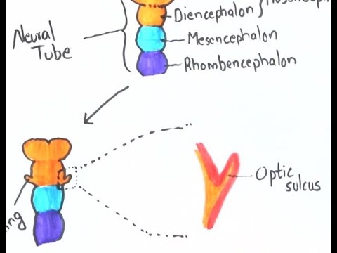
What are the three primary vesicles that develop from the cranial end of the neural tube?
-The three primary vesicles are the forebrain (prosencephalon), midbrain (mesencephalon), and hindbrain (rhombencephalon).
What is the most common cause of congenital abnormalities related to the development of the neural tube?
-One of the most common causes of congenital abnormalities related to the development of the neural tube is neural tube defects.
What is the condition known as anencephaly and why is it incompatible with life?
-Anencephaly is the failure of the neural tube to spontaneously close at the cranial end, preventing the brain from developing. It is incompatible with life due to the absence of a developed brain.
What is spina bifida and how does it differ from spina bifida occulta?
-Spina bifida is the failure of the neural tube to spontaneously close at the caudal end, causing the vertebral arch to remain open. Spina bifida occulta is an asymptomatic defect caused by the failure of the two halves of the vertebral arch to fuse at the midline, often only indicated by a small tuft of hair over the defect.
How do the levels of alpha-fetoprotein during pregnancy indicate neural tube defects?
-Increased alpha-fetoprotein levels during pregnancy can indicate neural tube defects such as anencephaly and spina bifida with meningomyelocele. However, spina bifida occulta does not cause an increase in alpha-fetoprotein levels.
Outlines
🧠 Neural Tube Development and Defects
This paragraph discusses the critical process of neural tube formation and its development into the spinal cord and brain. It highlights the complexity of this process and how any complication can lead to neural tube defects. The summary includes the beginning of the nervous system's development during the third week of gestation, the formation of the neural plate, and the eventual fusion into a closed neural tube. It also describes the role of neural crest cells and the development of the brain into primary and secondary vesicles. The paragraph concludes with information on neural tube defects, their causes, and specific conditions like anencephaly and spina bifida, including their impact on alpha-fetoprotein levels and pregnancy.
🩺 Types of Spina Bifida and Their Impacts
The second paragraph delves into the different types of spina bifida, a neural tube defect affecting the spinal cord. It explains spina bifida occulta, a mild form with no spinal cord involvement, and spina bifida with meningeal and myelomeningocele, where the meninges and spinal cord protrude through the vertebral defect. The summary outlines the physical manifestations of these conditions, their association with hydrocephalus, and the impact on alpha-fetoprotein levels during pregnancy. It emphasizes the severity of spina bifida with myelomeningocele, which is externally visible and associated with increased alpha-fetoprotein levels.
Mindmap
Keywords
💡Neural Tube
💡Neural Tube Defects
💡Ectoderm
💡Neural Crest
💡Folic Acid
💡Anencephaly
💡Spina Bifida
💡Alpha-fetoprotein
💡Meninges
💡Cranial and Caudal Neuropores
💡Secondary Vesicles
Highlights
The development of the nervous system begins during the third week of gestation.
Primary neurulation is the process that forms the functional central nervous system.
By day 18 of fetal development, the ectodermal germ layer forms a disc shape with cranial and caudal ends.
The central nodal cord induces the overlying ectoderm to thicken and form the neural plate.
The neural plate folds to form the neural tube by the end of the third week.
Neural folds grow towards each other to fuse at the midline, converting the neural groove into a closed, hollow neural tube.
Neural crest cells, which separate from the tips of the neural folds, migrate throughout the body to form various cell types.
The cephalic and caudal ends of the neural tube communicate with the amniotic cavity through neuropores before fusion.
The cranial neural pore closes on day 25, followed by the development of three primary vesicles from the cranial end of the neural tube.
The forebrain, midbrain, and hindbrain develop into five secondary vesicles.
Failure of the neural tube to close properly during early embryonic stages results in neural tube defects.
Neural tube defects are common causes of congenital abnormalities and can be associated with chromosomal disorders or environmental exposures.
Folic acid deficiency during pregnancy and folic acid antagonists increase the risk of neural tube defects.
Anencephaly is a fatal condition where the neural tube fails to close at the cranial end, preventing brain development.
Spina bifida is characterized by the failure of the neural tube to close at the caudal end, with varying degrees of severity.
Spina bifida occulta is an asymptomatic defect caused by incomplete fusion of the vertebral arch halves.
In spina bifida with meningocele, the meninges protrude through the vertebral defect without spinal cord protrusion.
Spina bifida with myelomeningocele involves both the meninges and spinal cord protruding through the vertebral defect.
Spina bifida with myelomeningocele is the most severe type, often associated with Chiari malformation and hydrocephalus, and leads to increased alpha-fetoprotein levels during pregnancy.
Transcripts
new relation is the process of neural
tube formation and its development into
the spinal cord and the brain any
complication during this complex process
can result in neural tube defects
the development of the nervous system
begins during the third week of
gestation primary neural ation is the
process that forms the functional
central nervous system let's begin with
the development of the embryonic nervous
system
by day 18 of fetal development
the ectodermal germ layer has the shape
of a disc with a cranial and caudal end
the cross-section on the right
illustrates three germ layers the
ectoderm mesoderm and endoderm at this
point the central nodal cord has induced
the overlying ectoderm to thicken and
form the neural plate the cross-section
on the right will help visualize the
folding of the neural plate leading to
the formation of the neural tube by the
end of the third week the edges of the
neural plate extend upward to become
neural folds the depressed mid region is
now the neural groove each neural fold
then grows toward each other to fuse at
the midline converting the neural groove
into a complete and closed and hollow
neural tube at the same time as the
neural folds fuse a population of cells
separates from the tips of the neural
folds to become a mass of his ankle
cells called the neural crest the neural
crest cells migrate throughout the body
to form a wide variety of cell types
including Schwann cells the meninges
endocardial cushions power follicular
cells and the adrenal medulla
before the fusion of the neural tube is
complete the cephalic and caudal ends
communicate with the amniotic cavity
through the cranial and caudal neuro
pores the cranial neural pore closes on
day 25 after closure the cranial end of
the neuro tube develops into three
primary vesicles the forebrain or pros
and Cephalon the midbrain or
mesencephalon and the hindbrain or
rhombencephalon these three develop
later into five secondary vesicles the
forebrain develops into the
telencephalon and diencephalon the
midbrain develops into the mesencephalon
the hind brain develops into Menton
Cephalon and the myelin Cephalon the
caudal neuro poor then closes on day 27
during early embryonic stages of
development failure of the neuro tube to
close properly results in neural tube
defects one of the most common causes of
congenital abnormalities neural tube
defects can occur as part of syndromes
and associated with chromosomal
disorders or as a result of an
environmental exposure folic acid
deficiency during pregnancy and folic
acid antagonists such as carbamazepine
phenytoin and trimethoprim
increased the risk of neural tube
defects
anencephaly is the failure of the neuro
tube to spontaneously close at the
cranial end hence the brain does not
develop this condition is incompatible
with life anencephaly causes high
alpha-fetoprotein levels and
polyhydramnios during pregnancy
spina bifida is the failure of the
neural tube to spontaneously close at
the caudal end vertebra overlying the
defect do not fully develop causing the
vertebral arch to remain open spina
bifida occulta is an asymptomatic defect
caused by failure of the two halves of
the vertebral arch to fuse at the
midline the only evidence of its
presence may be a small tuft of hair
over the defect
on the right illustration we can see
that spina bifida occulta
only affects the vertebral arch leaving
the spinal cord intact it's important to
know that in spina bifida occulta
there is no increase of
alpha-fetoprotein levels during
pregnancy in spina bifida with been in
jail seal the meninges protrude through
the vertebral defect without protrusion
of the spinal cord in spina bifida with
meningeal Milo seal both the meninges
and the spinal cord protrude through the
vertebral defect notice that the skin
covers the neural tube defect it is
usually associated with Kyary -
malformation and hydrocephalus
alpha-fetoprotein levels will be
increased in spina bifida with milo
species the spinal cord can be seen
externally since the skin does not cover
the protruding defect this is the most
severe type and will also cause
alpha-fetoprotein levels to be increased
during pregnancy
you
تصفح المزيد من مقاطع الفيديو ذات الصلة
2-Minute Neuroscience: Early Neural development
Early embryogenesis - Cleavage, blastulation, gastrulation, and neurulation | MCAT | Khan Academy
Prenatal Vitamin: How Do You Choose? What Ingredients Should You Look For?
Development of the Face and Palate
Pregnancy: A Month-By-Month Guide | 3D Animation
Prenatal Brain Development
5.0 / 5 (0 votes)