Hand Muscles
Summary
TLDRThis tutorial delves into the intrinsic muscles of the hand, focusing on the thenar, hypothenar, lumbrical, and interosseous muscles. It explains their actions, innervation by the median and ulnar nerves, and the unique function of the lumbricals in flexing the metacarpophalangeal joints and extending the interphalangeal joints. The video script also covers the anatomy and function of the adductor pollicis and the interosseous muscles, highlighting their role in thumb movement and finger abduction/adduction, respectively.
Takeaways
- 🤚 The intrinsic hand muscles originate and insert in the hand, providing dexterity but less power compared to extrinsic muscles.
- 👍 The thenar muscles act on the thumb, including the abductor pollicis brevis, flexor pollicis brevis, and opponens pollicis, and are innervated by the recurrent branch of the median nerve.
- 🤞 The hypothenar muscles act on the pinky finger, with actions of abduction, flexion, and opposition, and are innervated by the ulnar nerve after it traverses Guyon's canal.
- 🤌 The lumbrical muscles arise from the flexor digitorum profundus tendons and have a unique action of flexing the metacarpophalangeal (MCP) joint while extending the interphalangeal (IP) joints.
- 🔄 The median nerve provides innervation to the first two lumbrical muscles for the index and middle finger, while the ulnar nerve innervates the third and fourth lumbricals for the ring and pinky fingers.
- 🤏 The adductor pollicis has a transverse and an oblique head, both originating from the third metacarpal and carpal bones, and is responsible for thumb adduction.
- 👐 The interosseous muscles, with a dorsal and palmar set, are involved in the abduction and adduction of the fingers, respectively, and are innervated by the ulnar nerve.
- 🔑 The acronym 'PAD' stands for palmar interossei muscles that adduct the digits, while 'DAB' stands for dorsal interossei muscles that abduct the metacarpophalangeal joints.
- 🛤️ The median nerve courses through the carpal tunnel and sends branches to the thenar muscles and the first two lumbricals, while the ulnar nerve traverses Guyon's canal to innervate the hypothenar muscles and the third and fourth lumbricals.
- 🔍 The knowledge of the actions and innervation of these hand muscles is crucial for understanding conditions like claw hand and for medical board preparation.
Q & A
What are the intrinsic hand muscles?
-The intrinsic hand muscles are muscles that originate and insert in the hand. They are responsible for fine motor movements and dexterity, unlike the extrinsic muscles which provide power but less dexterity.
What are the three main muscles that act on the thumb?
-The three main muscles that act on the thumb are the abductor pollicis brevis, flexor pollicis brevis, and opponens pollicis. These muscles are responsible for thumb abduction, flexion, and opposition, respectively.
Which nerve provides motor innervation to the thenar muscles?
-The median nerve provides motor innervation to the thenar muscles. It sends a branch called the recurrent branch of the median nerve after traversing the carpal tunnel.
What is the role of the hypothenar muscles in hand function?
-The hypothenar muscles act on the pinky finger. They include the abductor digiti minimi, flexor digiti minimi, and opponens digiti minimi, and are responsible for abduction, flexion, and opposition of the pinky finger.
How does the median nerve innervate the hypothenar muscles?
-The median nerve innervates the hypothenar muscles after traversing the carpal tunnel and sending a branch called the recurrent branch of the median nerve.
What are the lumbar muscles and what is their function?
-The lumbar muscles are four in number and arise from the flexor digitorum profundus tendons. They help in flexing the metacarpophalangeal (MCP) joints and extending the interphalangeal (IP) joints, contributing to fine hand movements.
Which nerve innervates the lumbar muscles?
-The median nerve innervates the first two lumbar muscles (for the index and middle fingers), while the ulnar nerve innervates the third and fourth lumbar muscles (for the ring and pinky fingers).
What is the function of the adductor pollicis muscle?
-The adductor pollicis muscle, which has a transverse head and an oblique head, is responsible for adducting the thumb towards the palm.
What are the interosseous muscles and how do they affect finger movements?
-The interosseous muscles are two sets: the dorsal and palmar interosseous muscles. The dorsal interosseous muscles abduct the fingers away from the midline, while the palmar interosseous muscles adduct the fingers towards the midline.
Which nerve innervates the interosseous muscles?
-The interosseous muscles are innervated by the ulnar nerve, which also provides innervation to the hypothenar muscles.
What is the significance of the extensor expansion hood in relation to the lumbar muscles?
-The extensor expansion hood is significant because the pull of the lumbar muscles on this hood helps in flexing the MCP joints and extending the IP joints, contributing to the unique action of the lumbar muscles.
Outlines

هذا القسم متوفر فقط للمشتركين. يرجى الترقية للوصول إلى هذه الميزة.
قم بالترقية الآنMindmap

هذا القسم متوفر فقط للمشتركين. يرجى الترقية للوصول إلى هذه الميزة.
قم بالترقية الآنKeywords

هذا القسم متوفر فقط للمشتركين. يرجى الترقية للوصول إلى هذه الميزة.
قم بالترقية الآنHighlights

هذا القسم متوفر فقط للمشتركين. يرجى الترقية للوصول إلى هذه الميزة.
قم بالترقية الآنTranscripts

هذا القسم متوفر فقط للمشتركين. يرجى الترقية للوصول إلى هذه الميزة.
قم بالترقية الآن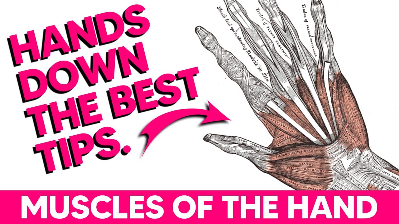


5.0 / 5 (0 votes)
