Digital Radiography: Image Post Processing: PACS System and Quality Control-Assurance
Summary
TLDRThis video explores the significant impact of advances in computer technology on radiology, covering key concepts like digital image processing, post-processing activities, and imaging protocols. It highlights the use of DICOM, PACS, and HL7 systems in healthcare communication, quality control, and assurance procedures. The video also discusses techniques such as smoothing, edge enhancement, image stitching, and the role of digital subtraction in enhancing image quality. Additionally, it emphasizes the importance of following institutional protocols for radiographic imaging while adhering to ALARA standards.
Takeaways
- 💡 Advances in computer technology have significantly impacted healthcare, particularly in the field of radiology.
- 📊 The presentation aims to familiarize participants with 11 key objectives related to radiology and digital imaging.
- 🖼️ Post-processing activities like smoothing, edge enhancement, image stitching, and electronic masking are crucial for image optimization.
- 🆚 The difference between HIS (Hospital Information System) and RIS (Radiology Information System) is essential for understanding workflow in radiology.
- 💻 DICOM (Digital Imaging and Communications in Medicine) is a standard for medical imaging that facilitates the exchange, viewing, and storing of images.
- 🗂️ PACS (Picture Archiving and Communication System) is vital for managing and accessing medical images digitally within a healthcare setting.
- 🚨 Safety and emergency procedures specific to imaging and communications are necessary for maintaining patient and staff safety.
- 🔑 HL7 is a standard for electronic health record communication that plays a significant role in healthcare communication systems.
- 🔄 The discussion of short-term and long-term image archival highlights the importance of data management in radiology.
- 🔍 Quality control (QC) and quality assurance (QA) procedures are differentiated, with a focus on their roles in maintaining imaging standards.
- 🖥️ Digital imaging systems offer a greater dynamic range than film-screen systems, allowing for more detailed and adjustable images.
Q & A
How have advances in computer technology impacted radiology?
-Advances in computer technology have significantly transformed radiology by enabling digital imaging systems that offer greater dynamic range, allowing for more shades of gray to be captured, and providing capabilities for image manipulation to enhance anatomic structures, suppress noise, and adjust brightness and contrast.
What are the 11 objectives that participants will understand after the presentation?
-The 11 objectives include understanding the purpose of common post-processing activities, differentiating between HIS and RIS, discussing the use of DICOM, describing PACS components and functions, discussing safety and emergency procedures, defining HL7 and its significance, describing short-term and long-term image archival, differentiating between quality control and quality assurance procedures, discussing quality assurance procedures in digital imaging, and discussing imaging artifacts in computed radiography and digital radiography.
What is the purpose of post-processing activities such as smoothing, edge enhancement, image stitching, equalization, and electronic masking?
-Post-processing activities are used to manipulate digital image data to enhance anatomic structures, suppress noise, adjust brightness and contrast, and improve the overall quality and aesthetic appeal of the images for better interpretation by radiologists.
What is the difference between HIS and RIS in the context of radiology?
-HIS stands for Hospital Information System, which manages patient data and administrative tasks across a hospital, while RIS stands for Radiology Information System, which specifically handles the workflow, data, and management related to radiology within a healthcare setting.
What is DICOM and why is it important in radiology?
-DICOM (Digital Imaging and Communications in Medicine) is a standard protocol for the transmission and sharing of medical images and related information. It is important because it ensures interoperability between different medical imaging devices and systems, facilitating the sharing of images and reports across various healthcare providers.
Can you describe the components and functions of a Picture Archiving and Communication System (PACS)?
-A PACS is a system designed to store, retrieve, manage, and distribute medical images and reports. Its components include image acquisition devices, image storage devices, network systems, workstations, and software applications. PACS functions to improve workflow, reduce physical storage needs, and enhance accessibility of images for healthcare professionals.
What does HL7 stand for and what is its significance in healthcare communication systems?
-HL7 stands for Health Level Seven International, a not-for-profit organization that develops standards for the exchange, integration, sharing, and retrieval of electronic health information. Its significance lies in promoting interoperability and data exchange among different healthcare software systems, improving the efficiency and quality of patient care.
What are the differences between quality control (QC) and quality assurance (QA) procedures in radiology?
-Quality control (QC) refers to the specific tests and calibrations performed to ensure that equipment and processes are functioning correctly. Quality assurance (QA) is a broader concept that includes QC procedures as well as the overall management system to ensure consistent quality in the delivery of radiological services.
How does image shuttering enhance the appearance of radiographic images?
-Image shuttering places a black border around the original collimated edges of the image, eliminating any white areas seen around a collimated image, making the displayed image more aesthetically pleasing to the viewer without replacing the need for proper pre-exposure collimation.
What is the role of edge enhancement in digital imaging?
-Edge enhancement is a software function that uses an algorithm to increase the contrast at edges in an image by averaging the signal strength of adjacent pixels. This process results in images with increased contrast, making it easier to visualize anatomic structures.
How does image stitching work in radiology?
-Image stitching is a software function that allows radiographers to join separate, processed images into a single image format. This is particularly useful for studies like scoliosis and limb length assessments that require multiple exposures on separate image receptors.
Outlines

هذا القسم متوفر فقط للمشتركين. يرجى الترقية للوصول إلى هذه الميزة.
قم بالترقية الآنMindmap

هذا القسم متوفر فقط للمشتركين. يرجى الترقية للوصول إلى هذه الميزة.
قم بالترقية الآنKeywords

هذا القسم متوفر فقط للمشتركين. يرجى الترقية للوصول إلى هذه الميزة.
قم بالترقية الآنHighlights

هذا القسم متوفر فقط للمشتركين. يرجى الترقية للوصول إلى هذه الميزة.
قم بالترقية الآنTranscripts

هذا القسم متوفر فقط للمشتركين. يرجى الترقية للوصول إلى هذه الميزة.
قم بالترقية الآنتصفح المزيد من مقاطع الفيديو ذات الصلة
PACS Systems and Quality Control Part 1 - Image Post Processing
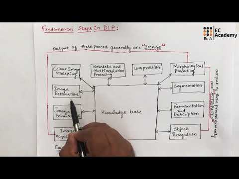
DIP#3 Fundamental steps in Digital image processing || EC Academy

Pertemuan 2 : Citra Digital, Sampling, dan Quantization - Part 1 : Apa itu citra digital ?

What is Image Processing? | Career Opportunities of Image Processing in 2020.

Pertemuan 1 - Digital Imaging - Raden Daniel Wisnu Wardhana

Pertemuan 2 : Citra Digital, Sampling, dan Quantization - Part 2 : Sejarah citra digital dan PCD
5.0 / 5 (0 votes)
