High Yield IM CARDIOVASCULAR Review for Step 2 CK & Shelf Exam
Summary
TLDRThis script offers an in-depth look at diagnosing and treating cardiovascular diseases, focusing on stable and unstable angina, stress tests, and pharmacological interventions. It covers various diagnostic methods like EKG, echo, and nuclear perfusion studies, and treatments including nitrates, aspirin, and beta blockers. The script also addresses acute coronary syndrome, differentiating unstable angina from STEMI, and outlines treatments for each. Additionally, it touches on Dressler syndrome, restrictive cardiomyopathy, and the management of hypertensive emergencies and peripheral vascular disease.
Takeaways
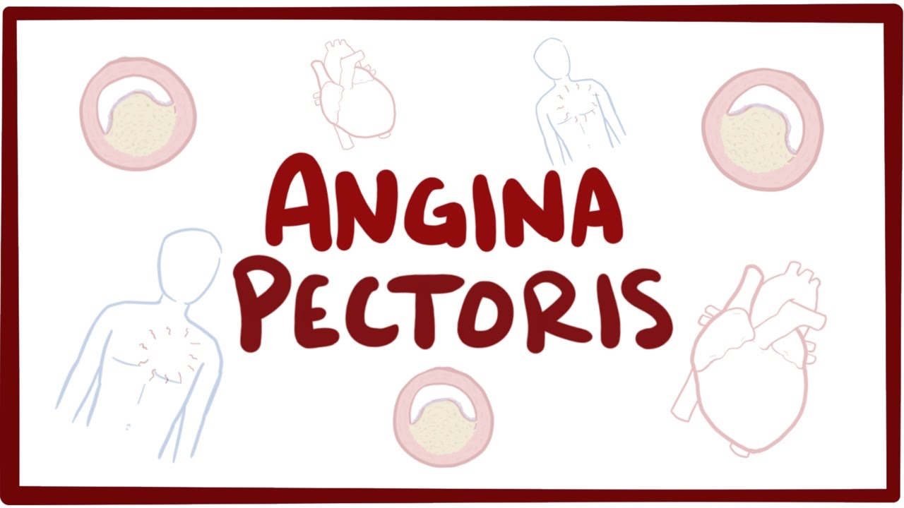
- 🏥 Stable angina is characterized by chest pain during exertion that improves with rest, and is assessed with stress tests like EKG, echo, or nuclear perfusion study.
- 💓 A positive stress test indicates ST depression, hypotension, pain, abnormal wall motion, or decreased nuclear isotope uptake.
- 🚑 Pharmacologic stress tests are used for those unable to exercise, utilizing drugs like adenosine to induce cardiac stress.
- 🩺 Angiography is the definitive diagnostic method for coronary artery disease but is invasive, hence stress tests are conducted first.
- 💊 First-line treatments for stable angina include nitrates, aspirin, and beta blockers, while unstable angina is treated with a broader regimen including morphine and heparin.
- 🆘 Acute coronary syndrome encompasses unstable angina, NSTEMI, and STEMI, and is initially assessed with EKG and cardiac enzymes.
- 🩹 In STEMI, cardiac enzymes aren't necessary for diagnosis if there's ST elevation or new left bundle branch block with chest pain.
- 🩩 Unstable angina is differentiated from stable by rest pain and worsening symptoms, without troponin elevations.
- 🌡️ Stress tests are indicated for chest pain assessment, with exercise tests preferred unless contraindicated by abnormal EKG or physical limitations.
- 🛑 Cardiac tamponade presents with Beck's triad (hypotension, JVD, muffled heart sounds) and is treated by draining the pericardial fluid.
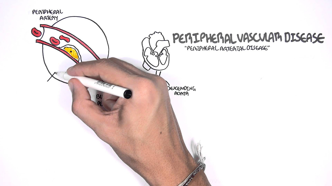
- 🩸 Peripheral artery disease is diagnosed with ankle-brachial index, with treatment ranging from exercise to interventional procedures based on severity.
Q & A
What is stable angina and how is it typically managed?
-Stable angina is a condition where substernal chest pain occurs with exercise or exertion and is relieved by rest. It is managed by stress tests such as EKG, echo, or nuclear perfusion study, and pharmacological stress tests if the patient is unable to exercise.
What are the three types of stress tests mentioned in the script?
-The three types of stress tests mentioned are EKG stress test, echo stress test, and nuclear perfusion study.
How is a positive stress test indicated in the script?
-A positive stress test is indicated by ST depression, hypotension, pain, abnormal wall motion in an echo, or decreased uptake of nuclear isotope in a nuclear perfusion study.
What is the first-line treatment for stable angina according to the script?
-The first-line treatment for stable angina is nitrates, aspirin, and beta blockers.
What is the difference between stable and unstable angina as described in the script?
-Stable angina occurs with exertion and is relieved by rest, while unstable angina is worsening, evolving, or occurs at rest without troponin elevations. Unstable angina with troponin elevations is considered NSTEMI.
What are the three types of acute coronary syndrome mentioned in the script?
-The three types of acute coronary syndrome mentioned are unstable angina, STEMI, and NSTEMI.
How is STEMI diagnosed in the script?
-STEMI is diagnosed with one millimeter ST elevations in two continuous leads or a new left bundle branch block with chest pain on an EKG.
What is the treatment for Dressler syndrome as per the script?
-Dressler syndrome is treated with aspirin, which is an NSAID used specifically for this autoimmune pericarditis that occurs after an MI.
What are the conditions associated with restrictive cardiomyopathy according to the script?
-The conditions associated with restrictive cardiomyopathy are hemochromatosis, amyloidosis, and sarcoidosis, which cause deposits in the myocardium leading to diastolic heart failure.
What is the first-line treatment for hypertensive emergency as mentioned in the script?
-The first-line treatments for hypertensive emergency are IV hydralazine, nitroprusside, or labetalol, with the necessity of end organ damage for it to be considered an emergency.
How is aortic dissection differentiated into Type A and Type B in the script?
-Aortic dissection is differentiated into Type A, which is anything proximal to the left subclavian, and Type B, which is distal to the left subclavian. Type A requires immediate surgery, while Type B is treated with beta blockers.
Outlines
🩺 Cardiology Overview: Stable Angina and Diagnostic Testing
This paragraph discusses stable angina, characterized by chest pain during exertion that subsides with rest. It emphasizes the importance of stress tests, which include EKG, echo, and nuclear perfusion studies. A positive stress test may show ST depression, hypotension, or pain. The necessity of a pharmacologic stress test for those unable to exercise is highlighted, using drugs like adenosine or dipyridamole. The paragraph also touches on the first-line treatments for stable and unstable angina, the latter being a more severe condition with pain at rest and no troponin elevations. The text transitions into the diagnosis of acute coronary syndrome, focusing on EKG and cardiac enzymes, and differentiates between unstable angina and NSTEMI based on troponin levels. It concludes with the importance of ruling out STEMI, which is diagnosed with specific EKG changes and chest pain.
🏥 Advanced Cardiac Conditions and Treatments
The second paragraph delves into the management of various cardiac conditions. It starts with the use of the TIMI score to determine the urgency of a stress test or a trip to the cath lab for patients with chest pain. The paragraph then explores specific conditions like Prinzmetal's angina, which involves coronary vasospasm, and the treatment of inferior wall MI, where nitrates are contraindicated due to right ventricular issues. It also discusses the use of drugs like dobutamine and atropine in cardiogenic shock and the importance of certain drugs in reducing mortality in MI. The paragraph further covers Dressler's syndrome, restrictive cardiomyopathy, and the treatment of different types of heart block. It ends with a discussion on CHF, highlighting the use of loop diuretics and the difference in treatment between supraventricular and ventricular tachycardia.
🩹 Cardiac Emergencies and Their Management
This section focuses on the diagnosis and treatment of acute cardiac conditions. It begins with the identification of cardiac tamponade through Beck's triad and pulsus paradoxus, and the use of imaging to detect pericardial fluid. The text then moves on to discuss mitral stenosis, its causes, and the diagnostic value of cardiac catheterization and echocardiograms. It also covers the complications of hypertension and aortic stenosis, including angina and syncope. The paragraph further addresses tricuspid valve issues related to IV drug use and carcinoid syndrome, and the diagnosis of infective endocarditis through fever, leukocytosis, and new murmurs. It concludes with the treatment of hypertensive emergencies with IV drugs and the differentiation between type A and type B aortic dissections, their symptoms, and appropriate treatments.
🚑 Peripheral Vascular Disease and Its Clinical Presentations
The fourth paragraph discusses peripheral vascular disease, emphasizing the importance of the ankle-brachial index for diagnosis. It describes the symptoms of claudication and the treatment options ranging from exercise to interventions like stenting or bypass surgery. The text also covers acute limb ischemia, its symptoms, and the use of heparin or embolectomy in treatment. It mentions LaRouche syndrome, a variation of peripheral vascular disease, and the use of IVC filters in cases where anticoagulants are contraindicated. The paragraph concludes with a discussion on the diagnosis and treatment of pulmonary embolism, highlighting the importance of heparin and CT angiography. It also touches on the signs of venous insufficiency and the differentiation from other conditions like CHF and cardiogenic shock.
🏋️♂️ Exercise and Interventional Treatments for Vascular Conditions
The final paragraph focuses on the role of exercise in managing peripheral vascular disease, especially in the early stages. It discusses the progression to more severe disease and the need for interventions like stenting or bypass surgery when patients experience pain at rest. The text also covers acute limb ischemia and its management with heparin and embolectomy. Additionally, it mentions LaRouche syndrome, its symptoms, and the importance of IVC filters in specific cases. The paragraph concludes with a discussion on the diagnosis and treatment of pulmonary embolism, emphasizing the use of heparin and CT angiography. It also addresses the signs of venous insufficiency and the differentiation from other conditions like CHF and cardiogenic shock.
Mindmap
Keywords
💡Stable Angina
💡Stress Test
💡EKG (Electrocardiogram)
💡Pharmacologic Stress Test
💡Angiography
💡Unstable Angina
💡Acute Coronary Syndrome
💡Cardiogenic Shock
💡Restrictive Cardiomyopathy
💡Congestive Heart Failure (CHF)
💡Supraventricular Tachycardia (SVT)
Highlights
Stable angina is indicated by substernal chest pain that occurs with exercise and is relieved by rest.
Three types of stress tests are available: EKG, echo, and nuclear perfusion study.
A positive stress test shows ST depression, hypotension, pain, or abnormal wall motion.
Pharmacologic stress tests are used when patients are unable to exercise.
Angiography is the definitive method to diagnose coronary artery disease, but it is invasive.
First-line treatment for stable angina includes nitrates, aspirin, and beta blockers.
Unstable angina is characterized by worsening chest pain at rest without troponin elevation.
STEMI is diagnosed with ST elevation on EKG and is a type of acute coronary syndrome.
Treatment for STEMI includes immediate cath lab intervention without waiting for cardiac enzymes.
Unstable angina treatment includes nitroglycerin, morphine, oxygen, aspirin, clopidogrel, beta blockers, ACE inhibitors, statins, and heparin.
For chest pain, an EKG is the first diagnostic step to rule out STEMI.
If no ST elevations are present, serial troponin and EKGs are used to diagnose unstable angina or NSTEMI.
Indications for cath lab include three-vessel disease, proximal left anterior descending disease, Prinzmetal angina, and inferior MI.
Treatment for inferior wall MI may include fluids instead of nitrates to avoid exacerbating hypotension.
Only three drugs have been shown to decrease mortality in MI: aspirin, beta blockers, and ACE inhibitors.
Dressler syndrome is an autoimmune pericarditis treated with aspirin, distinct from viral pericarditis.
Restrictive cardiomyopathy is associated with conditions like hemochromatosis, amyloidosis, and sarcoidosis.
Treatment for CHF includes loop diuretics, inotropes, and positioning, with loop diuretics being the first line.
Supraventricular tachycardia is treated with adenosine if stable, and with cardioversion if unstable.
Ventricular tachycardia is treated with amiodarone and cardioversion if unstable.
Cardiac tamponade presents with Beck's triad and is associated with pulsus paradoxus and electric alternans.
Mitral stenosis is often caused by previous rheumatic fever and can lead to left ventricular hypertrophy.
Aortic stenosis can be diagnosed with cardiac catheterization or echocardiogram and may require valve replacement.
Peripheral artery disease is diagnosed with the ankle-brachial index and is treated with exercise or interventions like stenting.
Acute limb ischemia in peripheral artery disease is treated with heparin or embolectomy.
Suspected PE is treated with heparin before diagnostic imaging with CT angio.
Transcripts
[Music]
so we're gonna start cardiovascular
disease so people who have stable angina
which means that they have substernal
chest pain that occurs with exercise or
exertion and is alleviated by rest then
this is a sign of stable angina because
it improves with rest people with this
you the next thing you want to do is a
stress test right and there's three
types of stress tests and EKG an echo or
a nuclear perfusion study so you would
do an echo if that or a nuclear
perfusion study if that person is uh has
any abnormalities on EKG so the actual
so a positive stress test would be
anything that shows ST depression or
hypotension or pain and then you know
the echo you might see abnormal wall
motion that would be a positive stress
test or and then nuclear perfusion
studies would show decreased uptake of
nuclear isotope and that'd be a positive
stress test as well so and then remember
if they are unable to exercise then
that's when you do a pharmacologic
stress test so there's two ways to
induce stress on the heart either
exercise or using drugs such as
adenosine or die period them all so most
of the time the correct answer will be
as exercise stress test using EKG if
they have an abnormal EKG which will
mask the results of that stress EKG then
you either do an echo or nuclear
perfusion studies if they can exercise
then they will exercise on the treadmill
if they can't exercise as an they are
wheelchair-bound
or have osteoporosis or some other thing
that is a contraindication to exercise
then that's when you do a pharmacologic
test
test and with the pharmacologic test it
can be observed either on EKG echo or
nuclear perfusion study as well so and
then remember that the definitive way to
actually diagnose coronary artery
disease is through angiography so the
reason why you do these stress tests
first is because angiography is very
invasive so that's why you do the others
first so um the first thing you want to
do first test you want to do with chest
pain as EKG the first-line treatment for
stable angina is nitrates aspirin and
beta blockers first-line treatment for
unstable angina is lemonis II - mnemonic
which is morphine oxygen nitrates
aspirin clopidogrel beta blockers ACE
inhibitors statin and heparin and
remember that the first thing you want
to give is aspirin and remember if the
person has unstable angina which means
if definition of unstable angina means
that their angina is worsening or
evolving or occurs at rest now which is
different than stable angina but
unstable angina also has no troponin
elevations if you have unstable angina
with troponin elevations and so
basically unstable angina becomes NSTEMI
as soon as there are any troponin if
there are opponents with st elevations
then this is what we call a STEMI what
alright so when someone comes in with
chest pain the first thing you want to
do is rule out acute coronary syndrome
acute coronary syndrome is unstable
angina and STEMI or STEMI so those are
the three types of acute coronary
syndrome so when someone comes in with
chest pain you want to roll that out so
we first you do an EKG and cardiac
enzymes but the thing is cardiac enzymes
can take a while to come
so first thing you want to do is what
the EKG is luck to see if this is STEMI
or not so it fits
dami if it's STEMI then to diagnose
Tammy you don't even need cardiac
enzymes all you need is one millimeter
st elevations and two continuous leads
or a new left bundle branch block with
chest pain and that's considered STEMI
you don't even need the enzymes if you
see those they go straight to cath lab
and then see you do the EKG but there
are no st elevations but they do have
the characteristic chest pain that they
were describing then you want to do
serial troponin and serial EKGs to see
if this is evolving or changing so then
it's either going to be unstable angina
or n STEMI if the troponin is come back
updated with elevated troponin then
that's now called an N STEMI as a non ST
elevation mi
if there are no troponin yet and after
serial troponin measurements and it
stays low then this is called unstable
angina and remember it the conditions
also have to be satisfied where the
chest pain has been evolving recently
and been getting worse and this person
has been having chest pain at rest this
is called unstable angina if they have
unstable angina or and STEMI then you
want to apply the Tammy's core if it's 0
to 2 this person will get a stress test
if it's 3 or more than this person will
go to cath lab anyone who has chest pain
who has has unstable vitals as well that
you suspect my they also go straight to
cath lab so those are some exceptions
main indications for a cabbage are three
vessel disease or proximal left anterior
descending disease with 70% plus
stenosis
next is prinzmetal angina which is
basically coronary vasospasm
so the angiography will show this so
spasm when given organ a vine or
Sedo : and you'll also see st-elevation
on EKG during these painful episodes and
you want to treat this with calcium
channel blockers or nitrates you only do
TPA and MMI if there's no access to PCI
Center inferior mi when you have an ro
cardial infarction of the inferior wall
which is 2/3 and a VF and this is the
only mi that has an exception where you
don't want to give nitrates because
because they have a right ventricular mi
it's already the heart is already having
problems pumping blood to the left side
of the heart so if you give nitrates
this will exacerbate the hypotension
so actually in an inferior wall mi you
actually want to give fluids sometimes
an inferior wall mi can because because
the right coronary artery supplies blood
to the SA node this can cause and then
that sinus bradycardia can cause
cardiogenic shock and usually first when
for cardiogenic shock is dobutamine
which is the beta 1 agonist but in the
case of inferior wall mi that has
bradycardia and cardiogenic shock this
is due to injury of the SA node so in
this special case you want to give
atropine remember that there's only
three drugs shown to decrease mortality
in MI and this is very high yield is
aspirin beta blockers and ACE inhibitors
and nitrates work in two ways but the
predominant way it works by for my eyes
is that it decreases preload it's a V no
die later and that decreases stress on
the myocardium due to excess blood so
when you minimize the preload there's
less stress on the heart muscle and also
it's secondary effect as it dilates the
coronary arteries so treatment of
first-degree and second-degree heart
block mobitz one is no treatment but mo
it's two and the complete heart block
you want to treat with pacemaker
Dressler syndrome is an autoimmune
pericarditis that happens two weeks
later after an mi host MI two weeks with
fever and symptoms of pericarditis with
leukocytosis you want to treat with
aspirin this is contrasted with other
causes of pericarditis such as viral
pericarditis those will be treated with
NSAIDs
this Dressler syndrome is specifically
treated with aspirin and then you have
restrictive cardiomyopathy I just
remember the o C's so hemochromatosis
amyloidosis sarcoidosis this creates a
diastolic heart failure with reduced
ejection fraction this is due to
deposits in the myocardium
so like amyloid deposits or granulomas
or iron deposits in the myocardium what
I'm trying to say is remember that humor
chrome mitosis amyloidosis and
sarcoidosis
or if associated with restrictive
cardiomyopathy and then remember
hemochromatosis is bronze diabetes and
iron overload so they'll have diabetes
bronze gaming don't have elevated liver
enzymes amyloidosis is think of like
protein deposits you're gonna have
deposits in the heart and the kidney and
in the joints and in the kidney you'll
see proteinuria versus sarcoidosis is
where you'll see heart and lung stuff so
bilateral hilar adenopathy a dry cough
uveitis erythema nodosum and also
restrictive cardiomyopathy there are
three CHF drugs shown to decrease
mortality and that's ace inhibitors beta
blockers and spironolactone which is a
potassium sparing their diuretic which
should be contrasted with the three
drugs that decrease mortality and mi
which is ace inhibitors as well and beta
blockers as well but the third is
aspirin remember met four
which is first-line treatment for type 2
diabetes remember its contraindications
which is it's contraindicated in renal
disease and CHF because it can cause
metabolic acidosis remember for CHF
acute decompensation of CHF which means
the heart failure is getting worse then
you want to treat it with them mnemonic
no lit nitrates oxygen loop diuretics
inotropes
and positioning such as elevating the
head of the bed but the first thing you
want to treat what is a loop diuretics
such as furiosa might you need to know
supraventricular tachycardia versus
ventricular tachycardia so a
supraventricular tachycardia will have
narrow QRS s you know it'll look like
QRS TQ r st qrst
and if they're stable you treat with
adenosine and if they're unstable then
you want to treat with cardioversion and
then ventricular tachycardia which has
wide bizarre qrs complexes after one
after another then you want to treat
with amiodarone and if they're unstable
then you want to treat with
cardioversion versus v-fib and pulseless
v-tach
first-line treatment for that is
different relation verses asystole and
pulseless electrical activity pulseless
electrical activity means that the EKG
shows any rhythm but when you feel for
the pulse there's no pulse that means P
e a and then to treat with that is CPR
and by the way remember that the
first-line treatment for a super
ventricular tachycardia before you
progress with an adenosine is vagal
maneuvers such as carotid massage so
torsades de pointes can lead to v-fib
and this is treated with IV magnesium
which stabilizes the cardiac membranes X
is constrictive pericarditis which is
idiopathic fibrous scarring replacing
the entire
pericardial space the key here I want
you to look for is when they do imaging
like a chest x-ray of the heart you'll
see calcifications calcifications is key
and it's usually caused by TB or lupus
and it can present similarly to
restrictive cardiomyopathy it can have
equal diastolic pressures and all
chambers and it can also have by atrial
enlargement and treatment is peri
cardiac t'me acute pericarditis causes
the main causes coxsackievirus and you
treat it with the NSAID versus Jess lair
which is treated with aspirin aspirin is
a type of NSAID but remember Dressler's
aspirin and on EKG you'll see diffuse St
elevations and it's improved with
leaning forward so cardiac tamponade is
just remember Beck's triad which is
hypotension jvd and muffled heart sounds
it's also associated with pulsus
paradoxus which means when you inspire
this increases filling to the right
ventricle which causes the
interventricular septum to bow over to
the left side which decreases the left
ventricular preload and because of this
the stroke volume is decreased and
because of this the systolic pressure
will drop by greater than 10 and that's
called pulsus paradoxus which means a
systolic pressure dropping by greater
than 10
upon inspiration you will see that in
cardiac tamponade it's also associated
with electric alternates which means the
QRS voltages kind of the amplitude kind
of becomes alternating between big and
small big and small big and small and
that's because the heart is literally
swinging within the pericardial fluid
which distorts the qrs measurements and
then you'll also see low-voltage QRS and
a KU small sign coos
sign which means when you inhale that
the jugular venous distention increases
because with cardiac tamponade shelling
of the right side of the heart is more
difficult because it's not as compliant
so then the venous blood tends to
overflow faster remember mitral stenosis
the majority of the causes of mitral
stenosis our previous episode of acute
rheumatic fever or rheumatic heart
disease hypertension or aortic stenosis
can over time lead to left ventricular
hypertrophy and if this is prolonged
this can become dilated cardiomyopathy
and people with hypertension or aortic
stenosis tend to get angina because of
decreased perfusion to the coronary
arteries another complication as syncope
due to decreased perfusion of the brain
another complication is left ventricular
hypertrophy because of increased
afterload another complication is
dilated cardiomyopathy from chronicity
and then you'll hear a soft s2 because
the valve doesn't move well and then
definitive diagnosis for aortic stenosis
is cardiac catheter to measure the valve
area what an echocardiogram can also
measure the valve diameter if it's less
than one square centimeter or if they
have any symptoms at all such as an
angina syncope or CHF then you want to
treat with valve replacement the
tricuspid valve remember that it's
associated with IV drug use and
carcinoid syndrome carcinoid syndrome is
a tumor that produces too much serotonin
and that creates bronchospasm flushing
diarrhea and right-sided heart murmurs
three causes of holosystolic murmur or
mitral regurg tricuspid regurge and VSD
so if someone has infective endocarditis
the easiest way to diagnose this is
one who has a fever with leukocytosis
and new onset of murmur and you don't
know the bugs it before you find out
from your blood culture which is the
first thing you want to do you treat it
empirically with vancomycin and an
aminoglycoside hypertensive emergency is
defined as 180 over 120 and the
first-line treatments for hypertensive
emergency is IV hydralazine
nitroprusside or labetalol and remember
that for it to be considered an
emergency there has to be evidence of
end organ damage
so encephalopathy or acute kidney injury
or liver injury where versus
hypertensive urgency is high blood
pressure over 180 over 120 but no end
organ damage so the difference is if
it's an emergency you treat IV but if
it's urgency you treat with oral
medications subarachnoid hemorrhage the
Thunder Clap headache worst headache of
your life first thing you want to do is
a CT head without contrast and if that's
negative and you still suspect oh so
brackenreid hemorrhage the next step is
lumbar puncture and you're gonna look
for positive xantho chromia which is the
presence of bilirubin in the CSF a or
DIC dissection is substernal chest pain
that is described as tearing and
radiates to the back and you have two
types type A and type B type B is
anything just fill to the left
subclavian and type a is anything
proximal to that and you treat them
differently a goes to surgery right away
and B you give beta blockers to treat it
and you diagnose an aortic dissection
with a CT angio or a transesophageal
echo and remember any type of CT imaging
make sure to always check the patient's
kidneys because anyone with kidney
disease it's contraindicated to you see
see with contrast which is the majority
of CT imaging next is peripheral
vascular disease or peripheral artery
disease and so the number one risk
factor is smoking and to diagnose it you
want to do something called the ankle
brachial index which is measuring the
differences in blood pressures from the
ankle and the arm and if the ratio in
the ankle to arm is less than 0.9 then
that's disease and if it's less than 0.4
then this is severe disease which will
most likely have pain at rest as well
and people with peripheral artery
disease will describe themselves as
having claudication and their legs while
walking so they'll walk a certain
distance and then feel pain in their
legs and then it improves with rest it's
sort of like stable angina of the legs
due to stenosis of the ephemeral or
popliteal arteries the ones that are
current rest would be synonymous to like
unstable angina and then sometimes they
can make clots which is called acute
limb ischaemia which would be synonymous
to like an MI so if someone has a ratio
between 0.4 to 0.9 which would be like
stable angina this is the initial stages
of peripheral vascular disease the
first-line treatment is an exercise
program
if it starts if they start to have
problems at rest and their ratio is
below point 4 now you have to do an
intervention such as a stand or a bypass
if if they have acute limb ischemia
which is due to some sort of thrombosis
that cuts off the circulation in the
legs where everything just still did
that starts getting cold and pulseless
and in a lot of pain then you want to
treat that with heparin or an
embolectomy
sometimes there's a variation of
peripheral vascular disease known as
LaRouche syndrome which is caused by
atherosclerosis proximal to the aortic
bifurcation if
or they become the iliac arteries and
this person will complain a bilateral
leg pain as well as the key here is
impotence and buttock pain and this is
us like a sub-type a variation of the
same thing
remember that IVC filters are placed if
contraindicated to heparin or warfarin
or if they've failed previous therapy
with heparin or warfarin if you suspect
a PE in a patient which is basically
acute sudden onset of tachypnea
tachycardia and hypoxemia the first
thing you want to do is give heparin
before you even do the CT angio so
heparin and then CT angio if you had to
pick what is the best next step and they
both of those are the options pick
heparin first and then low molecular
weight heparins remember they're
contraindicated and renal disease
someone who has venous insufficiency
looked for the medial malleolus ulster
which is a sign of venous insufficiency
which can be contrasted to other similar
presentations such as CHF cardiogenic
shock first-line treatment is the ina
trope such as dobutamine
septic shock first-line treatment is IV
antibiotics plus IV fluids and
potentially vasopressors neurogenic
shock remember everything is down
cardiac output is down heart rate is
down total peripheral resistance is down
wedge pressure is down and the jvd is
down and you treat this with IV fluids
تصفح المزيد من مقاطع الفيديو ذات الصلة
How Do Nitrates Work? (Nitroglycerin)
Myocardial Infarction[Heart Attack]; Causes, Pathogenesis, Signs & Symptoms, Diagnosis & Treatment
How To Treat Heart Failure (High-Yield Guide)
Coronary Artery Disease (CAD) Anatomy, Nursing, Heart Disease, Pathophysiology, Treatment Part 1
About Your Heart Attack | Nucleus Health
CCS 2
5.0 / 5 (0 votes)