Anatomy and physiology of the respiratory system
Summary
TLDRThis script explores the intricate process of respiration, detailing the journey of air from the nostrils through the nasal cavity, sinuses, and pharynx, to the lungs. It explains the roles of the diaphragm, chest muscles, and various airway structures in facilitating gas exchange. The script also delves into the function of the mucociliary escalator, the importance of alveoli, and the blood-gas barrier, highlighting the body's remarkable ability to oxygenate blood and expel carbon dioxide.
Takeaways
- 💨 The primary function of the lungs is to facilitate gas exchange, which involves taking in oxygen and expelling carbon dioxide.
- 🏗️ The diaphragm and chest muscles play a crucial role in the breathing process, contracting to inhale and relaxing to exhale.
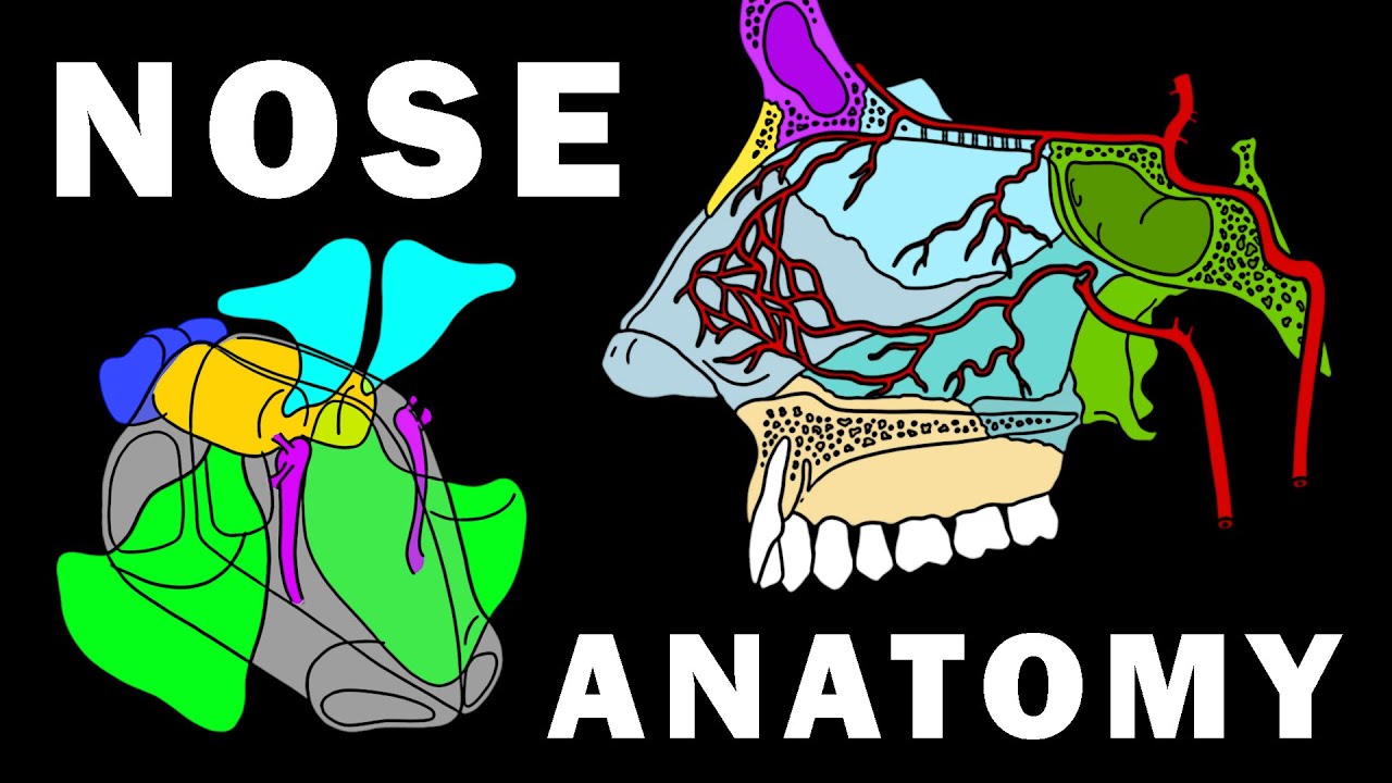
- 👃 The nasal cavity is lined with mucus-producing cells that help trap bacteria and other particles, while nose hairs filter out larger particles.
- 🕳️ The paranasal sinuses warm and moisten the air, and also contribute to voice resonance, becoming noticeable when congested.
- 🔄 The soft palate and uvula act as a valve to prevent food from entering the nasopharynx during swallowing.
- 🔒 The epiglottis seals off the airway during swallowing, ensuring food goes down the esophagus and not into the larynx.
- 🌪️ The trachea, or windpipe, splits into two bronchi, with the right bronchus being wider and more vertical, making it a common entry point for inhaled foreign objects.
- 🌀 The bronchi continue to divide into smaller airways supported by cartilage rings and lined with ciliated cells and goblet cells, which help move mucus and trapped particles out of the lungs.
- 🚶♂️ The autonomic nervous system, with its sympathetic and parasympathetic nerves, regulates the diameter of the airways, affecting breathing during different activities.
- 🌬️ Bronchioles are smaller airways that do not require cartilage for support and are lined with various cell types, including club cells that protect and regenerate the bronchiolar epithelium.
- 🌀 The alveoli are tiny air sacs where gas exchange occurs, surrounded by a thin barrier that allows for the diffusion of oxygen into the blood and carbon dioxide out of the blood.
Q & A
What is the primary function of the lungs?
-The primary function of the lungs is gas exchange, which involves pulling oxygen into the body and expelling carbon dioxide.
How do the diaphragm and chest muscles contribute to the breathing process?
-During inhalation, the diaphragm contracts to pull downward and the chest muscles contract to open the chest, creating a vacuum that sucks in air. During exhalation, these muscles relax, allowing the lungs to spring back and push the air out.
What is the role of mucus in the nasal cavity?
-Mucus in the nasal cavity is salty, sticky, and contains lysozymes, enzymes that help kill bacteria. It helps trap particles and pathogens, providing a first line of defense against inhaled substances.
What are the paranasal sinuses and how do they assist in the breathing process?
-The paranasal sinuses are air-filled spaces inside the bones surrounding the nose. They help circulate inspired air, allowing it to become warm and moist, and also act as echo chambers to amplify the sound of the voice.
How does the epiglottis prevent food from entering the trachea during swallowing?
-The epiglottis is a spoon-shaped flap of cartilage that acts as a lid, sealing off the airway when you're eating, ensuring that food only goes down the esophagus towards the stomach.
What is the significance of the carina in the respiratory system?
-The carina is the point where the trachea splits into the two mainstem bronchi. It is an important landmark in the respiratory system, marking the division into the bronchi that lead to the lungs.
Why is the right mainstem bronchus more prone to foreign object obstruction compared to the left?
-The right mainstem bronchus is wider and more vertical than the left, making it more likely for inhaled foreign objects, like a peanut, to enter and lodge in the right lung rather than the left.
What is the function of the autonomic nervous system in the airways?
-The autonomic nervous system, composed of sympathetic and parasympathetic nerves, regulates the diameter of the airways. Sympathetic nerves increase airway diameter during 'fight or flight' situations, while parasympathetic nerves decrease it during 'rest and digest' modes.
Can you explain the mucociliary escalator and its role in the respiratory system?
-The mucociliary escalator is a mechanism where mucus secreted by goblet cells traps particles, and ciliated columnar cells beat rhythmically to move the mucus and trapped particles towards the pharynx for expulsion or swallowing.
What are the functions of club cells in the bronchioles?
-Club cells secrete glycosaminoglycans, a material that protects the bronchiolar epithelium. They can also transform into ciliated columnar cells to regenerate and replace damaged cells in the bronchioles.
How does the respiratory system facilitate the exchange of oxygen and carbon dioxide?
-The respiratory system facilitates gas exchange through a series of airways ending in alveoli, where oxygen diffuses into the blood across the blood-gas barrier and carbon dioxide diffuses out from the blood into the alveoli to be exhaled.
Outlines
🌬️ The Mechanics of Breathing and Airflow in the Respiratory System
This paragraph details the primary function of the lungs—gas exchange—and describes the process of breathing, involving the diaphragm and chest muscles. It explains how air travels through the nasal cavity, where mucus and nose hairs filter out large particles and bacteria. The paragraph also covers the roles of the paranasal sinuses, pharynx, larynx, and epiglottis in directing air and food, and it explains the structure and function of the trachea and bronchi as air continues into the lungs. Key concepts include the different lobes of the lungs, the importance of smooth muscle and autonomic nervous system control in the airways, and the role of the mucociliary escalator in keeping the respiratory system clean.
🔬 The Role of Bronchioles and Alveoli in the Respiratory System
This paragraph focuses on the deeper structures of the respiratory system, specifically the bronchioles and alveoli. It describes how air travels through increasingly smaller bronchioles, highlighting the role of club cells in protecting and regenerating the bronchiolar epithelium. The passage details the transition from conducting bronchioles to respiratory bronchioles, where gas exchange occurs in the alveoli. It explains the importance of type I and type II pneumocytes, the production of surfactant to keep alveoli open, and the function of alveolar macrophages in removing particles. The paragraph concludes by summarizing the process of oxygen entering the blood and carbon dioxide being expelled, emphasizing the efficiency of the blood-gas barrier.
Mindmap
Keywords
💡Gas Exchange
💡Diaphragm
💡Mucus
💡Paranasal Sinuses
💡Epiglottis
💡Bronchi
💡Autonomic Nervous System
💡Mucociliary Escalator
💡Club Cells
💡Alveoli
💡Blood-Gas Barrier
Highlights
The primary function of the lungs is to facilitate gas exchange, extracting oxygen and expelling carbon dioxide.
Diaphragm and chest muscles play a crucial role in the inhalation and exhalation process, creating a vacuum for air intake and relaxing to expel air.
Mucus in the nasal cavity, containing lysozymes, helps kill bacteria and trap dust particles, forming boogers.
The paranasal sinuses warm and moisten the inspired air and contribute to voice amplification.
The soft palate and uvula act as a valve to prevent food from entering the nasopharynx during eating.
The epiglottis seals the airway during swallowing to ensure food goes down the esophagus.
The trachea and bronchi are supported by cartilage rings and have smooth muscle layers influenced by the autonomic nervous system.
Beta 2 adrenergic receptors in the airways dilate during 'fight or flight' responses, while muscarinic receptors constrict them during 'rest and digest'.
The mucociliary escalator is a mechanism where ciliated cells move mucus and trapped particles towards the pharynx.
Club cells in the bronchioles secrete glycosaminoglycans to protect the epithelium and can regenerate ciliated cells.
Terminal bronchioles transition to respiratory bronchioles which contain alveoli, the site of gas exchange.
Alveoli are lined by thin epithelial cells called pneumocytes, with type II pneumocytes secreting surfactant to reduce surface tension.
Alveolar macrophages in the lungs can engulf particles and transport them to the mucociliary escalator for expulsion.
The blood-gas barrier is the thin layer separating air in the alveoli from deoxygenated blood in the capillaries, allowing for efficient gas exchange.
Oxygen diffuses into the blood, and carbon dioxide diffuses out, completing the respiratory cycle.
The respiratory system's journey of oxygen and carbon dioxide is a vital process for sustaining life.
Support for the educational content can be provided through Patreon donations, channel subscriptions, or social media promotion.
Transcripts
The main job of the lungs is gas exchange, pulling oxygen into the body and getting rid
of carbon dioxide.
Normally, during an inhale - the diaphragm contracts to pull downward and chest muscles
contract to pull open the chest, which helps suck in air like a vacuum , and then during
an exhale - the muscles relax, allowing the lungs to spring back to their normal size
pushing that air out.
When you breathe in, air flows through the nostrils and enters the nasal cavity which
is lined by cells that release mucus.
That mucus is salty, sticky, and contains lysozymes, which are enzymes that help kill
bacteria.
Nose hairs at the entrance of the nasal cavity get coated with that mucus and are able to
trap large particles of dust and pollen as well as bacteria, forming tiny clumps of boogers.
The nasal cavity is connected to four sinuses which are air-filled spaces inside the bones
that surround the nose, there’s the frontal, ethmoid, sphenoid, and maxillary sinus.
The paranasal sinuses help the inspired air to circulate for a bit so it has time to get
warm and moist.
The paranasal sinuses also act like tiny echo-chambers that help amplify the sound of your voice,
which is why you sound so different when they’re clogged with mucus during a cold!
So the relatively clean, warm, and moist air goes from the nasal cavity into the pharynx
or throat, the region connecting the two is called the nasopharynx, and the part connecting
the pharynx to the oral cavity is called - you guessed it - the oropharynx.
The soft palate, the softer portion of the roof of your mouth behind the hard part that
you can feel with your tongue, and the pendulum-like uvula hanging at its end move together to
form a flap or valve that closes the nasopharynx off when you eat to prevent food from going
up into the nasopharynx.
Finally, there’s the laryngopharynx, the part of the pharynx that’s continuous with
the larynx or the voice box.
Up to this point, food and air share a common path.
But at the top of the larynx sits a spoon-shaped flap of cartilage called the epiglottis which
acts like a lid that seals the airway off when you’re eating, so that the food can
only go one way - down the esophagus and towards the stomach.
If anything other than air enters the larynx, then there’s a cough reflex to kick it right
out.
Now, once air makes it’s way into the larynx, it then continues down as the trachea or the
windpipe, which splits into the two mainstem bronchi.
The point at which they split is called the carina.
They then enter the lungs, and the right lung has three lobes - upper lobe, middle lobe,
and lower lobe, and the left lung has just an upper lobe and lower lobe.
The right mainstem bronchus is wider and more vertical than the left which is why if you
accidently inhale something big that can’t get coughed out like a peanut, then it’s
more likely to go into the right lung than the left.
The mainstem bronchi then divide into smaller and smaller bronchi.
The trachea and the first three generations of bronchi, are all pretty wide and use cartilage
rings for support.
Taking a look at a cross section chunk, there’s also a layer of smooth muscle which has nerves
of the autonomic nervous system within it.
The autonomic niervous system is made up of two basic types of nerves - sympathetic nerves
which are involved in ‘fight or flight’ mode like running from a turkey and parasympathetic
nerves which are involved in the ‘rest and digest’ mode - like eating ice cream on
the beach.
Smooth muscle along the trachea and the first few branches of bronchi have beta 2 adrenergic
receptors.
Going back to that turkey, when you’re running, the sympathetic nerves stimulate those beta
2 adrenergic receptors and increase the diameter of the airways.
But - those same airways also have muscarinic receptors which can get stimulated by parasympathetic
nerves, causing a decrease in the diameter of airways.
The large airways are lined mostly by ciliated columnar cells and a handful of goblet cells
which get their name from looking like a wine goblet or glass, and secrete mucus.
That mucus helps trap particles, and then the ciliated columnar cells beat rhythmically
together to move the mucus and any trapped particles from the air towards the pharynx
where they can either be spit out or swallowed- this mechanism is known as the mucociliary
escalator.
After the first three generations of bronchi, however, the airways become more narrow, called
bronchioles - ‘little bronchi’, and these can stay open without the need for cartilage.
Air is conducted through smaller and smaller bronchioles for about 15-20 generations, and
collectively they’re known as conducting bronchioles..
Now the walls of the conducting bronchioles are similarly lined by ciliated columnar cells
and mucus secreting goblet cells, as well as a new cell type called club cells because
they look like tiny clubs.
These club cells secrete glycosaminoglycans which is a material that protects the bronchiolar
epithelium.
These guys can transform into ciliated columnar cells, so they help regenerate and replace
damaged ciliated columnar epithelial cells if needed.
These conducting bronchioles receive oxygenated blood from the bronchial arteries
The last part of the conducting bronchioles are
the terminal bronchioles, and then after that air gets to the respiratory bronchioles, which
are unique because they have tiny outpouchings that bud off of their walls.
These outpouchings are called alveoli, and there are about 500 million of them within
the lungs.
Eventually the respiratory bronchioles ends when there are nothing but alveoli, and at
that point the airway is called an alveolar duct rather than a respiratory bronchiole.
This is the final destination of the inhaled air.
The alveolar wall has a completely different structure from the bronchioles - there are
no cilia or smooth muscle, and instead the wall is lined by thin epithelial cells called
pneumocytes.
Most are regular pneumocytes called type I pneumocytes, but some, called type II pneumocytes,
have the ability to secrete a substance called surfactant, which helps decrease the surface
tension within the alveoli and keeps them open.
Like the club cells, the type II pneumocytes are capable of transforming into type I pneumocyte,
so they can also help regenerate and replace damaged cells.
Finally, if a tiny particle ever makes it deep into the lungs, there are alveolar macrophages
that can gobble it up and then physically move up to the conducting bronchioles where
they can ride the mucociliary escalator all they way up to the pharynx to be either coughed
up or swallowed down.
Free from particles, the inhaled air is now in the alveolus surrounded by mostly type
I pneumocytes.
On the other side of the pneumocytes are endothelial cells that line the capillary walls - which
is where that sweet sweet blood is.
This time, though, that blood comes from the pulmonary arteries, carrying deoxygenated
blood.
The pneumocytes and the capillaries are glued together with a protein layer called basement
membrane.
So the alveolar wall, the basement membrane, and the capillary wall is really all that
separates the air from the blood, and this is called the blood-gas barrier.
At this point, carbon dioxide diffuses out from the deoxygenated blood and into the air
of the alveoli, which then gets breathed out.
And with each breath in, oxygen enters the alveoli and freely diffuses into the blood.
That freshly oxygenated blood then heads off to the pulmonary veins, the heart, and then
to the body’s tissues!
All right, as a quick recap, the respiratory system facilitates gas-exchange.
Oxygen in the air is inhales and makes it’s way through the pharynx, larynx, trachea,
large upper airways, conducting bronchioles, respiratory bronchioles, the alveoli, and
finally the capillary to be sent to the body’s tissue.
Then Carbon dioxide makes the reverse journey to eventually be exhaled into
the world.
Thanks for watching, you can help support us by donating
on patreon, or subscribing to our channel, or telling your friends about us on social
media.
浏览更多相关视频
5.0 / 5 (0 votes)