Anatomy Series, Male Urethra, Vas Deferens and Ejaculatory Duct by Dr. Shakti Chandra

MUNmedicine
31 Mar 201408:49

Summary

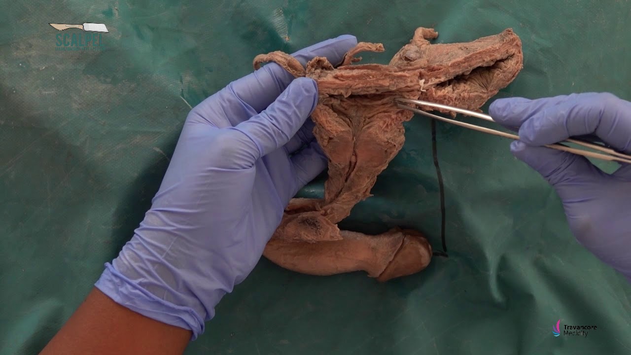
TLDRThis educational video offers an in-depth exploration of the male urethra and its associated structures. It visually demonstrates the course of the vas deferens, the location of ejaculatory ducts, and the function of bulbourethral glands. The video dissects the anatomy of the male reproductive system, highlighting the prostatic, membranous, and penile urethra, and explains the formation of the ejaculatory duct. It also provides a clear view of the vas deferens, seminal vesicles, and prostate, along with the surrounding pelvic structures, to deepen the understanding of the male urogenital system.

Takeaways

  • 📚 The video demonstrates the male urethra and associated structures using specimens.
  • 🔍 It points out the location of the openings of the ejaculatory ducts and the bulbourethral glands.
  • 🌟 The video provides an unobstructed view of the vas seminal vesicles and prostate.
  • 📏 The urethra is approximately 18 to 20 centimeters long and is divided into three parts: prostatic, membranous, and spongy.
  • 📍 The urethra begins at the base of the bladder, passes through the prostate and penis, and ends at the external urethral meatus.
  • 🧵 The video shows the course of the vas deferens from its beginning at the tail of the epididymis to its end at the prostate.
  • 💧 The membranous urethra is the shortest and least part of the urethra, where the vas deferens joins with the duct of the seminal vesicle to form the ejaculatory duct.
  • 🦵 The gluteal region is shown with landmarks such as the ischial tuberosity and pubic ramus, with the pelvic outlet clearly visible.
  • 🧬 The video explains the structure of the spermatic cord and the presence of the vas deferens, which has a thick wall and narrow lumen.
  • 💉 The testicular vessels, which traverse the inguinal canal, are identified in the video.
  • 🔪 In a sectioned specimen, the prostate is shown with the urethra and ejaculatory ducts, and the seminal vesicle and vas deferens are also visible.

Q & A

  • What structures does the video demonstrate in the male urethra and associated areas?

    -The video demonstrates the entire male urethra, ejaculatory ducts, bulbourethral glands, vas deferens, seminal vesicles, prostate, and the external urethral meatus.

  • What is the length of the male urethra as mentioned in the video?

    -The male urethra is about 18 to 20 centimeters long.

  • How many parts is the urethra divided into and what are they called?

    -The urethra is divided into three parts: the prostatic urethra, the membranous urethra, and the spongy or penile urethra.

  • What is the narrowest part of the urethra?

    -The external urethral meatus is the narrowest part of the urethra.

  • What is the purpose of the membranous urethra and where is it located?

    -The membranous urethra extends from the prostate to the bulb of the penis, passing through the urogenital diaphragm and the perineal membrane.

  • What are the structures that form the pelvic diaphragm?

    -The pelvic diaphragm is formed by muscles and connective tissue that support the pelvic organs.

  • What is the function of the vas deferens as described in the video?

    -The vas deferens transports sperm from the epididymis to join with the duct of the seminal vesicle to form the ejaculatory duct.

  • What is the location of the colliculus in relation to the urethra?

    -The colliculus is a midline swelling in the urethra where the openings of the ejaculatory ducts are located.

  • What is the Tunica vaginalis and its relation to the testes?

    -The Tunica vaginalis is a serous membrane that covers the testes and is a remnant of the peritoneum.

  • What are the testicular vessels and their function?

    -The testicular vessels are blood vessels that supply blood to the testes and traverse the inguinal canal.

  • What is the ampulla and its significance in the vas deferens?

    -The ampulla is a dilated part of the vas deferens where sperm are stored before ejaculation.

Outlines

00:00

🔍 Male Urethra and Associated Structures Overview

This paragraph introduces a video that provides a detailed look at the male urethra and its associated structures. The speaker plans to demonstrate the location of various openings, including the ejaculatory ducts and the bulbourethral glands, even though the latter may not be visible. Viewers are reminded of structures seen in previous videos and are promised an unobstructed view of the vas deferens, seminal vesicles, and prostate. The video begins with an anatomical orientation, identifying key landmarks such as the anterior abdominal wall, pubic symphysis, sacrum, and coccyx. It then describes the length and parts of the urethra, including the prostatic, membranous, and spongy sections, and their respective functions and anatomical positions.

05:03

📐 Deeper Exploration of Male Reproductive Anatomy

The second paragraph continues the exploration of male reproductive anatomy, focusing on the deeper structures not visible in the initial overview. The speaker discusses the pelvic outlet and the pelvic diaphragm, which have been removed to expose deeper structures. The inferior rectal nerves and their function are mentioned, as well as the relationship between the prostate, seminal vesicles, vas deferens, and bladder. The vas deferens is highlighted as it travels from the epididymis tail, through the spermatic cord, and across the midline, joining with the seminal vesicle duct to form the ejaculatory duct. The speaker also touches on the tunica vaginalis, tunica albuginea, and the location of the deep inguinal ring, inviting viewers to identify certain vessels. The paragraph concludes with a demonstration of the bladder and prostate, showing the urethra and ejaculatory ducts, and ends with questions for the viewers to consider.

Mindmap

Keywords

💡Urethra

The urethra is a tube that carries urine from the bladder to the outside of the body and also serves as a conduit for semen in males. In the video, the entire male urethra and its associated structures are demonstrated, emphasizing its length of about 18 to 20 centimeters and its division into three parts: prostatic, membranous, and spongy or penile urethra.

💡Ejaculatory ducts

Ejaculatory ducts are the passageways through which sperm from the vas deferens and seminal fluid from the seminal vesicles enter the urethra. The script points out their location where they open into the prostatic urethra, highlighting their role in the male reproductive system.

💡Bulbourethral glands

Also known as Cowper's glands, bulbourethral glands are exocrine glands that secrete a clear fluid into the urethra to facilitate lubrication during sexual arousal. Although not visible in the video, their function and location are discussed in relation to the male urethra.

💡Vas deferens

The vas deferens is a muscular tube that transports sperm from the epididymis to the ejaculatory ducts during ejaculation. The script follows the course of the vas deferens from its beginning at the tail of the epididymis to its end where it joins the seminal vesicle to form the ejaculatory duct.

💡Seminal vesicles

Seminal vesicles are glands that produce a significant portion of the seminal fluid, which nourishes and transports sperm. The video provides an unobstructed view of the seminal vesicles and their connection to the vas deferens, illustrating their importance in the reproductive process.

💡Prostate

The prostate is a gland that contributes to the production of seminal fluid. It is located behind the bladder and surrounds the urethra. The script describes the prostate's position relative to other structures and its role in the male reproductive system.

💡Membranous urethra

The membranous urethra is the middle part of the male urethra that passes through the urogenital diaphragm. It is the shortest and least part of the urethra, as mentioned in the script, and is crucial for the passage of semen during ejaculation.

💡Corpus spongiosum

The corpus spongiosum is a part of the penile tissue that surrounds the spongy or penile urethra. It is responsible for the erection and contains the openings of the bulbourethral glands. The script locates the corpus spongiosum as part of the third section of the urethra.

💡Tunica albuginea

The tunica albuginea is a thick, fibrous capsule that surrounds the testes and provides support. In the video, it is mentioned in the context of examining the testes, emphasizing its structural role in the male reproductive anatomy.

💡Epididymis

The epididymis is a coiled tube attached to the back of the testes where sperm matures and is stored. The script describes the epididymis as being loosely attached to the testes and continuous with the vas deferens, indicating its function in sperm maturation and storage.

💡Colliculus

The colliculus is a midline swelling in the prostatic urethra where the ejaculatory ducts open. The script explains the significance of the colliculus in the context of the urethral anatomy and the process of ejaculation.

Highlights

Demonstration of the male urethra and associated structures using specimens.

Identification of the openings of the ejaculatory ducts and the bulbourethral glands.

Unobstructed view of the vas seminal vesicles and prostate in situ.

Course of the vas deferens from its beginning to its end.

Description of the urethra's length and its narrowest part, the external urethral meatus.

Division of the urethra into three parts: prostatic, membranous, and spongy.

Location of the openings of the bulbourethral glands in the spongy urethra.

Anatomy of the gluteal region with focus on the pelvic outlet.

Removal of the pelvic diaphragm to expose deeper structures.

Formation of the ejaculatory duct from the vas deferens and duct of the seminal vesicle.

Examination of the unplastered specimen with focus on the testes, spermatic cord, and prostate.

Physical characteristics of the vas deferens, including its cord-like structure.

Junction of the vas deferens with the seminal vesicle to form the ejaculatory duct.

Identification of the Tunica vaginalis and its relation to the testes.

Location and function of the epididymis in relation to the testes.

Identification of the deep inguinal ring and associated testicular vessels.

Anatomical relationship between the ureter and vas deferens posterior to the peritoneum.

Sectioning of the prostate to expose the urethra and ejaculatory ducts.

Observation of fluid release from the seminal vesicle and ejaculatory ducts.

Description of the colliculus, a midline swelling in the urethra related to the ejaculatory ducts.

Transcripts

play00:01

[Music]

play00:23

in this video I will use a few specimens

play00:27

to demonstrate the entire male urethra

play00:30

and associated structures I will point

play00:34

out the location of the openings of the

play00:38

ejaculatory ducts and the bulb or

play00:41

urethral glands even though you cannot

play00:44

see the glands you may have seen parts

play00:48

of these structures in the video on the

play00:51

male urogenital system you will get an

play00:55

unobstructed view of the vas seminal

play00:59

vesicles and prostate in situ last but

play01:05

not least you will follow the course of

play01:08

the vas deferens from its beginning to

play01:11

its end we are looking at the right half

play01:16

of body 2 which was Hemis acted in the

play01:20

mid sagittal plane for orientation note

play01:23

the anterior abdominal wall the pubic

play01:27

symphysis the sacrum and the coccyx this

play01:33

is the bladder here is the prostate and

play01:37

this structure behind the prostate is

play01:41

the rectum the urethra is about 18 to 20

play01:47

centimeters long it begins at the base

play01:51

of the bladder passes through the

play01:53

prostate the penis and ends at the

play01:57

external urethral meatus that by the way

play02:01

is its narrowest part the urethra is

play02:04

divided into three parts the first part

play02:07

is the prostatic urethra and the

play02:10

ejaculatory ducts open here the second

play02:16

part of the urethra extends from the

play02:19

prostate to the bulb of the penis since

play02:23

it passes through the urogenital

play02:26

diaphragm and the perineal membrane it

play02:30

is called the membranous urethra this is

play02:34

the shortest and the least

play02:37

part of the urethra the third part of

play02:41

the urethra traverses the corpus

play02:44

spongiosum and his hands call the spongy

play02:47

or the penile urethra located here are

play02:51

the openings of the bellboy urethral

play02:54

glands we will now move on to body 1 we

play03:00

are looking at the gluteal region

play03:02

note the ischial tuberosity sacral

play03:06

tuberous ligament coccyx and the sto

play03:10

pubic ramus you can see the pelvic

play03:13

outlet clearly as I removed most of the

play03:17

pelvic diaphragm do you recall what

play03:20

forms the pelvic diaphragm i've also

play03:24

removed the rectum and the anal

play03:27

sphincter to expose these other deeper

play03:30

structures these by the way are the

play03:34

inferior rectal nerves which were going

play03:37

to the external anal sphincter here is

play03:41

the prostate the seminal vesicle and the

play03:46

vas deferens you can also see the

play03:49

bladder it's about at this point that

play03:53

the vas joins with the duct of the

play03:56

seminal vesicle to form the ejaculatory

play03:59

duct which opens into the prostatic

play04:02

urethra this is the membranous urethra

play04:08

we will now move on to an unplastered

play04:12

specimen this specimen was removed from

play04:18

an unembodied our note the testes

play04:22

spermatic cord the rectum bladder and

play04:29

the prostate

play04:33

in the spermatic cord i can feel this

play04:36

cord like structure which is the vas

play04:39

deferens

play04:40

it feels cord like because of a thick

play04:43

wall and narrow lumen the vas begins at

play04:49

the tail of the epididymis goes up in

play04:52

the spermatic cord goes through the

play04:55

inguinal canal changes direction and

play04:59

goes across towards the midline it

play05:03

crosses the ureter close to the base of

play05:06

the bladder and then it joins with the

play05:12

duct of the seminal vesicle to form the

play05:16

ejaculatory duct opening into the

play05:19

prostate as I press on the seminal

play05:22

vesicle and the prostate you can perhaps

play05:26

see little fluid losing out of the

play05:29

urethra reflecting the Tunica vaginalis

play05:38

which is remains of the peritoneum to

play05:41

expose the testers this part here thick

play05:45

membrane is the tunica albuginea this is

play05:49

the epididymis which is loosely attached

play05:52

to the testes not the head the body and

play05:58

the tail of the epididymis the tail is

play06:01

continuous with the vas you can note the

play06:06

narrow lumen of the vas also called the

play06:10

ductus deferens this here is the about

play06:18

the location of the deep and winel ring

play06:21

and at this point there are these two

play06:24

vessels you might be able to see them

play06:26

faintly which join on to the vas can you

play06:31

name those vessels give you a hint they

play06:34

traverse the inguinal canal those are

play06:39

the testicular vessels this other

play06:43

structure here

play06:45

the ureter note that the ureter as well

play06:49

as the vas both are posterior to the

play06:53

peritoneum I would like to show you one

play06:58

more specimen this is the bladder and

play07:02

here is an enlarged prostate I section

play07:07

the prostate to expose the urethra this

play07:13

is the urethra and you can see these two

play07:17

ejaculatory ducts opening into the

play07:20

prostatic urethra this is the seminal

play07:24

vesicle and here is the vas deferens

play07:28

this dilated part of the vas is called

play07:32

the ampulla it is here that the sperms

play07:35

are stalled as I press on the seminal

play07:40

vesicle may be able to see some fluid

play07:44

come out through this ejaculatory ducts

play07:47

which are cut the openings of these

play07:50

ducts are located on either side of a

play07:53

midline swelling in the urethra that

play07:56

swelling is called the colliculus here

play08:02

are a few questions for you

play08:12

you

Rate This

5.0 / 5 (0 votes)

相关标签
Male AnatomyUrethraProstateVas DeferensSeminal VesiclesDissectionMedical EducationPelvic RegionSurgical AnatomyUrogenital System
您是否需要英文摘要?