FULL VIDEO: Main muscles of the upper limb - Human Anatomy | Kenhub
Summary
TLDRThis tutorial delves into the complex anatomy of the upper limb's muscles, categorizing them into shoulder, arm, forearm, and hand regions. It highlights the deltoid and rotator cuff muscles of the shoulder, emphasizing their role in abduction and rotation. Moving to the arm, it distinguishes between the flexor muscles like biceps brachii and the extensor triceps brachii. The forearm's muscles are detailed, with the anterior compartment's flexors and the posterior's extensors, including the supinator's unique action. The hand's intrinsic muscles are also explored for their role in fine motor skills. The script concludes with insights on rotator cuff injuries, their symptoms, and treatment.
Takeaways
- 💪 The upper limb muscles are crucial for movement and can be divided into regions: shoulder, arm, forearm, and hand.
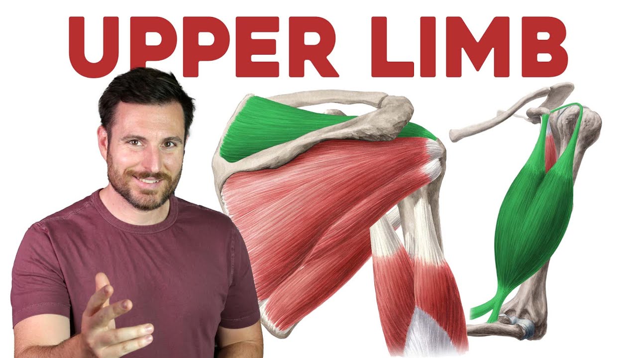
- 🏋️♀️ The shoulder region muscles include the deltoid and the rotator cuff muscles, which are essential for abduction, rotation, and stabilization of the arm.
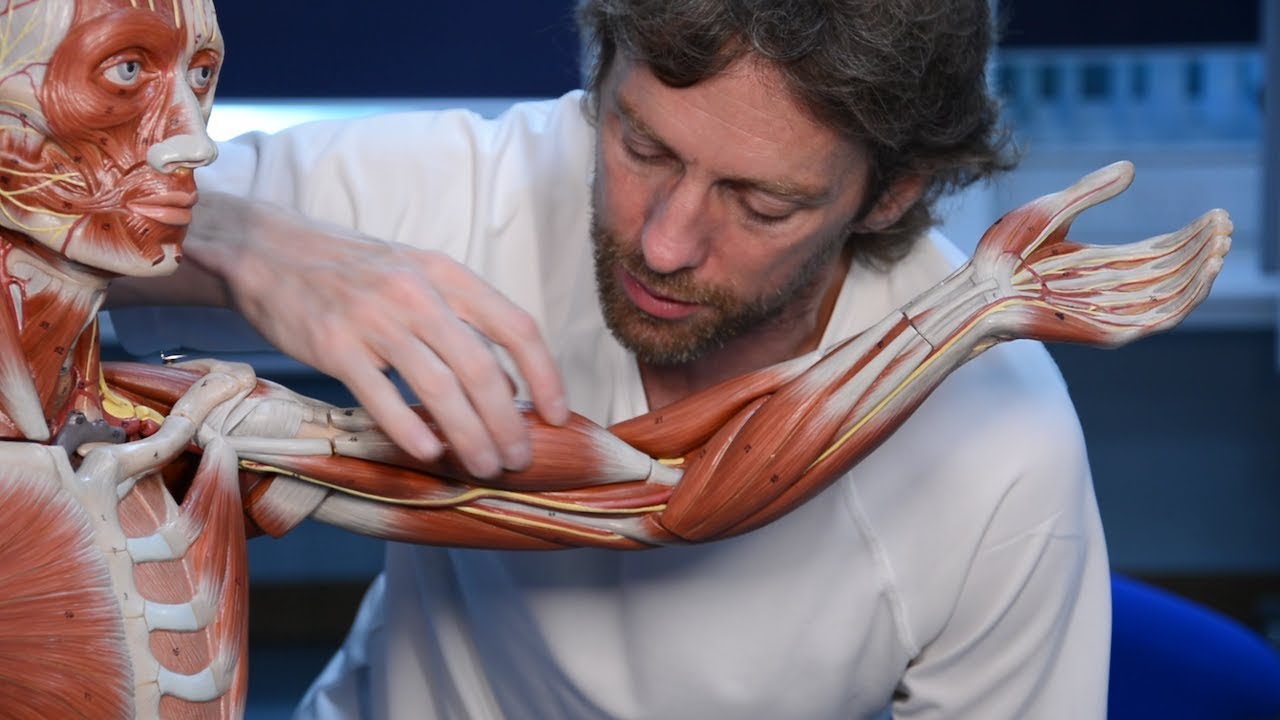
- 🤲 The arm muscles are divided into anterior (flexors) and posterior (extensors) groups, with the biceps brachii and triceps brachii being key players.
- 🙌 The forearm muscles are complex, with the anterior compartment muscles primarily responsible for flexion and the posterior for extension.
- 🤞 The intrinsic muscles of the hand, including the thenar and hypothenar muscles, are vital for fine motor movements and manipulation.
- 🤕 Rotator cuff injuries are common and can result from overuse or acute trauma, often requiring rest, ice, and sometimes surgery.
- 🏥 Clinically, it's important to maintain muscle balance around the shoulder joint to prevent injuries like rotator cuff strains.
- 🔍 The script provides a detailed anatomical review, highlighting the origins, insertions, and actions of various upper limb muscles.
- 📚 Learning about these muscles helps in understanding human movement and is beneficial for healthcare professionals and fitness enthusiasts.
- 📖 For further study, the script encourages visiting Kenhub.com for articles, quizzes, and anatomical images to deepen knowledge of anatomy.
Q & A
What are the main muscles of the shoulder region?
-The main muscles of the shoulder region include the deltoid muscle, which is superficial, and the rotator cuff muscles (supraspinatus, infraspinatus, teres minor, and subscapularis) which are deep muscles. The teres major is also a deep muscle of the shoulder but does not contribute to the rotator cuff.
What is the function of the deltoid muscle?
-The deltoid muscle is the major abductor of the arm at the shoulder joint, moving the arm away from the midline of the body.
Why are the rotator cuff muscles called 'rotator cuff'?
-The term 'rotator' comes from the rotational movement these muscles elicit when they contract, and 'cuff' originates from their positioning as they extend from the scapula and cuff the head of the humerus.
What is the SITS mnemonic for remembering the rotator cuff muscles?
-SITS is a mnemonic where S stands for supraspinatus, I for infraspinatus, T for teres minor, and the final S for subscapularis.
Which muscles are primarily responsible for the rotational movements of the arm at the shoulder joint?
-The deep muscles of the shoulder, specifically the rotator cuff muscles and the teres major, are responsible for the rotational movements of the arm at the shoulder joint.
What are the main muscles of the anterior compartment of the arm?
-The main muscles of the anterior compartment of the arm include the biceps brachii, coracobrachialis, and brachialis muscles.
What is the primary function of the biceps brachii muscle?
-The biceps brachii muscle is one of the major muscles of the arm and contributes to movement at both the shoulder and elbow joints.
How does the brachialis muscle differ from the biceps brachii muscle in terms of function?
-The brachialis muscle is a large and powerful muscle that acts only on the elbow joint, being the main and most powerful flexor of the forearm at the elbow.
What is the main muscle of the posterior compartment of the arm?
-The triceps brachii is the main muscle of the posterior compartment of the arm, acting at both the shoulder and elbow joints.
What are the main actions of the muscles in the anterior compartment of the forearm?
-The muscles in the anterior compartment of the forearm are predominantly flexors, with some contributing to other movements such as adduction of the arm and supination of the forearm.
What is the main action of the triceps brachii muscle?
-The triceps brachii muscle extends the arm and forearm posteriorly at the shoulder and elbow joints.
Which muscles are considered the main superficial muscles of the anterior forearm?
-The main superficial muscles of the anterior forearm include the pronator teres, flexor carpi radialis, palmaris longus, flexor carpi ulnaris, and flexor digitorum superficialis muscles.
What is the unique action performed by the supinator muscle in the posterior forearm?
-The supinator muscle produces supination of the forearm and wrist at the radioulnar joints.
How are the muscles of the hand divided?
-The muscles of the hand are divided into intrinsic and extrinsic muscles. Intrinsic muscles originate and insert within the hand, while extrinsic muscles originate in the arm or forearm and insert onto the palmar and dorsal surfaces of the hand and digits.
What are the five groups of intrinsic muscles of the hand?
-The five groups of intrinsic muscles of the hand are the dorsal interossei, palmar interossei, lumbricals, hypothenar muscles, and thenar muscles.
What is a rotator cuff strain and what are its symptoms?
-A rotator cuff strain occurs when the tendons of the rotator cuff muscles become overstretched or torn. Symptoms include pain on lifting or rotating the arm, swelling around the shoulder, and shoulder joint stiffness.
Outlines
💪 Introduction to Upper Limb Muscles
This paragraph introduces the tutorial on the main muscles of the upper limb, emphasizing the importance of understanding these muscles beyond just their physical appearance. It outlines the plan to discuss muscles by regions: shoulder, arm, forearm, and hand. It highlights the shoulder muscles, including the superficial deltoid and the deep rotator cuff muscles, which are crucial for shoulder joint movement. The paragraph also explains the origins and insertions of these muscles and their roles in abduction and rotation of the arm.
🤚 Muscles of the Arm and Forearm
The second paragraph delves into the muscles of the arm, divided into anterior and posterior compartments. It describes the biceps brachii, coracobrachialis, and brachialis in the anterior compartment, which are primarily flexors, and the triceps brachii in the posterior compartment, which is an extensor. The paragraph also touches on the anconius muscle and encourages viewers to explore further for a comprehensive understanding of arm muscles.
🤞 Complexities of Forearm Muscles
This section explores the muscles of the forearm, which are more complex and divided into anterior and posterior compartments with superficial and deep regions. It details the superficial muscles of the anterior forearm, including the pronator teres, flexor carpi radialis, palmaris longus, flexor carpi ulnaris, and flexor digitorum superficialis, explaining their actions and insertion points. The deep muscles of the anterior forearm are also mentioned, emphasizing their role in fine motor movements of the hand.
🤚🏻 Posterior Forearm Muscles and Their Movements
The fourth paragraph focuses on the posterior forearm muscles, which are primarily extensors. It describes the superficial muscles like brachioradialis, extensor carpi radialis longus, extensor digitorum, and extensor carpi ulnaris, and their roles in extending the wrist and fingers. The deep muscles of the posterior forearm are briefly mentioned, and the unique action of the supinator muscle in producing forearm rotation is highlighted.
🤲 Intrinsic Muscles of the Hand
The final paragraph discusses the intrinsic muscles of the hand, which are divided into dorsal and palmar interossei, lumbricals, hypothenar muscles, and thenar muscles. It explains how these muscles work together to create fine motor movements, such as abduction, adduction, flexion, extension, and rotation of the fingers and thumb. The paragraph concludes with a clinical perspective on rotator cuff injuries, their symptoms, causes, and treatment options.
🏋️♂️ Clinical Notes and Conclusion
This paragraph provides a brief clinical overview of rotator cuff injuries, focusing on strains and their treatment. It summarizes the tutorial by recapping the main muscles and actions discussed, from the shoulder to the hand. The paragraph ends with an encouragement to continue learning about anatomy through various resources, including articles, quizzes, and videos.
Mindmap
Keywords
💡Upper Limb
💡Deltoid Muscle
💡Rotator Cuff
💡Biceps Brachii
💡Triceps Brachii
💡Forearm
💡Intrinsic Muscles
💡Extrinsic Muscles
💡Supination
💡Rotator Cuff Injury
Highlights
Introduction to the muscles of the Upper Limb, emphasizing the importance of understanding them beyond just the visible biceps and triceps.
Division of the Upper Limb muscles into regions: shoulder, arm, forearm, and hand for easier understanding.
Identification of the deltoid muscle as the main superficial muscle of the shoulder region.
Explanation of the deltoid muscle's unique triangular shape, resembling the Greek letter Delta.
Introduction to the rotator cuff muscles, including supraspinatus, infraspinatus, teres minor, and subscapularis.
Description of the rotator cuff muscles' role in shoulder joint rotation and stability.
Discussion of the teres major muscle and its distinct role compared to the rotator cuff muscles.
Overview of the main muscles of the arm, including biceps brachii, coracobrachialis, and brachialis in the anterior compartment, and triceps brachii in the posterior compartment.
Detail on the biceps brachii muscle's dual function across the shoulder and elbow joints.
Explanation of the triceps brachii muscle's role in extending the arm and forearm.
Introduction to the muscles of the forearm, highlighting the differences between anterior and posterior compartments.
Description of the pronator teres muscle and its unique action of pronating the forearm.
Overview of the intrinsic muscles of the hand, including their roles in fine motor movements.
Clinical perspective on rotator cuff injuries, including tendonitis, bursitis, and strains.
Discussion on the treatment and prevention of rotator cuff injuries, emphasizing the importance of muscle balance and strength.
Summary of the tutorial, reinforcing the importance of understanding the complex muscle groups of the Upper Limb.
Transcripts
when you think of the muscles of the
Upper Limb you might think I know those
already
everyone knows where their bulging
biceps are we all know how to flex those
maybe you're even aware of your popping
triceps after doing some triceps tips
what about deltoids
you might have even heard of those
before
but if we look a little bit deeper
there's much more to the muscles of the
Upper Limb then it may seem at first
so rather than building up our muscles
at the gym let's build up our knowledge
on the main muscles of the Upper Limb
in today's tutorial we're going to take
a look at some of the main muscles of
the Upper Limb there are quite a lot of
them but don't worry we're going to
break them down into regions to make it
more manageable we will first begin with
muscles of the shoulder region moving
down to the arm the forearm and finally
the hand we will then identify the
different groups of muscles within each
region
as we go along we will discover the main
actions associated with each group of
muscles as well as their anatomical
relations to surrounding structures
so let's Dive Right In and begin with
the main muscles of the shoulder
we're going to focus on six of the main
muscles of the shoulder today most of
these muscles originate at the Bony
shoulder girdle and attach distally on
the humerus
that means most of these muscles cross
the shoulder joint
therefore when they contract movement of
the arm at the shoulder joint occurs
muscles of the shoulder can be divided
into superficial and deep layers
the main superficial muscle of the
shoulder is the well-known deltoid
muscle this large muscle is a bit like
an upside down triangle extending both
anteriorly and posteriorly its wide flat
base attaches at the shoulder with its
apex descending to attach to the arm the
anatomists who discovered this muscle
must have known their Greek alphabet
very well as they named this muscle
after the triangular-shaped Greek letter
Delta
if we were to peel back the deltoid
muscle we would find a group of smaller
but still very important muscles lying
just beneath these four deep muscles of
the shoulder are known as the rotator
cuff muscles you may be wondering why
these muscles are gripped together as
the rotator cuff muscles the term
Rotator comes from the rotational
movement these muscles elicit when they
contract whereas cuff originates from
their positioning and insertion as they
extend from the scapula and cuff the
head of the humerus
the rotator cuff muscles are also
sometimes known as sits muscles
sits is a handy little mnemonic that can
help you remember the rotator cuff
muscles where s stands for supraspinatus
I for infraspinatus t for Terrace minor
and the final s for subscapularis
the rotator cuff muscles all extend to
insert onto the head of the humerus but
it is their Origins that reveal their
differences
let's take a quick look at these muscles
individually
three of the rotated cuff muscles are
located on the posterior aspect of the
scapula the first of these muscles is
the supraspinatus muscle this muscle
sits on the posterior surface of the
scapula within a hollow known as the
supraspinous fossa this attachment is
where the supraspinatus gets its name
just below the supraspinatus we meet the
infraspinatus muscle sitting in the
infraspinous fossa are you starting to
see the pattern here
this is a triangular muscle with a wide
base and a narrow Apex
the last rotator cuff muscle on the
posterior surface of the scapula is the
teres minor muscle the Terrace minor is
smaller than both the supraspinatus and
infraspinatus muscles and sits inferior
to both at the lateral border of the
scapula
in order to see the final rotator cuff
muscle we need to flip the scapula
around so that we can view its anterior
surface
here we find the subscapularis muscle
sitting in the subscapular fossa hence
its name this muscle is the largest of
the rotator cuff muscles and covers
essentially the whole anterior surface
of the scapula
you may have noticed that there is
another muscle on the posterior surface
of the scapula that we haven't mentioned
yet
sitting just inferior to the Terrace
minor muscle we meet the Terrace major
muscle
teres major is one of the deep muscles
of the shoulder it inserts a little
lower on the shaft of the humerus and
therefore does not contribute to the
tenderness rotator cuff
let's take a look at the movements that
occur when some of the main muscles of
the shoulder contract
the large deltoid muscle is the major
abductor of the arm at the shoulder
joint it moves the arm away from the
midline of the body
the Deep muscles of the shoulder the
rotator cuff muscle and the teres major
are responsible for its rotational
movements they work together to
internally and externally rotate the arm
at the shoulder joint
let's now travel down and take a look at
the main muscles of the arm
muscles of the arm and forearm can be
divided into an anterior and a posterior
group
muscles of the anterior compartments of
the arm include the biceps brachii
the coracobrachialis
and the brachialis muscles
the main muscle of the posterior
compartment of the arm is the triceps
brachii
another small muscle that can be
considered both the muscle of the arm
and the forearm is the anconius muscle
however we'll not discuss it further in
this tutorial as this muscle is often
morphologically and functionally glassed
as a continuation of the triceps
if you want to find out more about the
anconius muscle and the other muscles of
the arm feel free to check out our more
in-depth videos on the muscles of the
arm
okay let's now take a look at the
muscles of the anterior arm in a little
more detail
the biceps brachii muscle is one of the
major muscles of the arm its name comes
from the Latin bi meaning two and kaput
meaning head
it has a long head and a short head the
biceps brachii muscle extends over two
joints the shoulder joint and the elbow
joint meaning that it contributes to
movement at both of them
the karako brachialis muscle is a small
muscle that Lies Beneath the biceps
brachii its name is derived from its
origin and location coraco indicates
this muscle's origin at the coracoid
process of the scapula while brachialis
indicates that it is located within the
brachial or arm region this muscle only
acts on the shoulder joint
a final muscle in the anterior
compartment of the arm is the brachialis
muscle
this muscle sits tucked behind the lower
portion of the biceps brachii muscle and
can be quite hard to find
the brachialis muscle is a large and
powerful muscle that acts only on the
elbow joint it is the main and most
powerful flexor of the forearm at the
elbow
now that we are familiar with the
muscles of the anterior compartment of
the arm let's take a quick look at the
posterior compartment
the triceps brachii muscle is the main
muscle of the posterior compartment
as we saw with the biceps brachii which
is a two-headed muscle the triceps
brachii is given its name because it has
three heads here we can see the long
head and the lateral head and peeking
through we can just about make the
medial head the triceps brachii muscle
is a powerful muscle that acts at both
the shoulder and elbow joints
now that we've had a look at the
anterior and posterior muscles of the
arm let's see how they act on the
shoulder and elbow joint as they
contract
muscles of the anterior compartment of
the arm are predominantly flexors
depending on their origin these three
muscles will either Flex the arm at the
shoulder or Flex the forearm at the
elbow
some muscles will also contribute to
other movements such as adduction of the
arm and supination of the forearm
the movement that occurs in the
posterior arm is quite simple
the triceps brachii muscle extends the
arm and forearm posteriorly at the
shoulder and elbow joints
so now that we've had a look at the
muscles of the arm let's move distally
to take a look at the muscles of the
forearm the region between the elbow and
the wrist
muscles of the forearm are a little bit
more complex
just like the arm the muscles of the
forearm are divided into anterior and
posterior compartments
as well as superficial and deep regions
within each compartment
let's begin by taking a look at some of
the superficial muscles of the anterior
region of the forearm first
starting off with the pronator Terrace
the pronated terrorist muscle is one of
the most superficial muscles of the
forearm it has two heads a humeral and
ulna head we can only see the humeral
head here as the ulnar head is hidden
under the muscle belly unlike most
muscles of the forearm it does not
travel down to reach the hand but
instead inserts onto the radius of the
forearm
lying just medial and slightly posterior
to the pronator teres muscle is the
flexor carpi radialis muscle the names
of this muscle give us an idea of where
it's headed carpi in Latin refers to the
bones of the hand while radialis
indicates the radial or lateral aspect
of the hand therefore by this muscle's
name we can presume that flexor carpi
radialis is heading down to the lateral
metacarpal bones
this muscle travels down to insert onto
the ligamentous structures of the palm
of the hand this muscle is extremely
variable and in fact not everybody has
it
you might be able to find this muscle's
tendon if you flex your hand at the
wrist joint if you have this muscle you
should be able to spot three tendons on
the anterior surface of the wrist if
it's absent you'll only see two tendons
the flexor carpi radialis and ulnaris
tendons
the flexocarpi ulnaris muscle lies just
medial to the palmaris longus muscle
much like the flexor carpi radialis
muscle this muscle also inserts at the
metacarpal bones of the hand but at the
ulna or medial aspect
the last muscle of the superficial layer
that we're going to look at today is the
flexor digitorum superficialis muscle
this muscle is the deepest of all the
superficial muscles of the anterior
compartment and lies just behind the
flexor carpialnurus and radialis muscles
it is a large muscle with two heads
let's now move on to the Deep
compartment of the anterior arm
here we meet the flexor pollicis longus
and the flexor digitorum profundus
muscles these muscles are quite small
and their actions weak so they are not
considered main flexors by many sources
however we do cover them in detail in
our other videos
the final muscle that we will look at in
the anterior deep layer is the pronator
quadratus muscle it is the deepest
muscle of the anterior forearm and sits
next to the wrist it stretches between
the distal end of the radius and ulna
hidden underneath layers of deep and
superficial muscles quadratus in Latin
means Square which is reflected in the
shape of the muscle
before we move on to the posterior
muscles let's take a quick look at some
of the movements of the muscles of the
anterior forearm
the muscles of the anterior forearm are
predominantly all flexors as their name
suggests most of the superficial muscles
Flex the forearm at the elbow joint hand
at the wrist and digits at the
metacarpophalangeal and interphalangeal
joints
deep muscles will also Aid in flexing
the hand and digits but do not act on
the elbow joint
muscles positioned medially or laterally
such as flexor carpi ulnaris and flexor
carpi radialis will also contribute to
abduction and adduction of the hand at
the wrist
the pronated teres and pronator
quadratus muscles work together to
produce a twisting motion known as
pronation
so now we're finished with the anterior
group of the forearm let's flip that arm
around and take a look at some of the
main muscles of the posterior forearm
just like the anterior forearm the
muscles of the posterior forearm are
also divided into superficial and deep
regions muscles within this compartment
are known as the extensor muscles as
this is the main action they perform
let's begin by taking a look at the six
muscles of the superficial layer first
the most superficial muscle of the
posterior forearm is the brachioradialis
muscle this muscle can sometimes be
confused for a muscle of the anterior
compartment due to its positioning it
sits on the lateral aspect of the
forearm and is best identified from the
anterior View
it even behaves like a muscle of the
anterior compartment as it flexes the
forearm at the elbow
it is however considered to be a muscle
of the posterior compartment as it is
innervated by the radial nerve just like
its fellow posterior muscles
slightly inferior to the brachioradialis
muscle we meet the extensor carpi
radialis longus muscle whew that was
quite a mouthful
as this muscle's name suggests it is
located to the radial or lateral aspect
of the posterior forearm the longest in
the name might also give you a clue that
this muscle has a shorter counterpart
and that is the extensor carpi radialis
brevis which is almost completely hidden
by the more superficial longus
you might notice that the three muscles
we just mentioned all contain radial in
their names signifying their location
along the radius these three muscles are
the most lateral muscles of the forearm
and are collectively known as radial
muscles
at the mid portion of the posterior
forearm just medial to the extensor
cuffed by radialis longus we find the
extensor digitorum muscle this muscle is
similar to the flexor digitorum muscles
of the anterior forearm it lies
centrally but instead of inserting onto
the Palmer surface of the hand it
travels on the back or dorsal surface to
insert onto the digits
just medial to the extensive digitorum
muscle is the extensor digiti minimi
muscle this muscle travels right down to
the little finger
the last muscle of the superficial
compartment is the extensor carpi
ulnaris muscle much like the extensor
carpi radialis longus muscle the name of
this muscle also gives us a vague idea
of where it is headed carpi meaning the
carpal bones of the hand and ulnaris
indicates the ulnicide or the medial
aspect of the forearm along which it
travels this muscle suits just medial to
the extensor digitorum muscle along the
length of the forearm
we've made it all the way to the Deep
layer of the posterior forearm
generally these muscles are not
considered main muscles of the Upper
Limb as they are smaller and their
actions are weaker so we will only
quickly run through them there are six
muscles altogether
the extensor policy's longest muscle
and brevis
the abductor pollicis longus
the extensor indices
and the supinator muscle which is the
deepest muscle of the posterior forearm
if you want to find out more about the
muscles of the posterior forearm we have
a whole video dedicated to them
let's pause here to take a minute to
look at the movements created by the
muscles of the posterior forearm as you
may have guessed by their names muscles
of the posterior forearm are
predominantly extensors
most of the superficial muscles extend
the hand at the wrist joint and the
digits at the metacarpophalangeal and
interphalangeal joints
the Deep muscles extend the hand at the
wrist joint and the digits at the
metacarpophalangeal and interphalangeal
Joints but do not act at the elbow joint
muscles positioned medially or laterally
such as extensor carpi radialis longus
and extensor carpi al-naris will also
contribute to the abduction and
adduction of the hand at the wrist
there's one unique muscle however and
that's the supinator muscle
the supinator muscle produces supination
of the forearm and wrist at the radio
ulnar joints this movement can be hard
to visualize so why don't you try
producing these movements yourself
okay so we're nearly there
and we've reached our last destination
which is the hand
muscles of the hand can be divided into
intrinsic and extrinsic muscles
intrinsic muscles are the muscles that
originate and insert within the hand
while extrinsic muscles are the muscles
that originate within the arm or the
forearm and insert onto the Palmer and
dorsal surfaces of the hand and digits
we already discussed the extrinsic
muscles of the hand when we talked about
the muscles of the forearm so now let's
focus on the intrinsic muscles
as with the Almond forearm intrinsic
muscles of the hand are also divided
into groups of muscles
the intrinsic muscles of the hands are
divided into five different groups the
dorsal and Palmer interossei
the lumbricals
the hypothenia muscles
and the thinner muscles
let's begin with the dorsal interossei
notice we are now looking at a dorsal
view of the head otherwise known as the
back of the hand
the dorsal interossei muscles are four
short muscles between the metacarpal
bones
the dorsal winter Rossi are numbered one
to four starting at the thumb
the Palmer into rossii come next and as
you can see we have flipped the hand
around again and now we are looking at
the palm of the hand
the Palmer into Rossi are three muscles
located between the metacarpal bones
these muscles can also be numbered one
to three
the third digit does not have an
Associated Palma interosseous muscle
working our way to the next group of
muscles we meet the lumbricals they are
four short hand muscles located between
the digits of the hand deep in the palm
of fascia the lumbricals actually arise
from the tendons of one of the extrinsic
muscles of the hand as we can see here
moving on we come to the hypothena
muscle group
this group is made up of four small
muscles all situated on the medial
aspect of the Palm at the base of the
little finger the four hypothena muscles
are the abducted digiti minimi
the flexor digiti minimi
the opponent's digital minimi
and finally the palmaris brevis muscle
if you take a look at the palm of your
hand you might actually be able to see
an outline of this muscle group at the
base of your little finger this fleshy
prominence is the hypothena imminence
which marks the surface anatomy of these
muscles
we're on the home stretch here we've
arrived at the final muscle group of the
hand the thinner muscles
the thinner muscles are located on the
lateral aspect of the Palm towards the
base of the thumb
they form a fleshy muscle pad in this
area known as the thinner eminence
the then our muscle group also comprises
four muscles
the thenar muscles are the abductor
pollicis brevis
the adductor pollicis
the flexor pollicis brevis
and the opponent's pollicis
do you recognize the Latin word pollicis
that's right all these muscles act on
the thumb
the intrinsic muscles of the hand work
together to create fine motor movements
the lumbricals and interossei work
together to abduct
adduct
flex and extends the fingers
while the Thena and hypothena muscles
abduct adduct Flex extend and rotate the
thumb and the little finger
now that we've had a look at all the
main muscles of the Upper Limb and their
Associated movements let's take a look
at some of the muscles we saw today from
a clinical perspective
we learned earlier about the rotator
cuff muscle group and how these four
muscles were important in rotating and
stabilizing the shoulder if you are a
gym buff or frequently play sports
you're probably familiar with the term
rotator cuff injury
there are three main types of rotator
cuff injuries tendonitis
bursitis
and strains
today we're going to be focusing on
strands
a rotator cuff strain occurs when the
tendons of the rotator cuff muscles
become overstretched or torn symptoms of
the rotator cuff strain include pain on
lifting or rotating the arm
swelling around the shoulder and
shoulder joint stiffness
strains are usually caused by overuse of
the rotator cuff or may occur from acute
trauma to the shoulder joint
treatment of a rotator cuff sprain
includes rest
ice
and a non-steroidal anti-inflammatory
drugs to reduce inflammation
strengthening exercises may also be used
to restore active function and strength
to the tendon on complete rupture of the
tendon surgical intervention may be
necessary
daily shoulder stretches can Aid in
increasing flexibility and strength of
the shoulder joint when stretching it is
important to strengthen the muscles
around the shoulder blade and posterior
shoulder specifically to optimize the
muscle balance of the rotator cuff
we did it we made it to the end
before we finish up though I'm going to
quickly summarize what we learned today
we started today's tutorial by looking
at the main muscles of the shoulder
we learned about the large deltoid
muscle and the rotator cuff muscles
which include the supraspinatus and
infraspinatus the teres minor and the
subscapularis muscles finally we
identified the Terrace major muscle we
explored how these muscles assist in
creating movements of abduction
adduction and internal and external
rotation of the arm at the shoulder
we then move distally to the arm the
main muscles of the arm were grouped
into anterior and posterior groups
muscles of the anterior group consisted
of the biceps brachii the Coco
brachialis and brachialis muscles
we identified the anterior group to be
predominantly flexors of the arm and
forearm the main muscle of the posterior
region was the triceps brachii muscle
and we learned that its main function
was extension
the forearm came next the main
superficial muscles of the anterior
forearm that we had a look at today
included the pronated Terrace the flexor
carpi radialis
the palmaris longus
the flexocarpi alnaris and the flexor
digitorum superficialis muscles
the main muscles of the deep anterior
forearm included the flexor pollicis
longus the flexor digitorum profundus
and the prinator credatus muscles
muscles of the superficial and deep
compartment of the anterior forearm were
seen to be predominantly flexors of the
forearm hands and fingers
then we met the superficial and deep
posterior forearm muscles some of the
main superficial muscles of the
posterior forearm included the
brachioradialis muscle the extensor
carpi radialis longus muscle the extent
cell digitorum and the extensor
carpialnaris
the main muscles of the deep posterior
forearm include
the extensor policies longus muscle the
extensor indices and the supinator
muscle
we learned that the main action of the
posterior muscles of the forearm was to
extend the forearm hand and fingers
finally we came to the hand we grouped
all those little muscles into five
separate groups and explored how each
group created fine motor movements of
the hand we divided the muscles of the
hand into the dorsal interossei the
Palmer into Rossi
the lumbricals
the hypothena muscle group and the
thinner muscle group we learned that the
muscles of the hypothena group included
the abductor digital minimi the flexor
digiti minimi the opponent's digital
minimi and the palmaris brevis muscles
the phener muscle group included the
abductor pollicis brevis the adductor
pollicis
the flexopolysis brevis and the
opponent's pollicis muscles
once we looked at all the main muscles
of the Upper Limb and their Associated
actions we then had a quick look at some
clinical notes on rotator cuff injury
we learned about a strain injury to the
rotator cuff tendon and identified the
symptoms causes and treatment regime for
this injury depending on the severity of
the injury
that brings us to the end of this
tutorial we hope you enjoyed this
tutorial on the main muscles of the
Upper Limb but don't let your learning
stop there visit kenhub.com where you
can read interesting articles test your
knowledge with challenging quizzes
explore our Atlas with beautiful
anatomical images or watch more video
tutorials like this one yes you'll find
everything you need to master anatomy in
no time go on click the button you know
you want to
5.0 / 5 (0 votes)