Overview of the CNS (Pars, Neurons, Neuroglia, White & Grey Matter, Development) - Anatomy

Taim Talks Med
29 Aug 202111:32

Summary

TLDRThis video script offers an in-depth exploration of the Central Nervous System (CNS), focusing on its anatomy, structure, and function. It begins by outlining the CNS's two main components: the brain and the spinal cord, then delves into the microscopic structures, detailing neurons and neuroglia. The script explains the roles of different types of neurons and their functions, such as afferent, efferent, and interneurons. It also discusses the distribution of grey and white matter, the significance of nerve tracts, and the developmental stages of the CNS. The video promises to continue with more detailed examinations of the CNS's anatomy in subsequent episodes, starting with the spinal cord.

Takeaways

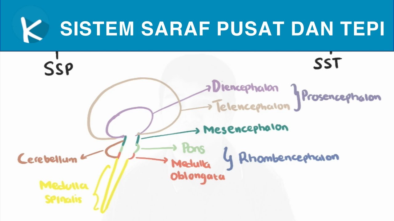
  • 🧠 The Central Nervous System (CNS) is composed of the Encephalon (brain) and the Spinal Cord.
  • 🌿 The brain is divided into five main parts: the brainstem (Medulla, Pons, Mesencephalon), Cerebellum, Diencephalon (including the hypothalamus), and Telencephalon.
  • 🔬 Neurons are the primary cells of the nervous system, consisting of dendrites, a cell body, and an axon, with variations like Multipolar, Pseudo-Unipolar, and Bipolar neurons.
  • ⚡ Neurons transmit signals through the CNS, with afferent neurons carrying sensory information to the CNS and efferent neurons sending motor commands from the CNS.
  • 🧬 Neuroglia, or glial cells, provide support, nutrients, and protection to neurons and include types like Astrocytes, Oligodendrocytes, Microglia, and Ependymal Cells.
  • 🤍 Grey matter in the CNS is rich in nerve cell bodies and dendrites, while white matter is rich in myelinated axons and glial cells.
  • 🔄 Nerve tracts are bundles of axons that connect different parts of the CNS, classified as association, commissural, or projection fibers.
  • 📈 The development of the CNS begins with the formation of the spinal cord and four brain vesicles, which later differentiate into the mature structures of the brain.
  • 🧐 The functions of the CNS are hierarchical, with basic functions like respiration and heart rate controlled by the hindbrain, and higher cognitive functions by the cerebral cortex.
  • 🔬 The CNS development from a primitive brain to an adult brain involves significant changes, with the forebrain and hindbrain dividing into various structures that form the mature brain.

Q & A

  • What are the two main parts of the Central Nervous System?

    -The Central Nervous System (CNS) consists of the Encephalon, which is the brain, and the Spinal Cord.

  • What are the three main parts of the brainstem?

    -The brainstem consists of the Medulla Oblongata, the Pons, and the Mesencephalon.

  • What is the function of the Cerebellum in the CNS?

    -The Cerebellum is essential for muscle memory and coordination.

  • What is the primary cell type in the nervous system?

    -Neurons are the primary cell type in the nervous system.

  • What are the components of a neuron and their functions?

    -A neuron consists of Dendrites, which receive signals and send them to the Cell Body; the Cell Body contains the nucleus; and an Axon, which sends signals to other cells.

  • What is the role of the Myelin sheath in the nervous system?

    -The Myelin sheath, formed by Schwann cells or Oligodendrocytes, wraps around axons and helps transmit signals much faster.

  • How can you differentiate a dendrite from an axon under a microscope?

    -Under a microscope, dendrites can be differentiated from axons by the presence of granules within the neuron; axons lack these granules.

  • What are the three general functions a neuron can have?

    -A neuron can function as an afferent or sensory nerve, an interneuron, or an efferent or motor nerve.

  • What are the two main categories of cells in the CNS and their roles?

    -The two main categories of cells in the CNS are Neurons and Neuroglia. Neurons transmit signals, while Neuroglia provide support, nutrients, and protection to nerve cells.

  • What is the difference between grey matter and white matter in the CNS?

    -Grey matter is tissue rich in nerve cell bodies and dendrites, while white matter is tissue rich in myelinated axons and glial cells.

  • What are nerve tracts and their significance in the CNS?

    -Nerve tracts are bundles of axons that connect grey matter to grey matter or nuclei to nuclei, allowing for the transmission of signals throughout the CNS.

  • How does the development of the CNS from the primitive brain influence its structure and function?

    -The development of the CNS from the primitive brain, which includes the division of the forebrain, midbrain, and hindbrain, influences its structure and function by determining the areas responsible for basic and higher-order functions.

Outlines

00:00

🧠 Central Nervous System (CNS) Anatomy Overview

The video segment introduces the anatomy of the Central Nervous System (CNS), focusing on its structure and function. The CNS is composed of the brain (Encephalon) and the spinal cord. The brain is further divided into the brainstem (Medulla, Pons, Mesencephalon), Cerebellum, Diencephalon (including the hypothalamus), and Telencephalon. The discussion highlights the role of neurons, the primary cells of the nervous system, and their structure, including dendrites, cell bodies, axons, and axon terminals. The video also touches on the support provided by neuroglia cells, the significance of white and grey matter distribution, and the concept of nerve tracts that facilitate communication within the CNS. Lastly, it briefly mentions the development of the nervous system, setting the stage for further exploration in subsequent videos.

05:00

🌐 Neuron Structure and Function

This paragraph delves into the structure and function of neurons, which are the fundamental units of the nervous system. It explains the roles of dendrites, cell bodies, and axons in receiving, processing, and transmitting signals. The video describes the myelin sheath formed by Schwann cells and Oligodendrocytes, which speeds up signal transmission. The distinction between different neuron types, such as Multipolar, Pseudo-Unipolar, and Bipolar neurons, is discussed, along with their unique shapes and functions. The paragraph further clarifies the functions of afferent (sensory), efferent (motor), and interneurons, using the example of sensing and responding to a cup of coffee. The support role of neuroglia cells like Astrocytes, Oligodendrocytes, Microglia, and Ependymal cells in the CNS is also highlighted.

10:06

🧬 Development and Classification of CNS Structures

The final paragraph of the script discusses the embryonic development of the CNS, starting from the primitive brain's four humps: the spinal cord, Hindbrain, Midbrain, and Forebrain. It details how these structures evolve into the Myelencephalon, Metencephalon, Mesencephalon, Diencephalon, and Telencephalon. The video emphasizes the functional hierarchy of the CNS, with basic functions like respiration and cardiac function located closer to the spinal cord and higher cognitive functions like decision-making in the cerebral cortex. The classification of nerve tracts into association, comisural, and projection fibers is introduced, along with the concept of ascending (sensory) and descending (motor) tracts. The video concludes by setting the stage for upcoming videos that will explore the detailed anatomy and function of each CNS component.

Mindmap

Keywords

💡Central Nervous System (CNS)

The Central Nervous System (CNS) is the part of the nervous system consisting of the brain and spinal cord. It serves as the central processing unit of the body, interpreting and responding to signals from the peripheral nervous system. In the video, the CNS is the main focus, with discussions on its anatomy, function, and development.

💡Encephalon

The Encephalon, also known as the brain, is the central organ of the CNS. It is responsible for higher cognitive functions and controls most of the body's functions. The video script describes the brain as being divided into functionally different parts, such as the brainstem, cerebellum, and diencephalon.

💡Neuron

Neurons are the primary cells of the nervous system, responsible for transmitting signals between different parts of the body. The video script explains that neurons consist of dendrites, a cell body, and an axon, and are essential for the functioning of the CNS.

💡Neuroglia

Neuroglia, or glial cells, are non-neuronal cells that provide support and protection for neurons in the CNS. The video mentions several types of neuroglia, including astrocytes, oligodendrocytes, microglia, and ependymal cells, each with specific roles in the CNS.

💡White Matter

White matter in the CNS consists of myelinated axons and glial cells. It appears white due to the lipid-rich myelin sheath surrounding the axons. The video script explains that white matter is essential for the fast transmission of nerve signals.

💡Grey Matter

Grey matter is composed mainly of nerve cell bodies and dendrites. It is darker in color compared to white matter and is found in areas such as the cerebral cortex. The video script uses grey matter as an example to illustrate the distribution of nerve cell bodies in the CNS.

💡Nerve Tracts

Nerve tracts are bundles of axons that connect different parts of the CNS. They are classified as association, commissural, or projection fibers. The video script discusses how nerve tracts facilitate communication between different areas of the brain and spinal cord.

💡Afferent Neurons

Afferent neurons, also known as sensory neurons, carry signals from sensory receptors towards the CNS. The video script uses the example of seeing and smelling coffee to illustrate how afferent neurons transmit sensory information to the brain.

💡Efferent Neurons

Efferent neurons transmit signals from the CNS to the muscles or glands, causing a response. The video script explains that efferent neurons exit the CNS and are involved in motor functions, such as picking up a coffee cup.

💡Myelin Sheath

The myelin sheath is a multilayered membrane that wraps around the axons of neurons and helps in the rapid transmission of nerve impulses. The video script mentions that myelin sheaths are formed by glial cells and are crucial for the speed of neural communication.

💡Development of CNS

The development of the CNS is a complex process that begins with the formation of the neural tube and proceeds through various stages of differentiation. The video script outlines the early development of the CNS, from the primitive brain's four humps to the mature structure of the adult brain.

Highlights

Introduction to the Central Nervous System (CNS) anatomy

CNS comprises the Encephalon (brain) and Spinal Cord

Brain is divided into the brainstem, Cerebellum, Diencephalon, and Telencephalon

Nerves transmit signals to the brain and generate responses through neurons

Neurons are the primary cells of the Nervous System, consisting of Dendrites and Axons

Myelin sheath formed by Schwann cells in PNS and Oligodendrocytes in CNS

Differentiation between dendrites and axons in microscopic examination

Neurons are characterized by their shape: Multipolar, Pseudo-Unipolar, and Bipolar

Neurons have three general functions: Afferent, Interneuron, and Efferent

Neuroglial cells provide support, nutrients, and protection to neurons

Types of Neuroglial cells: Astrocytes, Oligodendrocytes, Microglia, and Ependymal Cells

Distribution of CNS tissue into Grey Matter (cell bodies and dendrites) and White Matter (myelinated axons)

Nerve tracts are bundles of axons connecting grey matter to grey matter

Classification of nerve tracts: Association, Comisural, and Projection fibers

Ascending and Descending tracts, and their role in sensory and motor functions

Development of the CNS from primitive brain structures during early embryonic stages

Functional hierarchy in the CNS, from basic functions in the hindbrain to higher cognitive functions in the cerebral cortex

Upcoming detailed anatomy of the Spinal Cord in the next video

Transcripts

play00:03

What up. Meditay here. Let's talk about  the anatomy of the Central Nervous System.  

play00:08

In this segment, we will go through the base in  understanding how the anatomy of the CNS is built.  

play00:14

And to do that, we'll first go through the Parts  of the CNS, then we're going to go through the  

play00:20

microscopic structures of the central nervous  system, basically understand what Neurons and  

play00:23

Neuroglia are and how they're distributed in  the CNS. After that, we'll be talking about  

play00:29

the distribution of white and grey matter and  talk about nerve tracts. And then end by talking  

play00:34

about the general nervous system development. Alright, so the central nervous system consists  

play00:39

of two main parts. There's the Encephalon  or the brain. And then the Spinal Cord.  

play00:45

But the brain is also divided  into functionally different parts,  

play00:49

so if we look here, we have the spinal  cord. And then, above the spinal cord,  

play00:53

we'll find a structure called the brainstem And the brainstem consists of the Medulla,  

play00:57

or Medulla Oblongata, The Pons, and the  Mesencephalon. Behind the brainstem,  

play01:03

we'll find the Cerebellum, which is an essential  part of the brain for muscle memory. Above that,  

play01:08

there's the Diencephalon, which's the area you'll  find the hypothalamus. And then we have the  

play01:14

Telencephalon, which is what we call the highest  order in our brain where our personality is. 

play01:20

And so the way all of this works is that Nerves  pass signals towards the higher senses of the  

play01:25

brain, then there are nerves that interpret the  signals, which then generate an impulse, basically  

play01:30

activating neurons that send signals towards  a muscle or an organ to activate a response. 

play01:36

And so I say neurons because that's the primary  type of cells in our Nervous System. If we take  

play01:42

a segment of the spinal cord and look at  it underneath a microscope, you'll see  

play01:46

that they're composed of nerve tissue. And if  we take a small segment of the nerve tissue,  

play01:51

you'll find a lot of these cells we call a Neuron. Let's talk about the neuron a little bit. Here  

play01:57

you see a simple animated neuron. They consist of  Dendrites. Dendrites are what receive signals and  

play02:03

send them towards the Cell Body, which contains  a nucleus, of course, since it's a cell. The  

play02:08

signals are then sent through an axon, which are  long fibers that can extend at a large distance.  

play02:14

There are nerves that begin in your lower back  and extend all the way to the tip of your toes,  

play02:19

thanks to the length of the axon. And at  the end there, that's the axon terminal. 

play02:29

Now, axons can either be wrapped around by many  Shwan cells or Oligodendrocytes. If they're  

play02:35

in the Brain or Spinal cord, they're called  Oligodendrocytes. If they're outside the CNS,  

play02:41

they're called Schwann cells. These cells form  a myelin sheath around each segment it covers.  

play02:47

So axons can either be wrapped around in a  myelin sheath, or they can be free axons that  

play02:53

aren't covered by these myelin sheath. The Myelin  sheath help transmit the signal much, much faster. 

play03:01

And so this is how a general nerve impulse look  like. A signal is sent from one cell through the  

play03:06

axon into a dendrite of the next cell, which then  travels towards the cell body. And then through  

play03:12

the axon and into the dendrite of the next neuron. So if we go back to this picture. You'll now see  

play03:18

the Dendrites here, the cell body, and  an axon going out from the cell body.  

play03:23

You'll study this more in histology, but the way  you can differentiate a dendrite from an axon  

play03:28

underneath a microscope is by looking at the  granules within the neuron. The axon doesn't  

play03:34

have these granules, and you can see a clear  margin between the axon and the body here. 

play03:40

and so do all nerve cells look like  this? the answer is no, unfortunately 

play03:45

Nerves cells are actually characterized by  their shape. We have a Multipolar Neuron  

play03:50

with one axon and many dendrites that  give the cell a star-like shape. These  

play03:55

are widespread in the central nervous system. Then we have Pseudo-Unipolar Neuron. It's unipolar  

play04:01

because the axon and dendrite emerge from the  same place from the cell body. And it's Pseudo;  

play04:06

Pseudo means false or fake. It's false Because  the Signal still has to go through the cell body  

play04:12

to reach the axon. That's why it's fake.  It's not a straight line that has to go  

play04:16

through the body and then to the axon. And we also have bipolar nerve cells  

play04:21

where one axon and one dendrite emerge  from either side of the cell body. 

play04:26

So nerve cells differ in structure depending on  where you find them, but neurons also differ in  

play04:32

function. And there are 3 general functions a  neuron can have. A nerve can be an afferent or  

play04:38

sensory nerve. It can be an Interneuron,  or it can be an efferent or motor nerve. 

play04:44

Let me give you a simplified example of how this  works. Let's say you wake up in the morning and  

play04:48

see a coffee, and you don't just see it. You can  also smell the coffee or hear the coffee machine  

play04:55

working. All of those neurons being stimulated  will lead the signal towards the central nervous  

play05:00

system as sensory neurons or afferent neurons.  Then, these signals are interpreted in your brain  

play05:07

through interneurons. And suppose  you've decided that you want the coffee.  

play05:11

In that case, the brain is going to engage  motor neurons, or efferent neurons, to activate  

play05:17

muscles in order to pick up the coffee cup. So  Remember, Afferent neurons Arrive into the CNS,  

play05:24

Efferent Neurons Exit the CNS. Ok, so we now understand what a neuron is.  

play05:32

But you also need to visualize the fact that  they're not alone in the CNS. There are countless  

play05:38

cells we call Neurogllia that give mechanical  support and give nutrients and protection  

play05:43

to the nerve cells, as you see here. And so we  got many different types of NeuroGlia in the CNS 

play05:49

We got Astrocytes, which  are the largest neuroglia.  

play05:52

These astrocytes have long projections that  wrap around the blood vessels within your CNS,  

play05:57

and they form a so-called Blood-brain barrier. There are Oligodendrocytes, which are responsible  

play06:03

for the myelination of nerves in the CNS.  Remember, in the peripheral nervous system,  

play06:09

there are Shwan cells, and in the  CNS, there are Oligodendrocytes.  

play06:14

So that's that one, forming a myelin sheath. We  got Microglia, which are the smallest neuroglia.  

play06:20

These are basically the immune cells of the  CNS. They can do everything a macrophage does,  

play06:25

like phagocytosis, and migrate between the tissue.  And lastly, we also have Ependymal Cells, which  

play06:32

line all the cavities within our central nervous  system. So that was the two um main categories of  

play06:38

cells in the CNS. Neurons and Neuroglia. But the tissue in our CNS is distributed  

play06:45

as either white or grey matter. Grey matter is  tissue rich in Nerve cell Bodies and Dendrites.  

play06:52

White matter is tissue rich in  myelinated axons and glial cells.  

play06:56

And if you look at this neuron. In reality, the  whole Neuron is gray in color. They're all gray  

play07:02

underneath the microscope without any significant  staining. The Axons with myelin sheath around  

play07:08

are white because they're so rich in lipid that  they appear white underneath the microscope.  

play07:13

So cell bodies and Dendrites are grey matter,  and myelinated axons are white matter.  

play07:20

And as we study the CNS, we often need to look  at cross-sections to study the tracts and nuclei  

play07:26

within each segment of it. Like in the spinal  cord and the brain. In the spinal cord,  

play07:31

you'll find the gray matter centrally and  the white matter around it. And in the brain,  

play07:36

you'll find the gray matter at the external  border, we call it the cerebral cortex, and you’ll  

play07:41

find grey matter in some places within the brain  itself. Everywhere else is gonna be the white  

play07:47

matter. So gray matter, cell bodies, and  dendrites. White matter, myelinated axons. Cool. 

play07:54

Now. Here's something you'll see a lot when you  study the CNS anatomy. Its Nerve Tracts. Nerve  

play08:01

tracts are a bundle of axons that connect  Gray matter to Gray matter. Or fibers that  

play08:06

connect Nuclei to Nuclei. So imaging a hand that  either touches something, senses temperature or  

play08:12

gets pinched. All of those will activate specific  nerves that lead impulses towards the spinal cord,  

play08:19

leading the signals through specific places in  the brain and spinal cord in order to understand  

play08:24

what happened and react to it. What I want you  to know is that in Grey matter, we got nuclei,  

play08:30

and in white matter, you got Tracts. Now there are certain ways to classify  

play08:35

these Tracts. You can either classify them  as association fibers, connecting adjacent  

play08:40

structures, Comisural fibers, connecting  one part of the brain to the other side,  

play08:45

or projection fibers, leading tracts up and down  the spinal cord. I will talk more about this when  

play08:51

we talk about the internal structures of the  Cerebral Hemisphere because that's when this  

play08:55

classification becomes relevant to you.  But the most important thing to remember,  

play08:59

which you'll hear about a lot, are Ascending  tracts, leading sensory fibers. Descending tracts,  

play09:05

leading motor fibers, and Indirect Tracts that  interconnect certain parts of the brain. This  

play09:11

is another way to classify nerve tracts. So  that’s all I had about nerve tracts for now. 

play09:16

Lastly, let us understand the principle of how  the CNS is developed. Once you understand that,  

play09:22

you'll also understand why the CNS is built  like it is and why the adjacent structures  

play09:28

often have the same function. So if you look at the real primitive brain,  

play09:32

we find that we have these four humps at  a time of 4th week after fertilization.  

play09:37

And we call those. Well, the first  one is not a hump but the spinal cord. 

play09:42

We then have the Hindbrain, the midbrain  and then we finally have the forebrain.  

play09:46

Or in Latin, the Rhombencephalon, Mesencephalon  and the Procecephalon. But during development,  

play09:52

your brain changes drastically. So already  during the 5th week, you'll notice these  

play09:56

humps are starting to form shapes. You gonna see that the Rhombencephalon  

play10:00

and the Prosencephalon are gonna divide. The  Rhombencephalon divides into the Myelencephalon  

play10:05

and the metencephalon, where they're later  on gonna become the Pons, Cerebellum, and  

play10:09

the Medulla oblongata. The mesencephalon is just  gonna stay like that. It's called the midbrain.  

play10:13

And then, the prosencephalon will divide  into the telencephalon and the Diencephalon. 

play10:18

And so this is what an adult brain looks like. The  spinal cord is down here. And again, the medulla  

play10:23

oblongata, pons and the Cerebellum are all formed  by the Myelencephalon and the Metencephalon.  

play10:29

The mesencephalon is a synonym for  the midbrain since it doesn't divide.  

play10:33

The Diencephalon will become all the thalamus  structures like the hypothalamus and the thalamus.  

play10:38

And the Telencephalon is the actual  brain cortex and its fibers here in blue. 

play10:43

-- One thing you should remember is that the closer  

play10:45

we are to that spinal cord, the more basic the  functions are. And so down here at the hindbrain,  

play10:51

they're responsible for simple functions. So  they’ll regulate the respiratory frequence when  

play10:56

you're not thinking about it, cardiac function,  vasodilation, and reflexes like vomiting,  

play11:02

coughing, sneezing, and even swallowing  are considered basic functions.  

play11:06

And if you have any deep thoughts about  something or you decide to do a simple act,  

play11:11

that's going to be your cerebral cortex  giving orders to the rest of your body. 

play11:15

And so this was an overview of how the CNS is  distributed and its function and development. 

play11:21

In the next videos, we'll be looking detailed into  the anatomy of each of these parts and understand  

play11:22

how they function. So the next video will be going  through the whole anatomy of the Spinal Cord.

Rate This

5.0 / 5 (0 votes)

相关标签
CNS AnatomyNeurologyNeuron TypesBrain DevelopmentNeuroscienceCentral Nervous SystemMedical EducationNeurogliaNerve TractsHistology
您是否需要英文摘要?