How to see with sound - Jacques S. Abramowicz
Summary
TLDRThis script explores how bats use echolocation with ultrasound to navigate in darkness, inspiring human applications like SONAR and medical ultrasound imaging. It explains the science behind ultrasound, its use in detecting submarines and imaging internal body structures, including fetal development. The script highlights the advantages of ultrasound, such as being non-invasive and safe, and its ability to provide detailed, real-time images without harmful side effects.
Takeaways
- 🦇 Bats navigate in the dark using echolocation, a method that relies on their ears rather than eyes.
- 🔊 Echolocation involves the use of ultrasound, sound waves with frequencies above 20,000 Hz, which humans cannot hear.
- 🌊 Ultrasound waves are effective for detecting objects because they bounce off surfaces, creating echoes that carry information.
- 🗺️ Bats create an internal map of their environment by sensing the nuances in the echoes of their emitted ultrasound waves.
- 🇫🇷 French scientists during WWI used ultrasound to detect enemy submarines, demonstrating the practical application of echolocation.
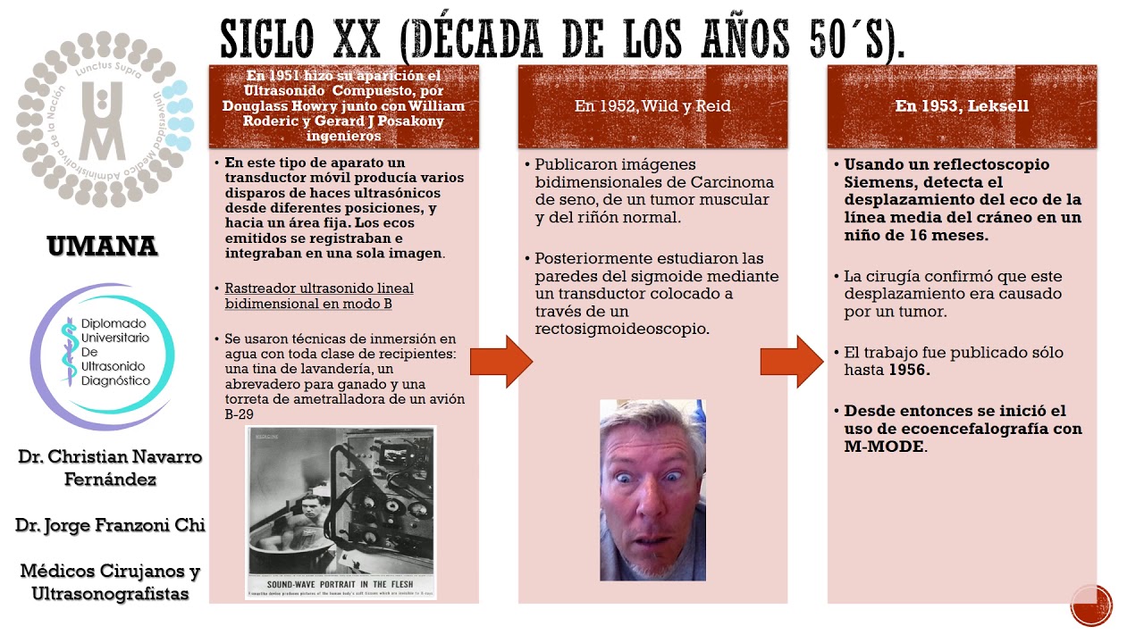
- 🏥 In the 1950s, medical professionals began using ultrasound as a non-invasive method to visualize internal body structures.
- 👶 Ultrasound imaging is widely used in fetal ultrasounds to evaluate fetal development and detect abnormalities.
- 🧪 Conductive gel is used in ultrasound procedures to ensure an airtight seal for sound wave transmission and to prevent loss of clarity.
- 🌐 Ultrasound waves pass through liquids without creating echoes, but bounce back when they encounter solid structures, forming images on the screen.
- 🔍 Multiple frequencies are used in ultrasound imaging to penetrate different depths and create a composite, life-like image.
- 🚑 Medical ultrasound is advantageous as it has no known negative side effects when used properly and is portable for field use.
Q & A
How do bats navigate in the dark?
-Bats navigate in the dark using echolocation, which involves emitting ultrasound waves and interpreting the echoes that bounce back from nearby surfaces.
What is the definition of ultrasound in the context of sound waves?
-Ultrasound refers to sound waves that exceed 20,000 cycles per second, which is beyond the range of human hearing.
How does the frequency of a sound wave relate to the number of cycles it completes over time?
-The frequency of a sound wave, measured in hertz, indicates the number of cycles it completes per second; a higher frequency wave completes more cycles over the same amount of time compared to a lower frequency wave.
What is the role of conductive gel in ultrasound imaging?
-Conductive gel is used to create an airtight seal between the body and the ultrasound wand, ensuring that sound waves do not lose speed or clarity when traveling from the wand to the body.
How do ultrasound waves interact with different types of tissues in the body during imaging?
-Ultrasound waves pass through liquids without creating echoes, but when they encounter solid structures, they bounce back, producing echoes that are rendered as dots on the imaging screen.
What is the significance of using multiple frequencies in ultrasound imaging?
-Multiple frequencies are used together in ultrasound imaging to penetrate different depths in the body. Longer, low-frequency waves penetrate deeper than shorter, high-frequency ones, allowing for a more comprehensive and detailed image.
How does ultrasound imaging help in medical diagnostics?
-Ultrasound imaging helps in diagnosing various conditions by providing detailed images of internal organs, evaluating organ damage, measuring tissue thickness, and detecting abnormalities such as gallbladder stones, tumors, and blood clots.
What is the historical context of using ultrasound for detecting submarines during World War One?
-During World War One, French scientists used ultrasound in the form of SONAR to detect enemy submarines by sending ultrasound beams into the ocean, leveraging the fact that sound waves travel faster in water.
How does the fetal ultrasound process work?
-The fetal ultrasound process involves applying conductive gel to the skin, using an ultrasound wand to send beams into the body, and capturing the echoes that bounce off the fetus and other structures to create an image on the screen.
What are some advantages of medical ultrasound over other imaging technologies?
-Medical ultrasound has advantages such as being non-invasive, having no known negative side effects when used properly, and being portable, which allows it to be used in various settings, including field medical emergencies.
Why are ultrasound waves with frequencies ranging from 2 million to 10 million hertz used in medical imaging?
-These high frequencies create detailed images that allow doctors to diagnose even the smallest developmental deviations in the brain, heart, spine, and more.
Outlines
🦇 Echolocation and Ultrasound
This paragraph explains how bats use echolocation to navigate in the dark. Bats emit ultrasound waves that bounce off surfaces, creating echoes that provide them with detailed environmental information. The concept of ultrasound, sound waves with frequencies above 20,000 hertz, is introduced, which is beyond human hearing range. The paragraph also touches on the historical and medical applications of ultrasound, including its use in detecting submarines during World War One and in modern medicine for non-invasive imaging of internal body structures.
🛸 The Evolution of Ultrasound Imaging
This section delves into the development and practical use of ultrasound imaging in medicine. It describes how ultrasound imaging works, starting with the application of conductive gel on the skin to facilitate the transmission of sound waves. The paragraph details how ultrasound waves interact with different body tissues, with solid structures producing echoes that form images on the screen. It explains the use of multiple frequencies to create detailed images and how operators can manipulate the image to focus on specific areas. The capabilities of ultrasound imaging in visualizing movement and its advantages over other imaging technologies due to the absence of harmful side effects are also highlighted.
Mindmap
Keywords
💡Echolocation
💡Ultrasound
💡Frequency
💡SONAR
💡Ultrasound Imaging
💡Conductive Gel
💡Echo
💡Hertz
💡Fetal Ultrasound
💡Medical Ultrasound
💡Non-Invasive
Highlights
Bats navigate in the dark using echolocation, a process that involves emitting ultrasound waves and interpreting the echoes.
Ultrasound is sound waves with frequencies exceeding 20,000 cycles per second, which are inaudible to humans.
Bats create an internal map of their environment by sensing the nuances in the echoes of their emitted ultrasound waves.
The concept of echolocation inspired the development of SONAR, which was used in World War One to detect submarines.
Sound waves travel faster in mediums with tightly packed molecules, such as water, which is why SONAR was successful.
In the 1950s, medical professionals began using ultrasound as a non-invasive method to visualize inside the human body.
Ultrasound imaging is used to evaluate organ damage, measure tissue thickness, and detect health issues like gallbladder stones, tumors, and blood clots.
Fetal ultrasound is a well-known application of ultrasound imaging, providing a detailed view of a developing fetus.
Conductive gel is used in ultrasound imaging to ensure an airtight seal between the body and the ultrasound wand, improving wave transmission.
Ultrasound waves pass through liquids without creating echoes, but bounce back when encountering solid structures, forming images on the screen.
Different frequencies of ultrasound waves are used to penetrate different depths within the body, creating a composite image.
Ultrasound machines can visualize movement in real time, allowing for dynamic imaging of the body's internal structures.
Medical ultrasound uses frequencies ranging from 2 million to 10 million hertz, providing highly detailed images for diagnosis.
Ultrasound imaging has no known negative side effects when used properly, unlike radiation-based imaging or invasive procedures.
High levels of ultrasound can generate heat that may damage tissues, but technicians use the lowest effective levels to mitigate this risk.
Modern ultrasound machines are portable, enabling doctors to use them in various settings, including medical emergencies.
Transcripts
In a pitch-black cave, bats can’t see much.
But even with their eyes shut,
they can navigate rocky topography at incredible speeds.
This is because a bat’s flight isn’t just guided by its eyes,
but rather, by its ears.
It may seem impossible to see with sound,
but bats, naval officers, and doctors do it all the time,
using the unique properties of ultrasound.
All sound is created when molecules in the air, water,
or any other medium vibrate in a pulsing wave.
The distance between each peak determines the wave’s frequency,
measured as cycles per second, or hertz.
This means that over the same amount of time,
a high frequency wave will complete more cycles than a low frequency one.
This is especially true of ultrasound,
which includes any sound wave exceeding 20,000 cycles per second.
Humans can't hear or produce sounds with such high frequencies,
but our flying friend can.
When it’s too dark to see, he emits an ultrasound wave with tall peaks.
Since the wave cycles are happening so quickly,
wave after wave rapidly bounces off nearby surfaces.
Each wave’s tall peak hits every nook and cranny,
producing an echo that carries a lot of information.
By sensing the nuances in this chain of echoes,
our bat can create an internal map of its environment.
This is how bats use sound to see,
and the process inspired humans to try and do the same.
In World War One, French scientists sent ultrasound beams into the ocean
to detect nearby enemy submarines.
This early form of SONAR was a huge success,
in large part because sound waves travel even faster through mediums
with more tightly packed molecules, like water.
In the 1950s, medical professionals began to experiment with this technique
as a non-invasive way to see inside a patient’s body.
Today, ultrasound imaging is used to evaluate organ damage,
measure tissue thickness, and detect gallbladder stones, tumors,
and blood clots.
But to explore how this tool works in practice,
let’s consider its most well-known use— the fetal ultrasound.
First, the skin is covered with conductive gel.
Since sound waves lose speed and clarity when traveling through air,
this gooey substance ensures an airtight seal
between the body and the wand emitting ultrasound waves.
Then the machine operator begins sending ultrasound beams into the body.
The waves pass through liquids like urine, blood, and amniotic fluid
without creating any echoes.
But when a wave encounters a solid structure, it bounces back.
This echo is rendered as a dot on the imaging screen.
Objects like bones reflect the most waves,
appearing as tightly packed dots forming bright white shapes.
Less dense objects appear in fainter shades of gray,
slowly creating an image of the fetus’s internal organs.
To get a complete picture,
waves need to reach different depths in the patient’s body,
bypassing some tissues while echoing off others.
Since longer, low frequency waves actually penetrate deeper
than short, high frequency ones,
multiple frequencies are often used together
and composited into a life-like image.
The operator can then zoom in and focus on different areas.
And since ultrasound machines send and receive cascades of waves in real time,
the machine can even visualize movement.
The waves used for medical ultrasound range from 2 million to 10 million hertz—
over a hundred times higher than human ears can hear.
These incredibly high frequencies create detailed images
that allow doctors to diagnose the smallest developmental deviations
in the brain, heart, spine, and more.
Even outside of pre-natal care,
medical ultrasound has huge advantages over similar technologies.
Unlike radiation-based imaging or invasive surgical procedures,
ultrasound has no known negative side effects when used properly.
At very high levels, the heat caused by ultrasound waves
can damage sensitive tissues,
but technicians typically use the lowest levels possible.
And since modern ultrasound machines can be small and portable,
doctors can use them in the field—
allowing them to see clearly in any medical emergency.
浏览更多相关视频
04 History of Ultrasound: Lecture by Dr. Eric Blackwell on History and Physics
1 1 2 HISTORIA DE LA ULTRASONOGRAFIA
Ultrasound Beam | Ultrasound Physics | Radiology Physics Course #15
Neonatal Neurosonography | Anatomy and Protocol
Ultrasound Physics with Sononerds Unit 2
KULIAH MAHASISWA: KEHAMILAN DENGAN KELAINAN AMNION - KELAINAN JUMLAH CAIRAN KETUBAN
5.0 / 5 (0 votes)