The Heart, Part 1 - Under Pressure: Crash Course Anatomy & Physiology #25
Summary
TLDRThis script explores the heart's function as the body's powerful pump, maintaining blood circulation through pressure gradients. It clarifies the heart's anatomy, including its chambers, valves, and connection to the circulatory system. The script also explains the pulmonary and systemic circulation loops, systolic and diastolic blood pressures, and the importance of these for overall health, debunking romanticized notions of the heart.
Takeaways
- 💓 The heart is a vital organ, often symbolized in culture, but it is essentially a muscular pump that powers the circulatory system.
- 🎼 Despite its cultural significance, the heart does not govern emotions or love; it is the brain that primarily controls these aspects.
- 🌊 The heart's function is to maintain blood pressure by creating a pressure gradient, similar to how fluids flow from high to low pressure areas.
- 📏 The average adult human heart is about the size of two clasped fists and is situated in the center of the chest, slightly to the left.
- 🛡️ The heart is protected by a double-walled sac called the pericardium, which reduces friction during its constant beating.
- 🏗️ The heart wall consists of three layers: the epicardium, myocardium, and endocardium, each serving different functions.
- 🔄 The heart is divided into four chambers—two atria and two ventricles—which work together to circulate blood through a system of valves.
- 🚫 Heart valves ensure unidirectional blood flow, preventing backflow into the previous chamber.
- 🔊 The sounds heard during a heartbeat ('lub-DUB') are caused by the opening and closing of the heart valves.
- 🔄 The circulatory system includes a pulmonary loop (heart to lungs and back) and a systemic loop (heart to body and back), forming a figure-eight pattern.
- 📉 High or low blood pressure, or any disruption to blood flow, can be dangerous and may lead to damage in various organs and systems.
Q & A
What is the primary function of the heart in the human body?
-The primary function of the heart is to act as a pump, maintaining blood circulation by generating high and low pressure to transport nutrients, oxygen, waste, heat, hormones, and immune cells throughout the body.
Why does the heart get more cultural recognition than other organs?
-The heart gets more cultural recognition due to its iconic status, often symbolized in holidays and media, despite other organs like the brain having a more direct role in emotions.
How does the heart create and maintain blood pressure?
-The heart maintains blood pressure by creating a pressure gradient, generating high hydrostatic pressure to pump blood out while also creating low pressure to draw it back in, similar to how fluids flow from high to low pressure areas.
What is the average size and weight of an adult human heart?
-The average adult human heart is about the size of two fists clasped together, weighing approximately 250 to 350 grams.
Where is the heart located in the human body?
-The heart is located in the center of the chest, nestled in the mediastinum cavity between the lungs, with most of its mass resting slightly to the left of the midsternal line.
What are the two main layers of the pericardium and their functions?
-The pericardium has two main layers: the fibrous pericardium, which protects the heart and anchors it to surrounding structures, and the serous pericardium, which consists of an inner visceral layer (epicardium) and an outer parietal layer, separated by fluid that acts as a lubricant.
Describe the three layers that make up the wall of the heart.
-The wall of the heart is composed of the epicardium (outer layer), the myocardium (middle layer made of cardiac muscle tissue), and the endocardium (innermost layer of squamous epithelial tissue).
What are the four chambers of the heart and their respective functions?
-The heart has four chambers: two atria (upper chambers for receiving blood) and two ventricles (lower chambers for pumping blood out). The right side handles deoxygenated blood, while the left side handles oxygenated blood.
How does the heart's anatomy contribute to its function of circulating blood?
-The heart's anatomy, with its chambers and valves, ensures unidirectional blood flow. The atria receive blood under low pressure, while the ventricles, with thicker walls, generate high pressure to pump blood out through the arteries.
What are the two main loops of blood circulation described in the script?
-The two main loops are the pulmonary circulation loop, which oxygenates blood in the lungs, and the systemic circulation loop, which distributes oxygenated blood to the body and returns deoxygenated blood to the heart.
What do the 'lub-DUB' sounds heard during a heartbeat represent?
-The 'lub-DUB' sounds represent the closing of the heart valves. 'Lub' is the sound of the mitral and tricuspid valves closing during systole, while 'DUB' is the sound of the aortic and pulmonary valves closing at the start of diastole.
How are systolic and diastolic blood pressures related to the heart's function?
-Systolic blood pressure is the peak pressure produced by the contracting ventricles, while diastolic blood pressure is the pressure when the ventricles are relaxed. These measurements indicate the health of the heart's pumping action and arterial condition.
What potential health risks can abnormal blood pressure indicate?
-Abnormal blood pressure, either too high or too low, can indicate issues with blood volume, hydration, or arterial health, and can lead to damage to the heart, lungs, brain, kidneys, and other organs if not addressed.
Outlines
💓 The Heart as a Pump
This paragraph delves into the heart's role as the body's central pump, responsible for circulating blood throughout the body. It clarifies misconceptions about the heart's emotional symbolism and emphasizes its true function: maintaining blood pressure through a pressure gradient. The heart's anatomy is introduced, including its size, location, and the protective pericardium sac. The structure of the heart's walls, consisting of the epicardium, myocardium, and endocardium, is also explained, setting the stage for understanding the heart's mechanics in the circulatory system.
🔁 The Circulation Loops and Blood Pressure
The second paragraph explains the process of blood circulation, detailing the pulmonary and systemic loops. It describes how deoxygenated blood is pumped from the right ventricle to the lungs to be oxygenated and then returned to the heart through the pulmonary veins. The systemic loop is then outlined, starting from the left ventricle, which pumps oxygen-rich blood through the aorta to the rest of the body. The return of oxygen-poor blood to the heart via the vena cava and its subsequent path through the heart's chambers is also described. The paragraph concludes with an explanation of systolic and diastolic blood pressure, the sounds produced by the heart valves, and the importance of maintaining healthy blood pressure for overall health.
Mindmap
Keywords
💡Heart
💡Circulatory System
💡Blood Pressure
💡Systole
💡Diastole
💡Valves
💡Aorta
💡Pulmonary Circulation
💡Systemic Circulation
💡Pericardium
💡Myocardium
Highlights
The heart is the most iconic organ, often symbolized in culture but scientifically it's a powerful pump for the circulatory system.
Despite emotional connotations, the heart's primary function is to maintain blood pressure, not to govern emotions.
The heart's action is similar to a pressure pump, creating a gradient necessary for blood flow.
Blood pressure is the measure of the strain on arteries as the heart circulates blood.
The heart beats approximately 100,000 times a day, illustrating its relentless work.
Anatomically, the heart is about the size of two clasped fists and is centrally located in the chest.
The heart is protected by a double-walled sac, the pericardium, which prevents friction during its beats.
The heart wall consists of three layers: the epicardium, myocardium, and endocardium, each with a distinct function.
The heart's chambers and valves work together to facilitate one-way blood flow through the body.
The sound of a heartbeat, 'lub-DUB', is caused by the opening and closing of the heart valves.
The heart's atria and ventricles have different wall thicknesses adapted to their roles in blood circulation.
Arteries carry blood away from the heart, while veins bring it back, contrary to a common misconception about blood color.
The pulmonary circulation loop is essential for oxygenating blood and removing carbon dioxide.
The systemic loop distributes oxygenated blood throughout the body and collects deoxygenated blood.
Blood pressure readings reflect the heart's performance during systole (contraction) and diastole (relaxation).
Abnormal blood pressure levels can indicate underlying health issues and have serious implications for the body.
The video was made possible by Patreon patrons and provides a comprehensive look at the heart's function and importance.
Transcripts
Your heart, that throbbing, beating muscle, is probably the most iconic organ in your body.
No other organ gets its own holiday, or as much radio play. And you’re not likely to
get a love note decorated with a kidney or a spleen, or even a brain, which is really what rules the emotions.
Don’t get me wrong, the heart does some great things -- namely, it powers the entire
circulatory system, transporting nutrients, oxygen, waste, heat, hormones, and immune
cells throughout the body, over and over.
But in the end, the heart does not make you love. It doesn’t break apart if you get
dumped by your boo. And it’s not a lonely hunter.
The truth is, the heart is really just a pump -- a big, wet, muscley brute of a pump.
And it doesn’t care about poetry or chocolate, or why you’re crying.
The heart only has one concern: maintaining pressure.
If you’ve ever squeezed the trigger on a squirt gun or opened up a shaken can of soda,
you’ve seen how fluids flow from areas of high pressure -- like inside the gun or the
can -- to areas of low pressure, like outside.
The heart’s entire purpose is to maintain that same kind of pressure gradient, by generating high
hydrostatic pressure to pump blood out of the heart, while also creating low pressure to bring it back in.
That gradient of force is what we mean when we talk about blood pressure.
It’s basically a measure of the amount of strain your arteries feel as your heart moves
your blood around -- more than five liters of it -- at about 60 beats per minute.
That’s about 100,000 beats a day, 35 million a year, 2 to 3 billion heart beats in a lifetime,
the basic physiology of which you can easily feel, just by taking your own pulse.
I don’t have a watch
Now, that might not inspire a lot of poetry, but it turns out, it’s still a a pretty good story.
Let us begin with a little anatomy.
Unless you happen to be of the Grinch persuasion, the average adult human heart is about the
size of two fists clasped together -- one of the few bits of trivia you often hear about
human anatomy that is actually true.
The heart is hollow, vaguely cone-shaped, and only weighs about 250 to 350 grams -- a
pretty modest size for your body’s greatest workhorse.
And although Americans tend to put their right hand over their left breast while pledging
allegiance, the heart is actually situated pretty much in the center of your chest, snuggled
in the mediastinum cavity between your lungs.
It sits at an angle, though, with one end pointing inferiorly toward the left hip, and
the other toward the right shoulder. So most of its mass rests just a little bit left of the midsternal line.
The heart is nestled in a double-walled sac called the pericardium.
The tough outer layer, or fibrous pericardium, is made of dense connective tissue and helps
protect the heart while anchoring it to some of the surrounding structures, so it doesn’t
like bounce all over the place while beating.
Meanwhile, the inner serous pericardium consists of an inner visceral layer, or epicardium
-- which is actually part of the heart wall -- and an outer parietal layer.
These two layers are separated by a thick film of fluid that acts like a natural lubricant,
providing a slippery environment for the heart to move around in so it doesn’t create friction as it beats.
The wall of the heart itself is made of yet more layers, three of them: that epicardium
on the outside; the myocardium in the middle, which is mainly composed of cardiac muscle
tissue that does all the work of contracting; and the innermost endocardium, a thin white
layer of squamous epithelial tissue.
Deeper inside, the heart has a whole lot of moving pieces that I’m not going to pick
apart here, because the really big thing to understand is how the general system of chambers,
and valves, veins, and arteries all work together to circulate blood around your body.
Of course fluid likes to move from areas of high pressure to areas of low pressure, and
the heart creates those pressures.
Form once again following function.
Your heart is divided laterally into two sides by a thin inner partition called the septum.
This division creates four chambers -- two superior atria, which are the low pressure
areas, and two inferior ventricles that produce the high pressures.
Each chamber has a corresponding valve, which acts like -- like a bouncer at a club at closing
time -- like he’ll let you out, but not back in.
When a valve opens, blood flows in one direction into the next chamber. And when it closes,
that’s it -- no blood can just flow back into the chamber it just left.
So if you put your ear against someone’s chest -- and yeah, ask for permission first
-- you’ll hear a “lub-DUB, lub-DUB”.
What you’re really hearing there are the person’s heart valves opening and closing.
It’s a relatively simple, but quite elegant set up, really.
Functionally, those atria are the receiving chambers for the blood coming back to the
heart after circulating through the body.
The ventricles, meanwhile, are the discharging chambers that push the blood back out of the heart.
As a result, the atria are pretty thin-walled, because the blood flows back into the heart under
low pressure, and all those atria have to do is push it down into the relaxed ventricles,
which doesn’t take a whole lot of effort.
The ventricles are beastly by comparison. They’re the true pumps of the heart, and
they need big strong walls to shoot blood back out of the heart with every contraction.
And the whole thing is connected to the rest of your circulatory system by way of arteries
and veins. We’ll go into a whole lot more detail about these later, but the thing to
remember first, if you don’t already remember it, is that arteries carry blood away from
the heart, and veins carry it back toward the heart.
To differentiate the two, anatomy diagrams typically depict arteries in red, while veins
are drawn in blue, which, incidentally, is part of what has led to the common misconception
that your blood is actually blue at some point.
But, it isn’t. It is always red. It’s just a brighter red when there’s oxygen in it.
So let’s look at how this all comes together, starting with a big burst of blood flowing out of your heart.
The right ventricle pumps blood through the pulmonary semilunar valve into the pulmonary
trunk, which is just a big vessel that splits to form the left and right pulmonary arteries.
From there -- and this is the only time in your body where deoxygenated blood goes through
an artery -- the blood goes straight through the pulmonary artery into the lungs, where
it can pick up oxygen.
It finds its way into very small, thin-walled capillaries, which allow materials to move
in and out of the blood stream. In the case of the lungs, oxygen moves in, and carbon dioxide moves out.
The blood then circles back to the heart by way of four pulmonary veins, where it keeps
moving to the area of lowest pressure -- because that is what fluids do -- and in this case
that’s inside the relaxed left atrium.
Then the atrium contracts, which increases the pressure, so the blood passes down through
the mitral valve into the left ventricle.
So the thing that just happened here, where a wave of blood was pumped from the right
ventricle to the lungs and then followed the lowest pressure back to the left atrium?
There is a name for that, it is the pulmonary circulation loop.
It’s how your blood unloads its burden of carbon dioxide into the lungs, and trades
it in for a batch of fresh oxygen. It’s short, it’s simple -- at least in the way
I have time to describe it -- and it’s just delightfully effective.
Of all of the substances you need to continue existing, oxygen is the most urgent -- the
one without which you will die in minutes instead of hours, or days, or weeks.
But it’s pretty useless unless the oxygen can actually reach your cells. And that hasn’t happened yet.
For that, your newly oxygenated blood needs to travel through the rest of your
organ systems and share the wealth.
And that fantastic journey -- known as the systemic loop -- begins in the left ventricle,
when it flexes to increase pressure. Now the blood would like to flow into the nice low
pressure left atrium where it just came from, but the mitral valve slams shut, forcing it
through the aortic semilunar valve into your body’s largest artery -- nearly as big around
as a garden hose -- the aorta, which sends it to the rest of your body.
And after all your various greedy muscles, and neurons, and organs, and the heart itself have had
their oxygen feast at the capillary-bed buffet, that now-oxygen-poor blood loops back to the
heart, entering through the big superior and inferior vena cava veins, straight into the right atrium.
And when the right atrium contracts, the blood passes through the tricuspid valve, into the
relaxed right ventricle, and right back to where we started.
This whole double-loop cycle plays out like a giant figure eight -- heart to lung to heart
to body to heart again -- and runs off that constant high-pressure, low-pressure gradient
exchange regulated by the heart valves.
So the first “lub” that you hear in that lub-DUB is made by the mitral and tricuspid
valves closing. And they do that because your ventricles contract to build up pressure and
pump blood out of the heart. This high pressure caused by ventricular contraction is called systole.
Now, the “DUB” sound -- and, just to be clear, I am not talking about dubstep sounds
-- that’s the aortic and pulmonary semilunar valves closing at the start of diastole. That’s
when the ventricles relax, to receive the next volume of blood from the atria.
When those valves close, the high-pressure blood that’s leaving the heart tries to
rush back in, but runs into the valves.
So you know when you get your blood pressure measured, and the nurse gives you two numbers,
like, 120 over 80?
The first number is your systolic blood pressure -- essentially the peak pressure, produced
by the contracting ventricles that push blood out to all of your tissues.
The second reading is your diastolic blood pressure, which is the pressure in your arteries
when the ventricles are relaxed.
These two numbers give your nurse a sense of how your arteries and ventricles are doing,
when they’re experiencing both high pressure -- the systolic -- and low pressure -- the diastolic.
So if your systolic blood pressure is too low, that could mean that, say, the volume
of your blood is too low -- like, maybe because you’ve lost a lot of blood, or you’re dehydrated.
And if your diastolic is too high, that could mean that your blood pressure is high,
even when it’s supposed to be lower.
Considering how much we’ve talked about the importance of homeostasis, it should come
as no surprise that blood pressure that’s too high or too low, or anything that affects
your blood’s ability to move oxygen around can be dangerous.
Prolonged high blood pressure can damage arterial walls, mess with your circulation and ultimately
endanger your heart, your lungs, brain, kidneys, and nearly every part of you.
So I guess you could say the best way to break a heart is to mess with its pressure.
But good luck trying to write a song about that.
Today you learned how the heart’s ventricles, atria, and valves create a pump that maintains
both high and low pressure to circulate blood from the heart to the body through your arteries,
and bring it back to the heart through your veins. We also talked about what systolic
and diastolic blood pressure are, and how they’re measured.
Thanks to our Headmaster of Learning, Thomas Frank, and to all of our Patreon patrons who
help make Crash Course possible, for free, through their monthly contributions. If you
like Crash Course and you want to help us keep making these videos and also maybe want
to get some cool stuff, you can check out patreon.com/crashcourse.
Crash Course is filmed in the Doctor Cheryl C. Kinney Crash Course Studio. This episode
was written by Kathleen Yale, edited by Blake de Pastino, and our consultant is Dr. Brandon
Jackson. It was directed by Nicholas Jenkins; the script supervisor and editor is Nicole
Sweeney; our sound designer is Michael Aranda, and the Graphics team is Thought Cafe.
Browse More Related Video
Sistema Cardiovascular - Toda Matéria
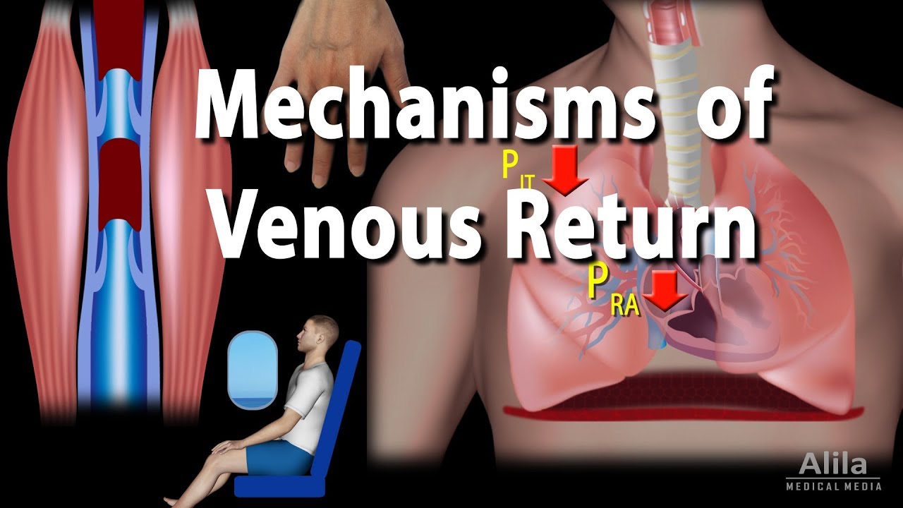
The Human Heart | #aumsum #kids #science #education #children
BIOLOGI Kelas 11 - Sistem Peredaran Darah (PART 2) | GIA Academy
Anatomy of the Heart - External & Internal Structures
(1/2) Anatomi Struktur Jantung : #1 BASIC CARDIOVASKULAR
Circulatory System I Animated I Grade 9 - Q1 l PART 2
5.0 / 5 (0 votes)