Case Discussion on Rheumatoid Nodule and Causes
Summary
TLDRThe video script focuses on the characteristics, diagnosis, and prognosis of rheumatoid arthritis. It covers key aspects such as the identification of rheumatoid nodules, laboratory investigations, and the significance of various tests like RF and anti-CCP antibodies. The speaker explains the interpretation of X-rays and the relevance of acute phase reactants. Additionally, the script discusses the importance of affordability and practicality in testing and imaging, particularly for patients in rural settings.
Takeaways
- π§ββοΈ Rheumatoid nodules are non-painful, subcutaneous, and commonly found on the extensor surfaces of the hands.
- π©Ί Rheumatoid nodules are a characteristic feature of rheumatoid arthritis and a marker of disease activity.
- π§ͺ For lab investigations in rheumatoid arthritis, important tests include CBC, ESR, CRP, and rheumatoid factor (RF).
- π¬ Rheumatoid factor (RF) recognizes the FC portion of the IgG antibody and is positive in 85% of RA patients within the first two years.
- π High titers of RF are associated with poorer prognosis and more extra-articular manifestations.
- π Other causes of positive RF include vasculitis, sarcoidosis, cryoglobulinemia, and chronic liver diseases.
- 𧫠Anti-CCP antibodies are highly specific (95%) and sensitive (65%) for diagnosing rheumatoid arthritis and have prognostic value.
- π¦ Inflammatory synovial fluid is identified through needle aspiration and is characterized by markers like elevated APRs and leukocytosis.
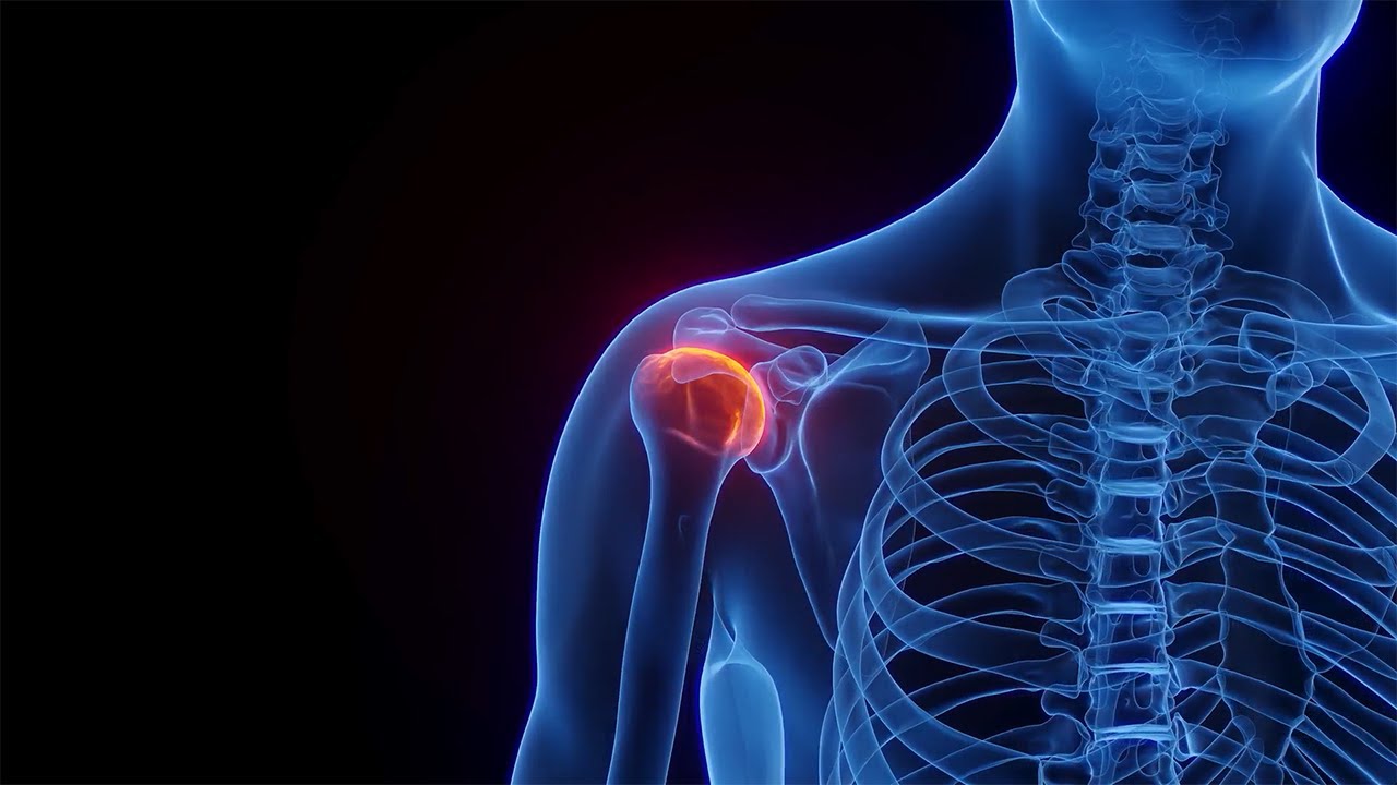
- π©» Imaging such as X-rays, CT scans, and MRIs are useful for diagnosing and assessing the extent of joint damage in rheumatoid arthritis.
- 𦴠Key radiographic findings in RA include osteopenia, symmetrical joint space narrowing, and marginal subchondral erosions.
Q & A
What are rheumatoid nodules and where are they typically found?
-Rheumatoid nodules are subcutaneous nodules that are characteristic features of rheumatoid arthritis, mainly found on the extensor surfaces of the hand. They are non-painful and can be present even if the features of rheumatoid arthritis are not evident.
What does the term 'neutropenia' refer to in the context of lab investigations?
-Neutropenia refers to a decrease in the number of neutrophils in the blood, which is a condition that can be assessed through a CBC (Complete Blood Count) and other lab tests like ESR and CRP.
What is the significance of the term 'GBP' in the context of lab investigations?
-In the script, 'GBP' seems to be a miscommunication. The correct term might be 'Globulin,' which is a part of blood proteins and can be checked as part of a blood test to evaluate various conditions including rheumatoid arthritis.
What is the role of Rheumatoid Factor (RF) in diagnosing rheumatoid arthritis?
-Rheumatoid Factor is an autoantibody that recognizes the Fc portion of IgG. It is present in about 85% of rheumatoid arthritis patients within the first two years of the disease and has prognostic value, with high titers often indicating a poor prognosis and more extra-articular manifestations.
What does the term 'acute phase reactants' refer to in lab tests?
-Acute phase reactants are proteins whose concentrations change significantly in response to inflammation. They include CRP (C-reactive protein), ESR (erythrocyte sedimentation rate), and fibrinogen, among others.
What is the significance
-null
Outlines
π©Ί Understanding Rheumatoid Nodules and Lab Investigations
This section discusses rheumatoid nodules, highlighting their appearance, prevalence, and characteristics. It emphasizes that these nodules are non-painful, subcutaneous, and commonly found on the extensor surfaces of the hand. The discussion then shifts to lab investigations necessary for diagnosing rheumatoid arthritis, such as complete blood count (CBC), erythrocyte sedimentation rate (ESR), and rheumatoid factor (RF). The importance of distinguishing between various antibodies, such as IgG and IgM, and understanding their roles in disease activity and progression is also covered.
𧬠Prognostic Value and Other Causes of Rheumatoid Factor
This paragraph explains the prognostic value of rheumatoid factor (RF) and its role in rheumatoid arthritis (RA). It notes that about 85% of RA patients become RF positive within the first two years, and high RF levels are associated with poor prognosis and extra-articular manifestations. Other causes of RF positivity, such as vasculitis, sarcoidosis, and chronic liver diseases, are mentioned. The importance of NTCCP antibodies in diagnosing and predicting the severity of RA is also highlighted, with emphasis on their diagnostic and prognostic value.
π¬ Synovial Fluid Analysis and Imaging for RA Diagnosis
This section covers the analysis of inflammatory synovial fluid, including how to aspirate it and its significance in diagnosing RA. It discusses the role of imaging, such as CT scans and MRIs, in detecting joint and bone abnormalities. The paragraph stresses the importance of considering patients' affordability before recommending expensive tests and highlights the basic investigations that should be performed initially.
𦴠Bone and Joint Space Abnormalities in RA
This paragraph focuses on identifying bone and joint space abnormalities in RA through X-rays. It describes osteopenia, marginal subchondral erosions, and symmetrical joint space narrowing. The importance of these features in diagnosing RA and understanding disease progression is emphasized. The section also mentions subluxation and its implications for joint health, particularly in the atlanto-axial joint, where ligament loosening can cause spinal cord compression.
Mindmap
Keywords
π‘Rheumatoid
π‘Subcutaneous nodules
π‘Neutropenia
π‘Acute phase reactants
π‘Rheumatoid factor (RF)
Highlights
Rheumatoid nodules are subcutaneous, non-painful, and mainly found on the extensor surfaces of the hand, seen in 20% of rheumatoid arthritis cases.
Lab investigations for rheumatoid arthritis include CBC, ESR, CRP, RF, and
Transcripts
foreign
[Music]
next is
as I told
what is uh
you said rheumatoid you can see that
what is rheumatoid see the picture
player carefully just
what do you see in a picture
what a remote you see it
these are the repression comes to you
with such characteristics
what kind of module it is what kind of
swelling it is tell me
so have you seen any patient with such
thing anyone have you seen a patient
with the rheumatoid nodule ever
foreign
these are uh form in nature okay seen in
like uh 20 percent of the cases
and these are non-painful these are not
painful okay there are uh these are
subcutaneous nodules and mainly found on
the extensor Services of the hand okay
it is a characteristic feature of the
rheumatoid arthritis okay and a marker
of a disease activity and can be present
even if the features of rheumatoid
arthritis are upset
so but so coming up to the lab
investigations okay as I told
neutropenia so for that you need to do a
cbct lcdlc check for HB ESR and GBP
what is GBP
what do you understand by GBP
anyone
foreign
was broken I guess
yes the presentation is not visible yeah
yeah it has gone actually that's why
sorry yeah divisible right yes it is
visible house okay
no no no no no government in peptide
yeah for the medicine party right
put a common sense in this I am asking
about lab investigation now why would
you write a gamma paint in peptide
just don't Google please
and uh what are the next things uh acute
phase reactants
pneumatized factors
okay RF and ntccb antibodies I'll tell
you in a quick uh like what is a
rheumatoid factor is that it recognizes
the FC portion of the IGG
does anyone know what is FC portion
what is the FC portion of the antibody
you must have read the antibody diagrams
AFC portion is the renewable antibody
diagram it is responsible that is for
the biological activity so just remember
it recognizes a fc portion of the IGG
immunoglobulin can be IGM IGG IGA and uh
85 percent see gross patients like 85
percent with ra over the first two years
become RF positive okay
the negative may be repeated four to six
months for the first two years of the
disease and may take 18 to 24 months to
become zero positive okay so keep in
mind that
so what is its prognostic valuation with
high titles of our average General tend
to have poor progresses more extra
articular manifestation
okay so 85 percent of people
you see there are there becomes RF
positive for the first two years and the
prognostic value is and with the high
titles of RF there is a poor prognosis
and most many like extraordicular
manifestations are presented with the
patient
so next is uh what do you see is
causes of the positive like what are the
other causes also
these rheumatoid arthritis as children
vasculitis sarcoidosis
cryoglobulemia chronic liver diseases
so you should know all these but other
things can cause pneumatite Factor
positive for the patient so for all this
is all for the exam point of view
so ntccp okay
so IGG against synovial membrane peptide
damage via inflam inflammation
and sensitivity is 65 and specificity is
95 so it is highly uh recommended and it
is both Diagnostic and have a prognostic
value so as I told patient you are
writing for an RI Factor so you should
always suggest energy ccb2 right away
and you should always
correlate it accordingly
so predictive of erosive disease
disease severity Radiology progression
and poor function what is uh what you
understand of erosive disease
in medical terms what does the meaning
of erosive
anyone
so erosive yes someone is right
destruction this is good very nice
so as you understand erosion means there
is a destruction
okay there is a bone erosion and there
is a uh it leads to what we call
functional decreased functional capacity
and the joint damage for the patients
next acute phase reactants you see
there is a mild elevation to moderate
and to marked elevation positive acute
phase Okay negative with albumin
transparent positive is mild elevation
you see celluloplasmin complement C3 C4
mild it comes to have to globulin
fibrinogen Alpha One and Alpha One
protease and acid lipoproteins marked
elevation you see is CRP serum alert a
protein
so uh see you can don't get all the
tests at all the centers some people
might be working in a government
periphery suppose I am work I am also
working it so what are the common tests
you can write
which which is available easily to
everyone
name a few tests
that you will write you suspect a
patient is suffering from osteoarthritis
oh sorry limited arthritis
crb ESR good
are effective good
three next
CRP ESR artifactor
IGG
uh is it available at your place IGG if
you're working at a hospital
artificial yes
CBC ESR CRP r a vector and
one one is missing
most important
as I told ntccp good these are the
common tests you should always write
anywhere you are okay so for a good
corporate setups you see everything
every test is available this test in
government setups are available for free
and you can suggest uh to patients for
the same
next
the other abnormalities elevated aprs
that
esrcrombocytosis leukocytosis Ana 30 to
40 percent
antinuclear antibodies inflammatory
synovial fluid and hypoalbum anemia so
how would you know it is an inflammatory
synovial fluid
how would you know that
you have written level normal it is how
how did I get to know what is
inflammatory cyanobic fluid what will I
do for that
the CRP okay
no no so synovial fluid
aspirations
you need to say word aspiration synovial
fluid with needle aspirations good yes
as period the synovial fluid so uh I'll
uh there's a question for you do you
think CT scan for a knee joint will be
helpful for this to know that a person
is suffering from any synovial fluid as
inflammation
the other cities can help for it or an
MRI
a CT an M or an MRI for the patient
what you will write for a patient
but you'll write a CT
uh
how many of you are working in a rural
setup
anyone working in a rural setup
well I'll suggest you people is before
writing a CT an MRI always always ask
for a affordability for the patient
first
you never know how what kind of a
patient is there who's coming to you ask
for the affordability not every doctor
can a spirit synovial fluid so basically
always go with the basic investigations
first and if you suspect that this
disease has worsened then you can
suggest for an author review then you
can go for a city or an MRI okay
so next
okay tell me you see ABC bonds what do
you see in a bone when you see in a b
and what do you see in a c
I'll I'll check what is the presence of
mind for you people you will get I'll
get to know now but you see in an a bone
b and a c
among the one
I'm gonna replied erosion in the sea
very nice
and what for a and b
so uh
and Monika tell me what kind of erosion
it is is it a marginal or it is complete
and what kind of
erosions
what kind of erosion do you see it is a
marginal or a complete erosion
yes good
see it is marginal you see that it is a
marginal and it is a subchondral erosion
always remember and for the a bone what
is in the a bone
what do you see in the a bone
anyone
osteopenia
very nice
how do you define it as an osteopenia
but how do you see it is an osteopenia
area okay see osteopenia what do you
understand there is a bone loss okay you
see there is a lot of bone loss you can
see good for the two you have replied
tell me for the B
what is what has happened in the B
for the B
and declamation
okay
see I'll tell you
see you can see there is a joint space
okay
you can see joint space here
what kind of joint space do you see here
is it symmetrical or asymmetrical first
of all
other bones symmetrical or asymmetrical
there is symmetry in them but there is
no symmetry in them
see the diagram carefully
there is no symmetry
anyone else
see the bone is symmetrical only see the
joint space you see that
but
what are the bone spaces narrowed or are
the bone spaces wide and tell me
what is a uh like joint species are
narrowed yes chord word so there is a
narrowing of bone spaces but it is in a
symmetrical way now you understand me
everyone
you understood all the ABC diagrams very
well so now you can easily check for the
X-ray and you can diagnose it well so
for the AUC there is an osteopenia
see what you see is in the bone B part
say there is a narrow what we say is
narrow
joint space symmetry but it is
symmetrical okay
and it is uniform
okay it is not on the one side or the
other side there is a symmetry of the
joint okay and it is uniform in nature
and also there is a
narrowing of the joint space okay
for the a it is ostominium for the B it
is uniform symmetric joint space
narrowing and for the C it is marginal
subchondral erosions
next okay what do you see in such
pictures
there is a
I'll be quick on this
see this you see that as I told
there is a subluxation you see that
understood everyone there is a
subluxation of the
joints
okay
yes
here you see that
this is Atlanta x equals axial joint
there is a loosening of the ligaments
and thickening which is causing the cord
compression you see
when they are compressing the card
see how beautiful it is written as you
can see that
foreign foreign
Browse More Related Video
5.0 / 5 (0 votes)