Heart Valves (Atrioventricular and Semilunar) Heart Valves | Physiology | Lecturio Nursing
Summary
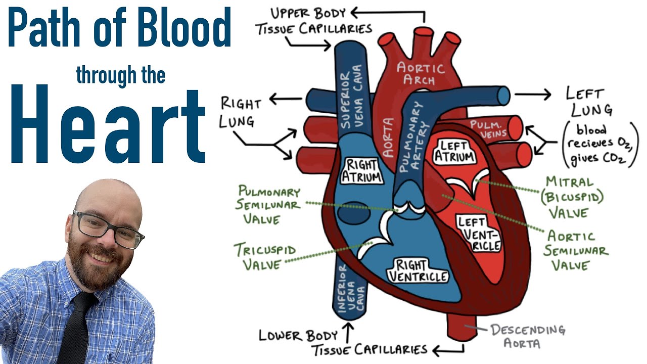
TLDRThe script delves into the anatomy and function of heart valves, crucial for unidirectional blood flow. It highlights two types: atrioventricular valves, including the tricuspid and mitral, which prevent backflow into atria, and semilunar valves, like the pulmonary and aortic, that prevent backflow from arteries. The tricuspid has three cusps, while the mitral has two. The script also explains the role of chordae tendineae in valve function, emphasizing their importance in maintaining heart health.
Takeaways
- 💓 The heart has valves to ensure blood flows in one direction through the heart, responding to pressure changes.
- 📍 There are two types of heart valves: atrioventricular valves and semilunar valves, each with a specific location and function.
- 🔍 The atrioventricular valves are between the atria and ventricles, preventing backflow when the ventricles contract.
- 🚫 The tricuspid valve is on the right side and has three cusps, while the mitral (bicuspid) valve on the left side has two cusps.
- 🌟 The semilunar valves are located between the ventricles and the major arteries, preventing backflow from arteries to the heart.
- 🔄 The pulmonary valve is on the right side, separating the right ventricle from the pulmonary trunk, and the aortic valve is on the left side, separating the left ventricle from the aorta.
- 🤝 The chordae tendineae anchor the valve cusps to papillary muscles, preventing the valve flaps from everting into the atria during pumping.
- 📈 The atrioventricular valves open due to increased pressure as blood fills the atria, allowing the ventricles to fill with blood.
- 🔄 The semilunar valves open when the ventricles contract and close when the ventricles relax, preventing blood from flowing back into the heart.
- 🌙 The semilunar valves have a half-moon shape, with three cusps that open and close in response to pressure changes in the heart.
- 🔍 The function of the heart valves is crucial for maintaining efficient blood circulation and preventing regurgitation of blood.
Q & A
What is the primary function of heart valves?
-The primary function of heart valves is to ensure unidirectional blood flow through the heart by opening and closing in response to pressure changes.
What are the two major types of valves found in the heart?
-The two major types of valves found in the heart are the atrioventricular valves and the semilunar valves.
Where are the atrioventricular valves located in the heart?
-The atrioventricular valves are located between the atria, which are the receiving chambers, and the ventricles, which are the pumping chambers.
Which valves are considered semilunar and where are they located?
-The semilunar valves are located between the ventricles and the major arteries, including the aorta and the pulmonary trunk.
What are the names of the atrioventricular valves and their respective locations?
-The atrioventricular valves are the tricuspid valve, located between the right atrium and right ventricle, and the mitral valve, located between the left atrium and left ventricle.
What is the role of the tricuspid valve?
-The tricuspid valve is an atrioventricular valve that prevents backflow into the right atrium when the right ventricle contracts.
What is the mitral valve also known as and what does it prevent?
-The mitral valve is also known as the bicuspid valve, and it prevents backflow into the left atrium when the left ventricle contracts.
What are the two semilunar valves and their respective locations?
-The two semilunar valves are the pulmonary valve, located between the right ventricle and the pulmonary trunk, and the aortic valve, located between the left ventricle and the aorta.
What is the purpose of the chordae tendineae in relation to the heart valves?
-The chordae tendineae anchor the cusp of the atrioventricular valves to the papillary muscles, helping to hold the valve flaps in a closed position and prevent them from everting back into the atria during increased pressure.
How do the semilunar valves function to prevent backflow from major arteries?
-The semilunar valves open when the ventricles contract and the intraventricular pressure rises, allowing blood to flow into the arteries. When the ventricles relax and the pressure decreases, the valves close, preventing blood from flowing back into the heart from the arteries.
What is the structural difference between the tricuspid and mitral valves?
-The tricuspid valve has three cusps and is located between the right atrium and ventricle, while the mitral valve has two cusps and is located between the left atrium and ventricle.
What are the internal ridges inside of the atria called and what is their relation to the heart valves?
-The internal ridges inside of the atria are called the trabeculae carneae. They are part of the structural support for the heart but are not directly related to the functioning of the valves.
Outlines
💓 Heart Valves and Their Functions
This paragraph provides an in-depth explanation of the heart's valves and their crucial role in ensuring unidirectional blood flow. It distinguishes between two main types of valves: the atrioventricular valves, including the tricuspid and mitral valves, which are located between the atria and ventricles, and the semilunar valves, namely the pulmonary and aortic valves, situated between the ventricles and the major arteries. The paragraph also describes the structure and function of the chordae tendineae and papillary muscles that support the atrioventricular valves, preventing backflow during ventricular contraction. The summary of the AV valves' operation during the cardiac cycle is included, illustrating how they open in response to pressure changes and close to prevent backflow.
🌀 Operation of Semilunar Valves
The second paragraph delves into the function of the semilunar valves, emphasizing their role in preventing blood from flowing back into the heart from the major arteries. It explains how these valves, with their characteristic half-moon shape, open when the ventricles contract and the intraventricular pressure increases, allowing blood to flow into the arteries. Conversely, when the ventricles relax and the pressure decreases, blood from the arteries pushes against the valve cusps, causing them to close and effectively stopping backflow. This mechanism is essential for maintaining the forward flow of blood in the circulatory system.
Mindmap
Keywords
💡Heart Valves
💡Atrioventricular Valves
💡Semilunar Valves
💡Tricuspid Valve
💡Mitral Valve
💡Pulmonary Valve
💡Aortic Valve
💡Chordae Tendineae
💡Papillary Muscles
💡Trabeculae Carnae
💡Ventricular Contraction
Highlights
Heart valves ensure unidirectional blood flow through the heart by opening and closing in response to pressure changes.
There are two major types of heart valves: atrioventricular valves and semilunar valves.
Atrioventricular valves are located between the atria and ventricles, preventing backflow when ventricles contract.
Semilunar valves are located between the ventricles and major arteries, such as the aorta and pulmonary trunk, to prevent backflow from arteries.
The tricuspid valve is an atrioventricular valve between the right atrium and right ventricle, with three cusps.
The mitral valve, also known as the bicuspid valve, is an atrioventricular valve between the left atrium and left ventricle, with two cusps.
The pulmonary valve is a semilunar valve on the right side of the heart, separating the right ventricle from the pulmonary trunk.
The aortic valve is a semilunar valve separating the left ventricle from the aorta.
Chordae tendineae anchor the cusps of atrioventricular valves to papillary muscles, preventing eversion during heart pumping.
Papillary muscles hold valve flaps closed and prevent backflow into atria during increased pressure.
Trabeculae carneae are internal ridges in the atria that support the structure and function of the heart.
AV valves open due to increased pressure as blood fills the atria and pushes against the valve.
Once the AV valve opens, the ventricle fills with blood as the valve flap hangs limply into the ventricle.
Atrial contraction forces additional blood from the atria into the ventricle through the open AV valve.
Semilunar valves prevent backflow from major arteries by opening and closing in response to pressure changes in the heart.
The pulmonary semilunar valve is located between the right ventricle and the pulmonary trunk.
The aortic semilunar valve is located between the left ventricle and the aorta, ensuring unidirectional blood flow.
Semilunar valves consist of three cusps resembling a half-moon, which allows them to open and close effectively.
As ventricles contract, blood pushes against semilunar valves, forcing them open for blood flow into arteries.
When ventricles relax, intraventricular pressure decreases, causing blood to flow back and close the semilunar valves.
Transcripts
So speaking of heart's valves, a heart valve ensures unidirectional blood flow through the heart.
These are going to open and close in response to pressure changes.
There are two major valves found in the heart.
We have the atrioventricular valves which are gonna be located between the atria which are our receiving chambers
and the ventricles which are our pumping chambers.
The other type of valve that we have in the heart is the semilunar valves.
These are going to be located between the ventricles which are going to be pumping blood out
and the major arteries including the aorta and the pulmonary trunk.
So let's take a closer look at these valves.
First we have the tricuspid valve.
The tricuspid valve is an atrioventricular valve that is going to be between the right atrium and the right ventricle
Next, we have our bicuspid valve the bicuspid valve.
The bicuspid valve is also an atrioventricular valve found on the left side of the heart
between the left atrium and the left ventricle.
The next valve is going to be the pulmonary valve.
The pulmonary valve is a semilunar valve on the right side of the heart
that separates the right ventricle from the pulmonary trunk.
We also have the aortic valve.
The aortic valve is also a semilunar valve and this is going to separate the left ventricle from the aorta.
So our two atrioventricular valves are going to prevent backflow into our atria when our ventricles contract.
The tricuspid valve on the right side of the heart is made up of three cusps that lie between the right atria and ventricles.
The mitral valve or the left atrioventricular valve is going to be made up of two cusps
and lies between the left atria and ventricles.
On these valves, we have these structures known as chordae tendineae
and these are going to anchor the cusp of these AV valves to papillary muscles
These muscles hold the valve flaps in a closed position and prevent the flaps from everting
back into the atria especially when we have this increased pressure in our heart during pumping.
Here, we have an image of the chordae tendineae which are the chords that are connected to the valve,
the papillary muscles that are controlling these chords
and as well the internal ridges inside of the atria known as the trabeculae carneae.
So how did these AV valves function?
Blood returning to the heart is going to fill the atria.
As the blood is filling the atria, it is pressing up against this AV valve.
This increases the pressure and eventually will force that AV valve open.
Once the AV valve is open, the ventricle is now going to start to be filled.
As the ventricle fills, the AV valve flap is going to hang limply into that ventricle.
Once the atria contracts, this is going to force any additional blood that is still in the atria into the ventricle.
The semilunar valves are going to have a little bit more activity.
These are gonna be responsible for preventing backflow from our major arteries back into the heart
On the pulmonary side, the pulmonary semilunar valve is going to be located
between our right ventricle and the pulmonary trunk.
And on the other side, on our aorta side, the aortic semilunar valve is gonna be located
between the left ventricle and the aorta.
Both of these valves are going to open and close in response to pressure changes in the heart.
The semilunar valves consist of three cusp that roughly resemble a half-moon
which is where they get their name.
So how did the semilunar valves function?
So as the ventricles contract and the intraventricular pressure rises,
blood is going to be pushed up against the semilunar valves.
This forces those valves open.
Subsequently, as the ventricles relax and the intraventricular pressure goes back down,
blood is going to flow back from the arteries such as the pulmonary trunk or the aorta
and instead of flowing back into the heart, they're going to fill the cusp of the semilunar valves,
forcing them to close.
5.0 / 5 (0 votes)